1 c3osqA_
100.0
15
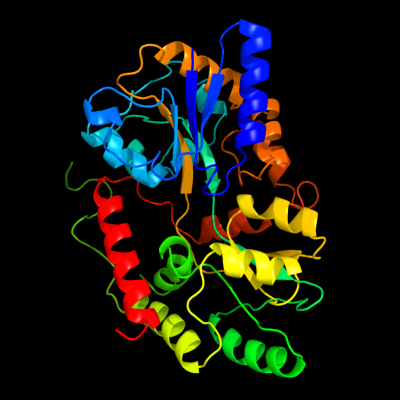
PDB header: fluorescent protein, transport proteinChain: A: PDB Molecule: maltose-binding periplasmic protein, green fluorescentPDBTitle: maltose-bound maltose sensor engineered by insertion of circularly2 permuted green fluorescent protein into e. coli maltose binding3 protein at position 175
2 c3ob4A_
100.0
15
PDB header: allergenChain: A: PDB Molecule: maltose abc transporter periplasmic protein, arah 2;PDBTitle: mbp-fusion protein of the major peanut allergen ara h 2
3 c3f5fA_
100.0
15
PDB header: transport, transferaseChain: A: PDB Molecule: maltose-binding periplasmic protein, heparanPDBTitle: crystal structure of heparan sulfate 2-o-sulfotransferase2 from gallus gallus as a maltose binding protein fusion.
4 c3py7A_
100.0
15
PDB header: viral proteinChain: A: PDB Molecule: maltose-binding periplasmic protein,paxillin ld1,protein e6PDBTitle: crystal structure of full-length bovine papillomavirus oncoprotein e62 in complex with ld1 motif of paxillin at 2.3a resolution
5 c3h4zC_
100.0
15
PDB header: allergenChain: C: PDB Molecule: maltose-binding periplasmic protein fused with allergenPDBTitle: crystal structure of an mbp-der p 7 fusion protein
6 c3dm0A_
100.0
15
PDB header: sugar binding protein,signaling proteinChain: A: PDB Molecule: maltose-binding periplasmic protein fused withPDBTitle: maltose binding protein fusion with rack1 from a. thaliana
7 c3o3uN_
100.0
15
PDB header: transport protein, signaling proteinChain: N: PDB Molecule: maltose-binding periplasmic protein, advanced glycosylationPDBTitle: crystal structure of human receptor for advanced glycation endproducts2 (rage)
8 c1y4cA_
100.0
14
PDB header: de novo proteinChain: A: PDB Molecule: maltose binding protein fused with designedPDBTitle: designed helical protein fusion mbp
9 c3d4cA_
100.0
15
PDB header: cell adhesionChain: A: PDB Molecule: maltose-binding periplasmic protein, linker, zona pellucidaPDBTitle: zp-n domain of mammalian sperm receptor zp3 (crystal form i)
10 c3mp6A_
100.0
15
PDB header: histone binding proteinChain: A: PDB Molecule: maltose-binding periplasmic protein, linker, saga-PDBTitle: complex structure of sgf29 and dimethylated h3k4
11 c1r6zA_
100.0
14
PDB header: gene regulationChain: A: PDB Molecule: chimera of maltose-binding periplasmic protein andPDBTitle: the crystal structure of the argonaute2 paz domain (as a mbp fusion)
12 c3osrA_
100.0
16
PDB header: fluorescent protein, transport proteinChain: A: PDB Molecule: maltose-binding periplasmic protein, green fluorescentPDBTitle: maltose-bound maltose sensor engineered by insertion of circularly2 permuted green fluorescent protein into e. coli maltose binding3 protein at position 311
13 c1hsjA_
100.0
15
PDB header: transcription/sugar binding proteinChain: A: PDB Molecule: fusion protein consisting of staphylococcusPDBTitle: sarr mbp fusion structure
14 c3c4mA_
100.0
14
PDB header: membrane proteinChain: A: PDB Molecule: fusion protein of maltose-binding periplasmic protein andPDBTitle: structure of human parathyroid hormone in complex with the2 extracellular domain of its g-protein-coupled receptor (pth1r)
15 d1a99a_
100.0
22
Fold: Periplasmic binding protein-like IISuperfamily: Periplasmic binding protein-like IIFamily: Phosphate binding protein-like16 c2vgqA_
100.0
14
PDB header: immune system/transportChain: A: PDB Molecule: maltose-binding periplasmic protein,PDBTitle: crystal structure of human ips-1 card
17 c2nvuB_
100.0
16
PDB header: protein turnover, ligaseChain: B: PDB Molecule: maltose binding protein/nedd8-activating enzymePDBTitle: structure of appbp1-uba3~nedd8-nedd8-mgatp-ubc12(c111a), a2 trapped ubiquitin-like protein activation complex
18 c3oaiB_
100.0
14
PDB header: membrane protein, cell adhesionChain: B: PDB Molecule: maltose-binding periplasmic protein, myelin protein p0;PDBTitle: crystal structure of the extra-cellular domain of human myelin protein2 zero
19 d1elja_
100.0
13
Fold: Periplasmic binding protein-like IISuperfamily: Periplasmic binding protein-like IIFamily: Phosphate binding protein-like20 c3csgA_
100.0
14
PDB header: de novo protein, sugar binding proteinChain: A: PDB Molecule: maltose-binding protein monobody ys1 fusion;PDBTitle: crystal structure of monobody ys1(mbp-74)/maltose binding2 protein fusion complex
21 d1pota_
not modelled
100.0
22
Fold: Periplasmic binding protein-like IISuperfamily: Periplasmic binding protein-like IIFamily: Phosphate binding protein-like22 c2zykA_
not modelled
100.0
17
PDB header: sugar binding proteinChain: A: PDB Molecule: solute-binding protein;PDBTitle: crystal structure of cyclo/maltodextrin-binding protein2 complexed with gamma-cyclodextrin
23 c2v84A_
not modelled
100.0
18
PDB header: transport proteinChain: A: PDB Molecule: spermidine/putrescine abc transporter, periplasmicPDBTitle: crystal structure of the tp0655 (tppotd) lipoprotein of2 treponema pallidum
24 c3ehuA_
not modelled
100.0
15
PDB header: membrane proteinChain: A: PDB Molecule: fusion protein of crfr1 extracellular domain and mbp;PDBTitle: crystal structure of the extracellular domain of human corticotropin2 releasing factor receptor type 1 (crfr1) in complex with crf
25 c3k02A_
not modelled
100.0
12
PDB header: transport proteinChain: A: PDB Molecule: acarbose/maltose binding protein gach;PDBTitle: crystal structures of the gach receptor of streptomyces glaucescens2 gla.o in the unliganded form and in complex with acarbose and an3 acarbose homolog. comparison with acarbose-loaded maltose binding4 protein of salmonella typhimurium.
26 c1mg1A_
not modelled
100.0
14
PDB header: viral proteinChain: A: PDB Molecule: protein (htlv-1 gp21 ectodomain/maltose-binding proteinPDBTitle: htlv-1 gp21 ectodomain/maltose-binding protein chimera
27 c2gh9A_
not modelled
100.0
15
PDB header: sugar binding proteinChain: A: PDB Molecule: maltose/maltodextrin-binding protein;PDBTitle: thermus thermophilus maltotriose binding protein bound with2 maltotriose
28 c2xd3A_
not modelled
100.0
13
PDB header: sugar binding proteinChain: A: PDB Molecule: maltose/maltodextrin-binding protein;PDBTitle: the crystal structure of malx from streptococcus pneumoniae2 in complex with maltopentaose.
29 c3oo6A_
not modelled
100.0
13
PDB header: sugar binding proteinChain: A: PDB Molecule: abc transporter binding protein acbh;PDBTitle: crystal structures and biochemical characterization of the bacterial2 solute receptor acbh reveal an unprecedented exclusive substrate3 preference for b-d-galactopyranose
30 c3uorB_
not modelled
100.0
15
PDB header: sugar binding proteinChain: B: PDB Molecule: abc transporter sugar binding protein;PDBTitle: the structure of the sugar-binding protein male from the phytopathogen2 xanthomonas citri
31 d1eu8a_
not modelled
100.0
15
Fold: Periplasmic binding protein-like IISuperfamily: Periplasmic binding protein-like IIFamily: Phosphate binding protein-like32 c3pu5A_
not modelled
100.0
12
PDB header: transport proteinChain: A: PDB Molecule: extracellular solute-binding protein;PDBTitle: the crystal structure of a putative extracellular solute-binding2 protein from bordetella parapertussis
33 c2fncA_
not modelled
100.0
15
PDB header: sugar binding proteinChain: A: PDB Molecule: maltose abc transporter, periplasmic maltose-bindingPDBTitle: thermotoga maritima maltotriose binding protein bound with2 maltotriose.
34 c3iouB_
not modelled
100.0
14
PDB header: signaling proteinChain: B: PDB Molecule: maltose-binding protein, huntingtin fusionPDBTitle: huntingtin amino-terminal region with 17 gln residues -2 crystal c94
35 c1svxB_
not modelled
100.0
14
PDB header: de novo protein/sugar binding proteinChain: B: PDB Molecule: maltose-binding periplasmic protein;PDBTitle: crystal structure of a designed selected ankyrin repeat2 protein in complex with the maltose binding protein
36 d1laxa_
not modelled
100.0
13
Fold: Periplasmic binding protein-like IISuperfamily: Periplasmic binding protein-like IIFamily: Phosphate binding protein-like37 c3rpwA_
not modelled
100.0
17
PDB header: transport proteinChain: A: PDB Molecule: abc transporter;PDBTitle: the crystal structure of an abc transporter from rhodopseudomonas2 palustris cga009
38 c1mh3A_
not modelled
100.0
14
PDB header: sugar binding, dna binding proteinChain: A: PDB Molecule: maltose binding-a1 homeodomain protein chimera;PDBTitle: maltose binding-a1 homeodomain protein chimera, crystal2 form i
39 c2z8fB_
not modelled
100.0
13
PDB header: sugar binding proteinChain: B: PDB Molecule: galacto-n-biose/lacto-n-biose i transporter substrate-PDBTitle: the galacto-n-biose-/lacto-n-biose i-binding protein (gl-bp) of the2 abc transporter from bifidobacterium longum in complex with lacto-n-3 tetraose
40 c3qufB_
not modelled
100.0
13
PDB header: transport proteinChain: B: PDB Molecule: extracellular solute-binding protein, family 1;PDBTitle: the structure of a family 1 extracellular solute-binding protein from2 bifidobacterium longum subsp. infantis
41 c2b3fD_
not modelled
100.0
11
PDB header: sugar binding proteinChain: D: PDB Molecule: glucose-binding protein;PDBTitle: thermus thermophilus glucose/galactose binding protein2 bound with galactose
42 c2uvgA_
not modelled
100.0
13
PDB header: sugar-binding proteinChain: A: PDB Molecule: abc type periplasmic sugar-binding protein;PDBTitle: structure of a periplasmic oligogalacturonide binding2 protein from yersinia enterocolitica
43 c1ursA_
not modelled
100.0
13
PDB header: maltose-binding proteinChain: A: PDB Molecule: maltose-binding protein;PDBTitle: x-ray structures of the maltose-maltodextrin binding2 protein of the thermoacidophilic bacterium alicyclobacillus3 acidocaldarius
44 d1ursa_
not modelled
100.0
13
Fold: Periplasmic binding protein-like IISuperfamily: Periplasmic binding protein-like IIFamily: Phosphate binding protein-like45 c2w7yA_
not modelled
100.0
13
PDB header: sugar-binding proteinChain: A: PDB Molecule: probable sugar abc transporter, sugar-bindingPDBTitle: structure of a streptococcus pneumoniae solute-binding2 protein in complex with the blood group a-trisaccharide.
46 c2i58B_
not modelled
100.0
15
PDB header: sugar binding proteinChain: B: PDB Molecule: sugar abc transporter, sugar-binding protein;PDBTitle: crystal structure of rafe from streptococcus pneumoniae complexed with2 raffinose
47 c3c9hB_
not modelled
100.0
13
PDB header: transport proteinChain: B: PDB Molecule: abc transporter, substrate binding protein;PDBTitle: crystal structure of the substrate binding protein of the abc2 transporter from agrobacterium tumefaciens
48 c2qryD_
not modelled
100.0
14
PDB header: transport proteinChain: D: PDB Molecule: thiamine-binding periplasmic protein;PDBTitle: periplasmic thiamin binding protein
49 d3thia_
not modelled
100.0
13
Fold: Periplasmic binding protein-like IISuperfamily: Periplasmic binding protein-like IIFamily: Phosphate binding protein-like50 c3i3vC_
not modelled
100.0
10
PDB header: transport proteinChain: C: PDB Molecule: probable secreted solute-binding lipoprotein;PDBTitle: crystal structure of probable secreted solute-binding2 lipoprotein from streptomyces coelicolor
51 d1y4ta_
not modelled
100.0
14
Fold: Periplasmic binding protein-like IISuperfamily: Periplasmic binding protein-like IIFamily: Phosphate binding protein-like52 d1q35a_
not modelled
100.0
13
Fold: Periplasmic binding protein-like IISuperfamily: Periplasmic binding protein-like IIFamily: Phosphate binding protein-like53 d1y9ua_
not modelled
100.0
14
Fold: Periplasmic binding protein-like IISuperfamily: Periplasmic binding protein-like IIFamily: Phosphate binding protein-like54 d1j1na_
not modelled
100.0
11
Fold: Periplasmic binding protein-like IISuperfamily: Periplasmic binding protein-like IIFamily: Phosphate binding protein-like55 c2pt1A_
not modelled
100.0
16
PDB header: metal transportChain: A: PDB Molecule: iron transport protein;PDBTitle: futa1 synechocystis pcc 6803
56 d1y3na1
not modelled
100.0
14
Fold: Periplasmic binding protein-like IISuperfamily: Periplasmic binding protein-like IIFamily: Phosphate binding protein-like57 d1xc1a_
not modelled
100.0
15
Fold: Periplasmic binding protein-like IISuperfamily: Periplasmic binding protein-like IIFamily: Phosphate binding protein-like58 d1xvxa_
not modelled
100.0
17
Fold: Periplasmic binding protein-like IISuperfamily: Periplasmic binding protein-like IIFamily: Phosphate binding protein-like59 d1xvya_
not modelled
100.0
16
Fold: Periplasmic binding protein-like IISuperfamily: Periplasmic binding protein-like IIFamily: Phosphate binding protein-like60 d1nnfa_
not modelled
100.0
16
Fold: Periplasmic binding protein-like IISuperfamily: Periplasmic binding protein-like IIFamily: Phosphate binding protein-like61 c2vozA_
not modelled
100.0
11
PDB header: metal-binding proteinChain: A: PDB Molecule: periplasmic iron-binding protein;PDBTitle: apo futa2 from synechocystis pcc6803
62 c3ombA_
not modelled
100.0
13
PDB header: transport proteinChain: A: PDB Molecule: extracellular solute-binding protein, family 1;PDBTitle: crystal structure of extracellular solute-binding protein from2 bifidobacterium longum subsp. infantis
63 d2onsa1
not modelled
100.0
9
Fold: Periplasmic binding protein-like IISuperfamily: Periplasmic binding protein-like IIFamily: Phosphate binding protein-like64 c3k6wA_
not modelled
100.0
10
PDB header: transport proteinChain: A: PDB Molecule: solute-binding protein ma_0280;PDBTitle: apo and ligand bound structures of moda from the archaeon2 methanosarcina acetivorans
65 c3cfzA_
not modelled
99.9
10
PDB header: transport proteinChain: A: PDB Molecule: upf0100 protein mj1186;PDBTitle: crystal structure of m. jannaschii periplasmic binding2 protein moda/wtpa with bound tungstate
66 c3cfxA_
not modelled
99.9
10
PDB header: transport proteinChain: A: PDB Molecule: upf0100 protein ma_0280;PDBTitle: crystal structure of m. acetivorans periplasmic binding protein2 moda/wtpa with bound tungstate
67 d1atga_
not modelled
99.9
15
Fold: Periplasmic binding protein-like IISuperfamily: Periplasmic binding protein-like IIFamily: Phosphate binding protein-like68 d1sbpa_
not modelled
99.9
13
Fold: Periplasmic binding protein-like IISuperfamily: Periplasmic binding protein-like IIFamily: Phosphate binding protein-like69 c3cg3A_
not modelled
99.9
9
PDB header: transport proteinChain: A: PDB Molecule: upf0100 protein ph0151;PDBTitle: crystal structure of p. horikoshii periplasmic binding2 protein moda/wtpa with bound tungstate
70 c3cg1A_
not modelled
99.9
7
PDB header: transport proteinChain: A: PDB Molecule: upf0100 protein pf0080;PDBTitle: crystal structure of p. furiosus periplasmic binding protein2 moda/wtpa with bound tungstate
71 d1amfa_
not modelled
99.9
16
Fold: Periplasmic binding protein-like IISuperfamily: Periplasmic binding protein-like IIFamily: Phosphate binding protein-like72 c2h5yC_
not modelled
99.9
16
PDB header: metal transportChain: C: PDB Molecule: molybdate-binding periplasmic protein;PDBTitle: crystallographic structure of the molybdate-binding protein of2 xanthomonas citri at 1.7 ang resolution bound to molybdate
73 c3fj7A_
not modelled
99.9
13
PDB header: protein bindingChain: A: PDB Molecule: major antigenic peptide peb3;PDBTitle: crystal structure of l-phospholactate bound peb3
74 c3lr1A_
not modelled
99.8
13
PDB header: transport proteinChain: A: PDB Molecule: tungstate abc transporter, periplasmic tungstate-PDBTitle: the crystal structure of the tungstate abc transporter from2 geobacter sulfurreducens
75 c3muqB_
not modelled
99.7
11
PDB header: structural genomics, unknown functionChain: B: PDB Molecule: uncharacterized conserved protein;PDBTitle: the crystal structure of a conserved functionally unknown protein from2 vibrio parahaemolyticus rimd 2210633
76 c3kn3C_
not modelled
99.6
12
PDB header: transcriptionChain: C: PDB Molecule: putative periplasmic protein;PDBTitle: crystal structure of lysr substrate binding domain (25-263) of2 putative periplasmic protein from wolinella succinogenes
77 c1twyG_
not modelled
99.4
10
PDB header: structural genomics, unknown functionChain: G: PDB Molecule: abc transporter, periplasmic substrate-binding protein;PDBTitle: crystal structure of an abc-type phosphate transport receptor from2 vibrio cholerae
78 d1twya_
not modelled
99.4
10
Fold: Periplasmic binding protein-like IISuperfamily: Periplasmic binding protein-like IIFamily: Phosphate binding protein-like79 d1pc3a_
not modelled
98.0
12
Fold: Periplasmic binding protein-like IISuperfamily: Periplasmic binding protein-like IIFamily: Phosphate binding protein-like80 d1ixha_
not modelled
97.5
12
Fold: Periplasmic binding protein-like IISuperfamily: Periplasmic binding protein-like IIFamily: Phosphate binding protein-like81 c3tqwA_
not modelled
97.4
12
PDB header: transport proteinChain: A: PDB Molecule: methionine-binding protein;PDBTitle: structure of a abc transporter, periplasmic substrate-binding protein2 from coxiella burnetii
82 c3k2dA_
not modelled
96.5
10
PDB header: immune systemChain: A: PDB Molecule: abc-type metal ion transport system, periplasmic component;PDBTitle: crystal structure of immunogenic lipoprotein a from vibrio vulnificus
83 c3ir1F_
not modelled
95.2
11
PDB header: protein bindingChain: F: PDB Molecule: outer membrane lipoprotein gna1946;PDBTitle: crystal structure of lipoprotein gna1946 from neisseria2 meningitidis
84 c3gxaA_
not modelled
94.4
11
PDB header: protein bindingChain: A: PDB Molecule: outer membrane lipoprotein gna1946;PDBTitle: crystal structure of gna1946
85 d1xs5a_
not modelled
93.7
9
Fold: Periplasmic binding protein-like IISuperfamily: Periplasmic binding protein-like IIFamily: Phosphate binding protein-like86 c3n5lA_
not modelled
93.3
8
PDB header: transport proteinChain: A: PDB Molecule: binding protein component of abc phosphonate transporter;PDBTitle: crystal structure of a binding protein component of abc phosphonate2 transporter (pa3383) from pseudomonas aeruginosa at 1.97 a resolution
87 c3cvgC_
not modelled
92.9
7
PDB header: metal binding proteinChain: C: PDB Molecule: putative metal binding protein;PDBTitle: crystal structure of a periplasmic putative metal binding protein
88 d1p99a_
not modelled
88.9
14
Fold: Periplasmic binding protein-like IISuperfamily: Periplasmic binding protein-like IIFamily: Phosphate binding protein-like89 c1p99A_
not modelled
88.9
14
PDB header: structural genomics, unknown functionChain: A: PDB Molecule: hypothetical protein pg110;PDBTitle: 1.7a crystal structure of protein pg110 from staphylococcus2 aureus
90 c2de4B_
not modelled
88.8
10
PDB header: hydrolaseChain: B: PDB Molecule: dibenzothiophene desulfurization enzyme b;PDBTitle: crystal structure of dszb c27s mutant in complex with biphenyl-2-2 sulfinic acid
91 c2dvzA_
not modelled
84.7
16
PDB header: transport proteinChain: A: PDB Molecule: putative exported protein;PDBTitle: structure of a periplasmic transporter
92 c2rejA_
not modelled
83.7
13
PDB header: choline-binding proteinChain: A: PDB Molecule: putative glycine betaine abc transporter protein;PDBTitle: abc-transporter choline binding protein in unliganded semi-2 closed conformation
93 c3hn0A_
not modelled
79.1
16
PDB header: transport proteinChain: A: PDB Molecule: nitrate transport protein;PDBTitle: crystal structure of an abc transporter (bdi_1369) from2 parabacteroides distasonis at 1.75 a resolution
94 c3un6A_
not modelled
74.4
7
PDB header: unknown functionChain: A: PDB Molecule: hypothetical protein saouhsc_00137;PDBTitle: 2.0 angstrom crystal structure of ligand binding component of abc-type2 import system from staphylococcus aureus with zinc bound
95 c3l6gA_
not modelled
69.6
16
PDB header: glycine betaine-binding proteinChain: A: PDB Molecule: betaine abc transporter permease and substrate bindingPDBTitle: crystal structure of lactococcal opuac in its open conformation
96 c3ix1A_
not modelled
68.0
14
PDB header: biosynthetic proteinChain: A: PDB Molecule: n-formyl-4-amino-5-aminomethyl-2-methylpyrimidine bindingPDBTitle: periplasmic n-formyl-4-amino-5-aminomethyl-2-methylpyrimidine binding2 protein from bacillus halodurans
97 c2x26A_
not modelled
64.7
13
PDB header: transport proteinChain: A: PDB Molecule: periplasmic aliphatic sulphonates-binding protein;PDBTitle: crystal structure of the periplasmic aliphatic sulphonate2 binding protein ssua from escherichia coli
98 c3ix1B_
not modelled
64.4
14
PDB header: biosynthetic proteinChain: B: PDB Molecule: n-formyl-4-amino-5-aminomethyl-2-methylpyrimidine bindingPDBTitle: periplasmic n-formyl-4-amino-5-aminomethyl-2-methylpyrimidine binding2 protein from bacillus halodurans
99 c3e4rA_
not modelled
64.2
13
PDB header: transport proteinChain: A: PDB Molecule: nitrate transport protein;PDBTitle: crystal structure of the alkanesulfonate binding protein2 (ssua) from the phytopathogenic bacteria xanthomonas3 axonopodis pv. citri bound to hepes
100 c3uifA_
not modelled
63.7
11
PDB header: transport proteinChain: A: PDB Molecule: sulfonate abc transporter, periplasmic sulfonate-bindingPDBTitle: crystal structure of putative sulfonate abc transporter, periplasmic2 sulfonate-binding protein ssua from methylobacillus flagellatus kt
101 c2f78A_
not modelled
61.6
7
PDB header: gene regulationChain: A: PDB Molecule: hth-type transcriptional regulator benm;PDBTitle: benm effector binding domain with its effector benzoate
102 c2h9bB_
not modelled
54.6
7
PDB header: transcriptionChain: B: PDB Molecule: hth-type transcriptional regulator benm;PDBTitle: crystal structure of the effector binding domain of a benm variant2 (benm r156h/t157s)
103 c2qpqC_
not modelled
52.3
14
PDB header: transport proteinChain: C: PDB Molecule: protein bug27;PDBTitle: structure of bug27 from bordetella pertussis
104 d2fyia1
not modelled
49.9
17
Fold: Periplasmic binding protein-like IISuperfamily: Periplasmic binding protein-like IIFamily: Phosphate binding protein-like105 c3ho7A_
not modelled
49.6
19
PDB header: transcriptionChain: A: PDB Molecule: oxyr;PDBTitle: crystal structure of oxyr from porphyromonas gingivalis
106 c2f7cA_
not modelled
49.1
11
PDB header: gene regulationChain: A: PDB Molecule: hth-type transcriptional regulator catm;PDBTitle: catm effector binding domain with its effector cis,cis-muconate
107 c3qslA_
not modelled
48.6
18
PDB header: structural genomics, unknown functionChain: A: PDB Molecule: putative exported protein;PDBTitle: structure of cae31940 from bordetella bronchiseptica rb50
108 d2v3qa1
not modelled
48.3
4
Fold: Periplasmic binding protein-like IISuperfamily: Periplasmic binding protein-like IIFamily: Phosphate binding protein-like109 c3n6uA_
not modelled
46.6
18
PDB header: transcription regulatorChain: A: PDB Molecule: lysr type regulator of tsambcd;PDBTitle: effector binding domain of tsar in complex with its inducer p-2 toluenesulfonate
110 c2h9qC_
not modelled
46.2
11
PDB header: transcriptionChain: C: PDB Molecule: hth-type transcriptional regulator catm;PDBTitle: crystal structure of the effector binding domain of a catm2 variant (r156h)
111 c3jv9B_
not modelled
46.2
7
PDB header: transcriptionChain: B: PDB Molecule: transcriptional regulator, lysr family;PDBTitle: the structure of a reduced form of oxyr from n. meningitidis
112 c2ql3G_
not modelled
43.7
14
PDB header: transcriptionChain: G: PDB Molecule: probable transcriptional regulator, lysr family protein;PDBTitle: crystal structure of the c-terminal domain of a probable lysr family2 transcriptional regulator from rhodococcus sp. rha1
113 c3tmgA_
not modelled
40.3
11
PDB header: transport proteinChain: A: PDB Molecule: glycine betaine, l-proline abc transporter,PDBTitle: crystal structure of glycine betaine, l-proline abc transporter,2 glycine/betaine/l-proline-binding protein (prox) from borrelia3 burgdorferi
114 d1i6aa_
not modelled
38.9
14
Fold: Periplasmic binding protein-like IISuperfamily: Periplasmic binding protein-like IIFamily: Phosphate binding protein-like115 c2x7pA_
not modelled
38.2
14
PDB header: unknown functionChain: A: PDB Molecule: possible thiamine biosynthesis enzyme;PDBTitle: the conserved candida albicans ca3427 gene product defines a new2 family of proteins exhibiting the generic periplasmic binding3 protein structural fold
116 d1ixca2
not modelled
34.2
18
Fold: Periplasmic binding protein-like IISuperfamily: Periplasmic binding protein-like IIFamily: Phosphate binding protein-like117 c3b50A_
not modelled
33.7
9
PDB header: transport proteinChain: A: PDB Molecule: sialic acid-binding periplasmic protein siap;PDBTitle: structure of h. influenzae sialic acid binding protein2 bound to neu5ac.
118 c2f5xC_
not modelled
30.6
11
PDB header: transport proteinChain: C: PDB Molecule: bugd;PDBTitle: structure of periplasmic binding protein bugd
119 c2yuiA_
not modelled
30.4
11
PDB header: apoptosisChain: A: PDB Molecule: anamorsin;PDBTitle: solution structure of the n-terminal domain in human2 cytokine-induced apoptosis inhibitor anamorsin
120 c1jrjA_
not modelled
27.7
8
PDB header: hormone/growth factorChain: A: PDB Molecule: exendin-4;PDBTitle: solution structure of exendin-4 in 30-vol% trifluoroethanol