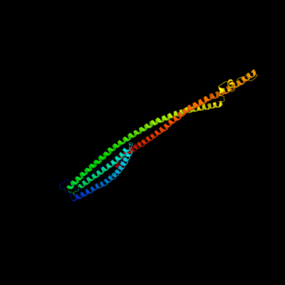
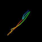
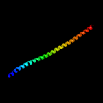
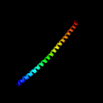
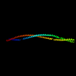
1 c1ciiA_
99.6
7
PDB header: transmembrane proteinChain: A: PDB Molecule: colicin ia;PDBTitle: colicin ia
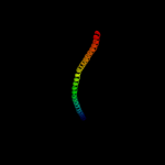
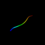
2 c3s4rB_
99.3
34
PDB header: structural proteinChain: B: PDB Molecule: vimentin;PDBTitle: crystal structure of vimentin coil1a/1b fragment with a stabilizing2 mutation
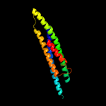
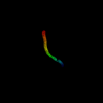
3 c1c1gA_
99.3
10
PDB header: contractile proteinChain: A: PDB Molecule: tropomyosin;PDBTitle: crystal structure of tropomyosin at 7 angstroms resolution2 in the spermine-induced crystal form
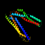
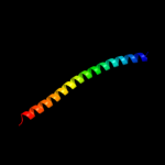
4 c3tnuA_
99.0
61
PDB header: cytosolic proteinChain: A: PDB Molecule: keratin, type i cytoskeletal 14;PDBTitle: heterocomplex of coil 2b domains of human intermediate filament2 proteins, keratin 5 (krt5) and keratin 14 (krt14)
5 c3tnuB_
98.9
31
PDB header: cytosolic proteinChain: B: PDB Molecule: keratin, type ii cytoskeletal 5;PDBTitle: heterocomplex of coil 2b domains of human intermediate filament2 proteins, keratin 5 (krt5) and keratin 14 (krt14)
6 c1gk4A_
98.8
31
PDB header: vimentinChain: A: PDB Molecule: vimentin;PDBTitle: human vimentin coil 2b fragment (cys2)
7 c1x8yA_
98.8
31
PDB header: structural proteinChain: A: PDB Molecule: lamin a/c;PDBTitle: human lamin coil 2b
8 c3swkB_
98.7
31
PDB header: structural proteinChain: B: PDB Molecule: vimentin;PDBTitle: crystal structure of vimentin coil1b fragment
9 c1yvlB_
98.7
10
PDB header: signaling proteinChain: B: PDB Molecule: signal transducer and activator of transcriptionPDBTitle: structure of unphosphorylated stat1
10 c3ojaB_
98.7
13
PDB header: protein bindingChain: B: PDB Molecule: anopheles plasmodium-responsive leucine-rich repeat proteinPDBTitle: crystal structure of lrim1/apl1c complex
11 c2oevA_
98.7
12
PDB header: protein transportChain: A: PDB Molecule: programmed cell death 6-interacting protein;PDBTitle: crystal structure of alix/aip1
12 c3movB_
98.6
27
PDB header: structural proteinChain: B: PDB Molecule: lamin-b1;PDBTitle: crystal structure of human lamin-b1 coil 2 segment
13 c3ol1A_
98.6
32
PDB header: structural proteinChain: A: PDB Molecule: vimentin;PDBTitle: crystal structure of vimentin (fragment 144-251) from homo sapiens,2 northeast structural genomics consortium target hr4796b
14 c1bf5A_
98.5
9
PDB header: gene regulation/dnaChain: A: PDB Molecule: signal transducer and activator of transcriptionPDBTitle: tyrosine phosphorylated stat-1/dna complex
15 c4jioA_
98.4
10
PDB header: protein bindingChain: A: PDB Molecule: bro1;PDBTitle: bro1 v domain and ubiquitin
16 c2ch7A_
98.4
8
PDB header: chemotaxisChain: A: PDB Molecule: methyl-accepting chemotaxis protein;PDBTitle: crystal structure of the cytoplasmic domain of a bacterial2 chemoreceptor from thermotoga maritima
17 c2efrB_
98.4
14
PDB header: contractile proteinChain: B: PDB Molecule: general control protein gcn4 and tropomyosin 1 alpha chain;PDBTitle: crystal structure of the c-terminal tropomyosin fragment with n- and2 c-terminal extensions of the leucine zipper at 1.8 angstroms3 resolution
18 c2d3eD_
98.3
14
PDB header: contractile proteinChain: D: PDB Molecule: general control protein gcn4 and tropomyosin 1PDBTitle: crystal structure of the c-terminal fragment of rabbit2 skeletal alpha-tropomyosin
19 c2xv5A_
98.3
35
PDB header: structural proteinChain: A: PDB Molecule: lamin-a/c;PDBTitle: human lamin a coil 2b fragment
20 c2oexB_
98.2
12
PDB header: protein transportChain: B: PDB Molecule: programmed cell death 6-interacting protein;PDBTitle: structure of alix/aip1 v domain
21 c1gk6B_
not modelled
98.2
29
PDB header: vimentinChain: B: PDB Molecule: vimentin;PDBTitle: human vimentin coil 2b fragment linked to gcn4 leucine2 zipper (z2b)
22 c3vkhA_
not modelled
98.2
9
PDB header: motor proteinChain: A: PDB Molecule: dynein heavy chain, cytoplasmic;PDBTitle: x-ray structure of a functional full-length dynein motor domain
23 c1ei3C_
not modelled
98.2
8
PDB header: blood clottingChain: C: PDB Molecule: fibrinogen;PDBTitle: crystal structure of native chicken fibrinogen
24 c3cwgA_
not modelled
98.2
10
PDB header: transcriptionChain: A: PDB Molecule: signal transducer and activator of transcriptionPDBTitle: unphosphorylated mouse stat3 core fragment
25 c3vkgA_
not modelled
98.1
12
PDB header: motor proteinChain: A: PDB Molecule: dynein heavy chain, cytoplasmic;PDBTitle: x-ray structure of an mtbd truncation mutant of dynein motor domain
26 c3ghgI_
not modelled
98.1
11
PDB header: blood clottingChain: I: PDB Molecule: fibrinogen gamma chain;PDBTitle: crystal structure of human fibrinogen
27 c1gk7A_
not modelled
98.1
47
PDB header: vimentinChain: A: PDB Molecule: vimentin;PDBTitle: human vimentin coil 1a fragment (1a)
28 c1jchC_
98.1
7
PDB header: ribosome inhibitor, hydrolaseChain: C: PDB Molecule: colicin e3;PDBTitle: crystal structure of colicin e3 in complex with its immunity protein
29 c2b9cA_
not modelled
98.1
15
PDB header: contractile proteinChain: A: PDB Molecule: striated-muscle alpha tropomyosin;PDBTitle: structure of tropomyosin's mid-region: bending and binding2 sites for actin
30 c1bg1A_
not modelled
98.1
11
PDB header: transcription/dnaChain: A: PDB Molecule: protein (transcription factor stat3b);PDBTitle: transcription factor stat3b/dna complex
31 c3vkgB_
not modelled
98.0
9
PDB header: motor proteinChain: B: PDB Molecule: dynein heavy chain, cytoplasmic;PDBTitle: x-ray structure of an mtbd truncation mutant of dynein motor domain
32 c3na7A_
not modelled
98.0
16
PDB header: gene regulation, chaperoneChain: A: PDB Molecule: hp0958;PDBTitle: 2.2 angstrom structure of the hp0958 protein from helicobacter pylori2 ccug 17874
33 c3o0zD_
not modelled
98.0
11
PDB header: transferaseChain: D: PDB Molecule: rho-associated protein kinase 1;PDBTitle: crystal structure of a coiled-coil domain from human rock i
34 c4a7fB_
not modelled
98.0
14
PDB header: structural protein/hydrolaseChain: B: PDB Molecule: tropomyosin 1 alpha;PDBTitle: structure of the actin-tropomyosin-myosin complex (rigor atm 3)
35 c2v71A_
not modelled
98.0
8
PDB header: nuclear proteinChain: A: PDB Molecule: nuclear distribution protein nude-like 1;PDBTitle: coiled-coil region of nudel
36 c1hciB_
not modelled
97.9
8
PDB header: triple-helix coiled coilChain: B: PDB Molecule: alpha-actinin 2;PDBTitle: crystal structure of the rod domain of alpha-actinin
37 c3ojaA_
not modelled
97.9
11
PDB header: protein bindingChain: A: PDB Molecule: leucine-rich immune molecule 1;PDBTitle: crystal structure of lrim1/apl1c complex
38 c2i1kA_
not modelled
97.8
10
PDB header: cell adhesion, membrane proteinChain: A: PDB Molecule: moesin;PDBTitle: moesin from spodoptera frugiperda reveals the coiled-coil domain at2 3.0 angstrom resolution
39 c2fxmB_
not modelled
97.8
16
PDB header: contractile proteinChain: B: PDB Molecule: myosin heavy chain, cardiac muscle beta isoform;PDBTitle: structure of the human beta-myosin s2 fragment
40 c1deqF_
not modelled
97.8
6
PDB header: blood clottingChain: F: PDB Molecule: fibrinogen (gamma chain);PDBTitle: the crystal structure of modified bovine fibrinogen (at ~42 angstrom resolution)
41 c3dtpA_
not modelled
97.8
12
PDB header: contractile proteinChain: A: PDB Molecule: myosin 2 heavy chain chimera of smooth andPDBTitle: tarantula heavy meromyosin obtained by flexible docking to2 tarantula muscle thick filament cryo-em 3d-map
42 c1ei3E_
not modelled
97.7
12
PDB header: blood clottingChain: E: PDB Molecule: fibrinogen;PDBTitle: crystal structure of native chicken fibrinogen
43 c1sjjB_
not modelled
97.7
8
PDB header: contractile proteinChain: B: PDB Molecule: actinin;PDBTitle: cryo-em structure of chicken gizzard smooth muscle alpha-2 actinin
44 c2ocyB_
not modelled
97.5
16
PDB header: endocytosis/exocytosisChain: B: PDB Molecule: rab guanine nucleotide exchange factor sec2;PDBTitle: complex of the guanine exchange factor sec2p and the rab2 gtpase sec4p
45 c4a55B_
not modelled
97.5
13
PDB header: transferaseChain: B: PDB Molecule: phosphatidylinositol 3-kinase regulatory subunit alpha;PDBTitle: crystal structure of p110alpha in complex with ish2 of p85alpha and2 the inhibitor pik-108
46 c3ghgK_
not modelled
97.5
8
PDB header: blood clottingChain: K: PDB Molecule: fibrinogen beta chain;PDBTitle: crystal structure of human fibrinogen
47 c1u4qB_
not modelled
97.5
11
PDB header: structural proteinChain: B: PDB Molecule: spectrin alpha chain, brain;PDBTitle: crystal structure of repeats 15, 16 and 17 of chicken brain2 alpha spectrin
48 c3q8tB_
not modelled
97.5
13
PDB header: apoptosisChain: B: PDB Molecule: beclin-1;PDBTitle: crystal structure of the coiled coil domain of beclin 1, an essential2 autophagy protein
49 c1deqO_
not modelled
97.5
9
PDB header: blood clottingChain: O: PDB Molecule: fibrinogen (beta chain);PDBTitle: the crystal structure of modified bovine fibrinogen (at ~42 angstrom resolution)
50 c3hizB_
not modelled
97.4
14
PDB header: transferase/oncoproteinChain: B: PDB Molecule: phosphatidylinositol 3-kinase regulatory subunitPDBTitle: crystal structure of p110alpha h1047r mutant in complex with2 nish2 of p85alpha
51 c2v66C_
not modelled
97.4
13
PDB header: structural proteinChain: C: PDB Molecule: nuclear distribution protein nude-like 1;PDBTitle: crystal structure of the coiled-coil domain of ndel1 (a.a.2 58 to 169)c
52 c2y3aB_
not modelled
97.3
9
PDB header: transferaseChain: B: PDB Molecule: phosphatidylinositol 3-kinase regulatory subunit beta;PDBTitle: crystal structure of p110beta in complex with icsh2 of p85beta and2 the drug gdc-0941
53 c1deqD_
not modelled
97.2
9
PDB header: blood clottingChain: D: PDB Molecule: fibrinogen (alpha chain);PDBTitle: the crystal structure of modified bovine fibrinogen (at ~42 angstrom resolution)
54 c3u59C_
not modelled
97.2
9
PDB header: contractile proteinChain: C: PDB Molecule: tropomyosin beta chain;PDBTitle: n-terminal 98-aa fragment of smooth muscle tropomyosin beta
55 c1f5nA_
not modelled
97.0
9
PDB header: signaling proteinChain: A: PDB Molecule: interferon-induced guanylate-binding protein 1;PDBTitle: human guanylate binding protein-1 in complex with the gtp2 analogue, gmppnp.
56 c2rd0B_
not modelled
97.0
9
PDB header: transferase/oncoproteinChain: B: PDB Molecule: phosphatidylinositol 3-kinase regulatory subunit alpha;PDBTitle: structure of a human p110alpha/p85alpha complex
57 c3kltB_
not modelled
96.8
35
PDB header: structural proteinChain: B: PDB Molecule: vimentin;PDBTitle: crystal structure of a vimentin fragment
58 c4gkwB_
not modelled
96.7
11
PDB header: structural proteinChain: B: PDB Molecule: spindle assembly abnormal protein 6;PDBTitle: crystal structure of the coiled-coil domain of c. elegans sas-6
59 c3l9oA_
not modelled
96.4
10
PDB header: hydrolaseChain: A: PDB Molecule: atp-dependent rna helicase dob1;PDBTitle: crystal structure of mtr4, a co-factor of the nuclear exosome
60 c3r6nA_
not modelled
96.4
11
PDB header: cell adhesionChain: A: PDB Molecule: desmoplakin;PDBTitle: crystal structure of a rigid four spectrin repeat fragment of the2 human desmoplakin plakin domain
61 c3ipkA_
not modelled
96.3
11
PDB header: cell adhesionChain: A: PDB Molecule: agi/ii;PDBTitle: crystal structure of a3vp1 of agi/ii of streptococcus mutans
62 c4l6yB_
not modelled
96.1
6
PDB header: structural proteinChain: B: PDB Molecule: protein regulator of cytokinesis 1;PDBTitle: structure of the microtubule associated protein prc1 (protein2 regulator of cytokinesis 1)
63 c4hpqC_
not modelled
96.1
13
PDB header: protein transportChain: C: PDB Molecule: atg17;PDBTitle: crystal structure of the atg17-atg31-atg29 complex
64 c4dylA_
not modelled
95.7
11
PDB header: transferaseChain: A: PDB Molecule: tyrosine-protein kinase fes/fps;PDBTitle: f-bar domain of human fes tyrosine kinase
65 c2gl2B_
not modelled
95.6
10
PDB header: cell adhesionChain: B: PDB Molecule: adhesion a;PDBTitle: crystal structure of the tetra muntant (t66g,r67g,f68g,2 y69g) of bacterial adhesin fada
66 c3hnwB_
not modelled
95.2
13
PDB header: structural genomics, unknown functionChain: B: PDB Molecule: uncharacterized protein;PDBTitle: crystal structure of a basic coiled-coil protein of unknown function2 from eubacterium eligens atcc 27750
67 c2qa7C_
not modelled
95.0
13
PDB header: actin bindingChain: C: PDB Molecule: huntingtin-interacting protein 1;PDBTitle: crystal structure of huntingtin-interacting protein 12 (hip1) coiled-coil domain with a basic surface suitable3 for hip-protein interactor (hippi)
68 c1y4cA_
not modelled
94.7
10
PDB header: de novo proteinChain: A: PDB Molecule: maltose binding protein fused with designedPDBTitle: designed helical protein fusion mbp
69 c2no2A_
not modelled
94.7
16
PDB header: cell adhesionChain: A: PDB Molecule: huntingtin-interacting protein 1;PDBTitle: crystal structure of the dllrkn-containing coiled-coil2 domain of huntingtin-interacting protein 1
70 c2nrjA_
not modelled
94.0
11
PDB header: toxinChain: A: PDB Molecule: hbl b protein;PDBTitle: crystal structure of hemolysin binding component from2 bacillus cereus
71 c3vkhD_
not modelled
93.9
6
PDB header: motor proteinChain: D: PDB Molecule: PDBTitle: x-ray structure of a functional full-length dynein motor domain
72 c3g67A_
not modelled
93.9
6
PDB header: signaling proteinChain: A: PDB Molecule: methyl-accepting chemotaxis protein;PDBTitle: crystal structure of a soluble chemoreceptor from thermotoga2 maritima
73 c1quuA_
not modelled
93.3
7
PDB header: contractile proteinChain: A: PDB Molecule: human skeletal muscle alpha-actinin 2;PDBTitle: crystal structure of two central spectrin-like repeats from2 alpha-actinin
74 c2v1yB_
not modelled
93.3
9
PDB header: transferaseChain: B: PDB Molecule: phosphatidylinositol 3-kinase regulatory subunit alpha;PDBTitle: structure of a phosphoinositide 3-kinase alpha adaptor-2 binding domain (abd) in a complex with the ish2 domain3 from p85 alpha
75 c1l8dB_
not modelled
92.3
13
PDB header: replicationChain: B: PDB Molecule: dna double-strand break repair rad50 atpase;PDBTitle: rad50 coiled-coil zn hook
76 c3kbtA_
not modelled
92.0
8
PDB header: structural proteinChain: A: PDB Molecule: spectrin beta chain, erythrocyte;PDBTitle: crystal structure of the ankyrin binding domain of human erythroid2 beta spectrin (repeats 13-15) in complex with the spectrin binding3 domain of human erythroid ankyrin (zu5-ank)
77 c2i1jA_
not modelled
92.0
7
PDB header: cell adhesion, membrane proteinChain: A: PDB Molecule: moesin;PDBTitle: moesin from spodoptera frugiperda at 2.1 angstroms resolution
78 c2dq3A_
not modelled
92.0
15
PDB header: ligaseChain: A: PDB Molecule: seryl-trna synthetase;PDBTitle: crystal structure of aq_298
79 c3lssA_
not modelled
91.9
8
PDB header: ligaseChain: A: PDB Molecule: seryl-trna synthetase;PDBTitle: trypanosoma brucei seryl-trna synthetase in complex with atp
80 c2lemA_
not modelled
91.8
5
PDB header: lipid transportChain: A: PDB Molecule: apolipoprotein a-i;PDBTitle: monomeric mouse apoai(1-216)
81 c3edvB_
not modelled
91.7
10
PDB header: structural proteinChain: B: PDB Molecule: spectrin beta chain, brain 1;PDBTitle: crystal structure of repeats 14-16 of beta2-spectrin
82 c2dfsA_
not modelled
91.4
10
PDB header: contractile protein/transport proteinChain: A: PDB Molecule: myosin-5a;PDBTitle: 3-d structure of myosin-v inhibited state
83 c2jeeA_
not modelled
91.4
16
PDB header: cell cycleChain: A: PDB Molecule: yiiu;PDBTitle: xray structure of e. coli yiiu
84 c1m1jA_
not modelled
91.3
8
PDB header: blood clottingChain: A: PDB Molecule: fibrinogen alpha subunit;PDBTitle: crystal structure of native chicken fibrinogen with two different2 bound ligands
85 c2yo3C_
not modelled
90.9
13
PDB header: membrane proteinChain: C: PDB Molecule: general control protein gcn4, putative inner membranePDBTitle: salmonella enterica sada 1185-1386 fused to gcn4 adaptors (sadak14)
86 c2v4hA_
not modelled
90.1
21
PDB header: transcriptionChain: A: PDB Molecule: nf-kappa-b essential modulator;PDBTitle: nemo cc2-lz domain - 1d5 darpin complex
87 c4etpA_
not modelled
89.8
22
PDB header: motor proteinChain: A: PDB Molecule: kinesin-like protein kar3;PDBTitle: c-terminal motor and motor homology domain of kar3vik1 fused to a2 synthetic heterodimeric coiled coil
88 c1g8xB_
not modelled
89.3
9
PDB header: structural proteinChain: B: PDB Molecule: myosin ii heavy chain fused to alpha-actinin 3;PDBTitle: structure of a genetically engineered molecular motor
89 c2j69D_
not modelled
89.0
7
PDB header: hydrolaseChain: D: PDB Molecule: bacterial dynamin-like protein;PDBTitle: bacterial dynamin-like protein bdlp
90 c2qihA_
not modelled
88.9
10
PDB header: cell adhesionChain: A: PDB Molecule: protein uspa1;PDBTitle: crystal structure of 527-665 fragment of uspa1 protein from2 moraxella catarrhalis
91 c1u5pA_
not modelled
88.7
10
PDB header: structural proteinChain: A: PDB Molecule: spectrin alpha chain, brain;PDBTitle: crystal structure of repeats 15 and 16 of chicken brain2 alpha spectrin
92 c2e7sM_
not modelled
88.7
15
PDB header: endocytosis/exocytosisChain: M: PDB Molecule: rab guanine nucleotide exchange factor sec2;PDBTitle: crystal structure of the yeast sec2p gef domain
93 c1qu7A_
not modelled
88.5
10
PDB header: signaling proteinChain: A: PDB Molecule: methyl-accepting chemotaxis protein i;PDBTitle: four helical-bundle structure of the cytoplasmic domain of a serine2 chemotaxis receptor
94 c4dvzA_
not modelled
88.2
9
PDB header: oncoproteinChain: A: PDB Molecule: cytotoxicity-associated immunodominant antigen;PDBTitle: crystal structure of the helicobacter pylori caga oncoprotein
95 c2wpqA_
not modelled
88.1
13
PDB header: membrane proteinChain: A: PDB Molecule: trimeric autotransporter adhesin fragment;PDBTitle: salmonella enterica sada 479-519 fused to gcn4 adaptors (2 sadak3, in-register fusion)
96 c2yy0D_
not modelled
87.6
25
PDB header: transcriptionChain: D: PDB Molecule: c-myc-binding protein;PDBTitle: crystal structure of ms0802, c-myc-1 binding protein domain2 from homo sapiens
97 c3q0xA_
not modelled
86.8
12
PDB header: structural proteinChain: A: PDB Molecule: centriole protein;PDBTitle: n-terminal coiled-coil dimer domain of c. reinhardtii sas-6 homolog2 bld12p
98 c3a7pB_
not modelled
86.6
12
PDB header: protein transportChain: B: PDB Molecule: autophagy protein 16;PDBTitle: the crystal structure of saccharomyces cerevisiae atg16
99 d2ap3a1
not modelled
86.2
6
Fold: Four-helical up-and-down bundleSuperfamily: MW0975(SA0943)-likeFamily: MW0975(SA0943)-like100 c2xzrA_
not modelled
86.2
15
PDB header: cell adhesionChain: A: PDB Molecule: immunoglobulin-binding protein eibd;PDBTitle: escherichia coli immunoglobulin-binding protein eibd 391-438 fused2 to gcn4 adaptors
101 c3u1aC_
not modelled
85.6
19
PDB header: contractile proteinChain: C: PDB Molecule: smooth muscle tropomyosin alpha;PDBTitle: n-terminal 81-aa fragment of smooth muscle tropomyosin alpha
102 c3cvfA_
not modelled
84.3
14
PDB header: signaling proteinChain: A: PDB Molecule: homer protein homolog 3;PDBTitle: crystal structure of the carboxy terminus of homer3
103 c2eqbC_
not modelled
83.6
14
PDB header: endocytosis/exocytosisChain: C: PDB Molecule: rab guanine nucleotide exchange factor sec2;PDBTitle: crystal structure of the rab gtpase sec4p, the sec2p gef2 domain, and phosphate complex
104 c1cz7C_
not modelled
83.3
10
PDB header: contractile proteinChain: C: PDB Molecule: microtubule motor protein ncd;PDBTitle: the crystal structure of a minus-end directed microtubule2 motor protein ncd reveals variable dimer conformations
105 c2xgjA_
not modelled
83.1
11
PDB header: hydrolase/rnaChain: A: PDB Molecule: atp-dependent rna helicase dob1;PDBTitle: structure of mtr4, a dexh helicase involved in nuclear rna2 processing and surveillance
106 c2odvA_
not modelled
82.3
8
PDB header: structural proteinChain: A: PDB Molecule: plectin 1;PDBTitle: crystal structure of a fragment of the plakin domain of plectin, cys2 to ala mutant.
107 c1junB_
not modelled
82.2
22
PDB header: transcription regulationChain: B: PDB Molecule: c-jun homodimer;PDBTitle: nmr study of c-jun homodimer
108 c2zvnF_
not modelled
81.9
15
PDB header: signaling protein/transcriptionChain: F: PDB Molecule: nf-kappa-b essential modulator;PDBTitle: nemo cozi domain incomplex with diubiquitin in p2121212 space group
109 c1wt6B_
not modelled
81.3
15
PDB header: transferaseChain: B: PDB Molecule: myotonin-protein kinase;PDBTitle: coiled-coil domain of dmpk
110 d1ykhb1
not modelled
81.2
12
Fold: Mediator hinge subcomplex-likeSuperfamily: Mediator hinge subcomplex-likeFamily: CSE2-like111 c3r2pA_
not modelled
79.4
14
PDB header: lipid transportChain: A: PDB Molecule: apolipoprotein a-i;PDBTitle: 2.2 angstrom crystal structure of c terminal truncated human2 apolipoprotein a-i reveals the assembly of hdl by dimerization.
112 c3ghgD_
not modelled
79.3
13
PDB header: blood clottingChain: D: PDB Molecule: fibrinogen alpha chain;PDBTitle: crystal structure of human fibrinogen
113 c3ajwA_
not modelled
79.1
15
PDB header: protein transportChain: A: PDB Molecule: flagellar flij protein;PDBTitle: structure of flij, a soluble component of flagellar type iii export2 apparatus
114 c1i84V_
not modelled
78.9
15
PDB header: contractile proteinChain: V: PDB Molecule: smooth muscle myosin heavy chain;PDBTitle: cryo-em structure of the heavy meromyosin subfragment of2 chicken gizzard smooth muscle myosin with regulatory light3 chain in the dephosphorylated state. only c alphas4 provided for regulatory light chain. only backbone atoms5 provided for s2 fragment.
115 c3pe0B_
not modelled
76.8
7
PDB header: structural proteinChain: B: PDB Molecule: plectin;PDBTitle: structure of the central region of the plakin domain of plectin
116 c3cveC_
not modelled
75.8
19
PDB header: signaling proteinChain: C: PDB Molecule: homer protein homolog 1;PDBTitle: crystal structure of the carboxy terminus of homer1
117 c2q6qB_
not modelled
75.2
30
PDB header: cell cycleChain: B: PDB Molecule: spindle pole body component spc42;PDBTitle: crystal structure of spc42p, a critical component of spindle pole body2 in budding yeast
118 c3a6mB_
not modelled
74.6
12
PDB header: chaperoneChain: B: PDB Molecule: protein grpe;PDBTitle: crystal structure of grpe from thermus thermophilus hb8
119 c4aniA_
not modelled
74.6
17
PDB header: chaperoneChain: A: PDB Molecule: protein grpe;PDBTitle: structural basis for the intermolecular communication between2 dnak and grpe in the dnak chaperone system from3 geobacillus kaustophilus hta426
120 c1ik9B_
not modelled
73.8
16
PDB header: gene regulation/ligaseChain: B: PDB Molecule: dna repair protein xrcc4;PDBTitle: crystal structure of a xrcc4-dna ligase iv complex