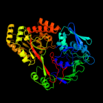
1 d1jeta_
100.0
86
Fold: Periplasmic binding protein-like IISuperfamily: Periplasmic binding protein-like IIFamily: Phosphate binding protein-like2 c3o9pA_
100.0
49
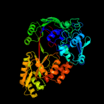
PDB header: peptide binding protein/peptideChain: A: PDB Molecule: periplasmic murein peptide-binding protein;PDBTitle: the structure of the escherichia coli murein tripeptide binding2 protein mppa
3 c3tpaA_
100.0
25
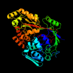
PDB header: heme binding proteinChain: A: PDB Molecule: heme-binding protein a;PDBTitle: structure of hbpa2 from haemophilus parasuis
4 d1dpea_
100.0
24
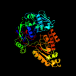
Fold: Periplasmic binding protein-like IISuperfamily: Periplasmic binding protein-like IIFamily: Phosphate binding protein-like5 c3m8uA_
100.0
23
PDB header: transport proteinChain: A: PDB Molecule: heme-binding protein a;PDBTitle: crystal structure of glutathione-binding protein a (gbpa) from2 haemophilus parasuis sh0165 in complex with glutathione disulfide3 (gssg)
6 c2wokA_
100.0
20
PDB header: peptide binding protein/peptideChain: A: PDB Molecule: clavulanic acid biosynthesis oligopeptidePDBTitle: clavulanic acid biosynthesis oligopeptide2 binding protein 2 complexed with bradykinin
7 d1xoca1
100.0
21
Fold: Periplasmic binding protein-like IISuperfamily: Periplasmic binding protein-like IIFamily: Phosphate binding protein-like8 d1zlqa1
100.0
23
Fold: Periplasmic binding protein-like IISuperfamily: Periplasmic binding protein-like IIFamily: Phosphate binding protein-like9 c3t66A_
100.0
23
PDB header: transport proteinChain: A: PDB Molecule: nickel abc transporter (nickel-binding protein);PDBTitle: crystal structure of nickel abc transporter from bacillus halodurans
10 d1uqwa_
100.0
23
Fold: Periplasmic binding protein-like IISuperfamily: Periplasmic binding protein-like IIFamily: Phosphate binding protein-like11 c3rqtA_
100.0
17
PDB header: unknown functionChain: A: PDB Molecule: putative uncharacterized protein;PDBTitle: 1.5 angstrom crystal structure of the complex of ligand binding2 component of abc-type import system from staphylococcus aureus with3 nickel and two histidines
12 c1ztyA_
100.0
19
PDB header: sugar binding protein, signaling proteinChain: A: PDB Molecule: chitin oligosaccharide binding protein;PDBTitle: crystal structure of the chitin oligasaccharide binding2 protein
13 c3ftoA_
100.0
20
PDB header: peptide binding proteinChain: A: PDB Molecule: oligopeptide-binding protein oppa;PDBTitle: crystal structure of oppa in a open conformation
14 c2o7jA_
100.0
19
PDB header: sugar binding proteinChain: A: PDB Molecule: oligopeptide abc transporter, periplasmicPDBTitle: the x-ray crystal structure of a thermophilic cellobiose2 binding protein bound with cellopentaose
15 d1vr5a1
100.0
21
Fold: Periplasmic binding protein-like IISuperfamily: Periplasmic binding protein-like IIFamily: Phosphate binding protein-like16 c2grvC_
100.0
18
PDB header: biosynthetic proteinChain: C: PDB Molecule: lpqw;PDBTitle: crystal structure of lpqw
17 c2d5wA_
100.0
20
PDB header: peptide binding proteinChain: A: PDB Molecule: peptide abc transporter, peptide-binding protein;PDBTitle: the crystal structure of oligopeptide binding protein from thermus2 thermophilus hb8 complexed with pentapeptide
18 c3ry3B_
100.0
19
PDB header: transport proteinChain: B: PDB Molecule: putative solute-binding protein;PDBTitle: putative solute-binding protein from yersinia pestis.
19 c3lvuB_
100.0
19
PDB header: transport proteinChain: B: PDB Molecule: abc transporter, periplasmic substrate-binding protein;PDBTitle: crystal structure of abc transporter, periplasmic substrate-binding2 protein spo2066 from silicibacter pomeroyi
20 c3pamB_
100.0
18
PDB header: transport proteinChain: B: PDB Molecule: transmembrane protein;PDBTitle: crystal structure of a domain of transmembrane protein of abc-type2 oligopeptide transport system from bartonella henselae str. houston-1
21 c3o6pA_
not modelled
100.0
24
PDB header: protein bindingChain: A: PDB Molecule: peptide abc transporter, peptide-binding protein;PDBTitle: crystal structure of peptide abc transporter, peptide-binding protein
22 c3chgB_
not modelled
96.4
18
PDB header: ligand binding proteinChain: B: PDB Molecule: glycine betaine-binding protein;PDBTitle: the compatible solute-binding protein opuac from bacillus2 subtilis in complex with dmsa
23 c3l6gA_
not modelled
95.5
19
PDB header: glycine betaine-binding proteinChain: A: PDB Molecule: betaine abc transporter permease and substrate bindingPDBTitle: crystal structure of lactococcal opuac in its open conformation
24 c3tmgA_
not modelled
94.5
12
PDB header: transport proteinChain: A: PDB Molecule: glycine betaine, l-proline abc transporter,PDBTitle: crystal structure of glycine betaine, l-proline abc transporter,2 glycine/betaine/l-proline-binding protein (prox) from borrelia3 burgdorferi
25 d1r9la_
not modelled
93.2
14
Fold: Periplasmic binding protein-like IISuperfamily: Periplasmic binding protein-like IIFamily: Phosphate binding protein-like26 c2rejA_
not modelled
92.2
10
PDB header: choline-binding proteinChain: A: PDB Molecule: putative glycine betaine abc transporter protein;PDBTitle: abc-transporter choline binding protein in unliganded semi-2 closed conformation
27 c3kn3C_
not modelled
81.5
21
PDB header: transcriptionChain: C: PDB Molecule: putative periplasmic protein;PDBTitle: crystal structure of lysr substrate binding domain (25-263) of2 putative periplasmic protein from wolinella succinogenes
28 c3r6uA_
not modelled
80.2
7
PDB header: transport proteinChain: A: PDB Molecule: choline-binding protein;PDBTitle: crystal structure of choline binding protein opubc from bacillus2 subtilis
29 c3nohA_
not modelled
72.3
13
PDB header: peptide binding proteinChain: A: PDB Molecule: putative peptide binding protein;PDBTitle: crystal structure of a putative peptide binding protein (rumgna_00914)2 from ruminococcus gnavus atcc 29149 at 1.60 a resolution
30 d1sw5a_
not modelled
67.3
2
Fold: Periplasmic binding protein-like IISuperfamily: Periplasmic binding protein-like IIFamily: Phosphate binding protein-like31 c3lr1A_
not modelled
65.8
18
PDB header: transport proteinChain: A: PDB Molecule: tungstate abc transporter, periplasmic tungstate-PDBTitle: the crystal structure of the tungstate abc transporter from2 geobacter sulfurreducens
32 c3ir1F_
not modelled
64.5
16
PDB header: protein bindingChain: F: PDB Molecule: outer membrane lipoprotein gna1946;PDBTitle: crystal structure of lipoprotein gna1946 from neisseria2 meningitidis
33 c3pppA_
not modelled
61.8
16
PDB header: transport proteinChain: A: PDB Molecule: glycine betaine/carnitine/choline-binding protein;PDBTitle: structures of the substrate-binding protein provide insights into the2 multiple compatible solutes binding specificities of bacillus3 subtilis abc transporter opuc
34 c1tvmA_
not modelled
61.4
10
PDB header: transferaseChain: A: PDB Molecule: pts system, galactitol-specific iib component;PDBTitle: nmr structure of enzyme gatb of the galactitol-specific2 phosphoenolpyruvate-dependent phosphotransferase system
35 c3ombA_
not modelled
58.1
11
PDB header: transport proteinChain: A: PDB Molecule: extracellular solute-binding protein, family 1;PDBTitle: crystal structure of extracellular solute-binding protein from2 bifidobacterium longum subsp. infantis
36 c3kzgB_
not modelled
54.5
7
PDB header: transport proteinChain: B: PDB Molecule: arginine 3rd transport system periplasmic bindingPDBTitle: crystal structure of an arginine 3rd transport system2 periplasmic binding protein from legionella pneumophila
37 c3muqB_
not modelled
50.2
20
PDB header: structural genomics, unknown functionChain: B: PDB Molecule: uncharacterized conserved protein;PDBTitle: the crystal structure of a conserved functionally unknown protein from2 vibrio parahaemolyticus rimd 2210633
38 c2e76D_
not modelled
48.1
4
PDB header: photosynthesisChain: D: PDB Molecule: cytochrome b6-f complex iron-sulfur subunit;PDBTitle: crystal structure of the cytochrome b6f complex with tridecyl-2 stigmatellin (tds) from m.laminosus
39 c2rc9A_
not modelled
47.2
12
PDB header: membrane proteinChain: A: PDB Molecule: glutamate [nmda] receptor subunit 3a;PDBTitle: crystal structure of the nr3a ligand binding core complex with acpc at2 1.96 angstrom resolution
40 c3gxaA_
not modelled
44.3
14
PDB header: protein bindingChain: A: PDB Molecule: outer membrane lipoprotein gna1946;PDBTitle: crystal structure of gna1946
41 d1xs5a_
not modelled
42.1
6
Fold: Periplasmic binding protein-like IISuperfamily: Periplasmic binding protein-like IIFamily: Phosphate binding protein-like42 c3hlyA_
not modelled
41.6
11
PDB header: flavoproteinChain: A: PDB Molecule: flavodoxin-like domain;PDBTitle: crystal structure of the flavodoxin-like domain from2 synechococcus sp q5mzp6_synp6 protein. northeast structural3 genomics consortium target snr135d.
43 d2p0la1
not modelled
41.6
14
Fold: Class II aaRS and biotin synthetasesSuperfamily: Class II aaRS and biotin synthetasesFamily: LplA-like44 c3k2dA_
not modelled
40.2
17
PDB header: immune systemChain: A: PDB Molecule: abc-type metal ion transport system, periplasmic component;PDBTitle: crystal structure of immunogenic lipoprotein a from vibrio vulnificus
45 c3ix1A_
not modelled
40.2
10
PDB header: biosynthetic proteinChain: A: PDB Molecule: n-formyl-4-amino-5-aminomethyl-2-methylpyrimidine bindingPDBTitle: periplasmic n-formyl-4-amino-5-aminomethyl-2-methylpyrimidine binding2 protein from bacillus halodurans
46 c3ix1B_
not modelled
40.2
10
PDB header: biosynthetic proteinChain: B: PDB Molecule: n-formyl-4-amino-5-aminomethyl-2-methylpyrimidine bindingPDBTitle: periplasmic n-formyl-4-amino-5-aminomethyl-2-methylpyrimidine binding2 protein from bacillus halodurans
47 d1e5da1
not modelled
37.9
11
Fold: Flavodoxin-likeSuperfamily: FlavoproteinsFamily: Flavodoxin-related48 c2zykA_
not modelled
35.7
12
PDB header: sugar binding proteinChain: A: PDB Molecule: solute-binding protein;PDBTitle: crystal structure of cyclo/maltodextrin-binding protein2 complexed with gamma-cyclodextrin
49 d1p99a_
not modelled
35.5
17
Fold: Periplasmic binding protein-like IISuperfamily: Periplasmic binding protein-like IIFamily: Phosphate binding protein-like50 c1p99A_
not modelled
35.5
17
PDB header: structural genomics, unknown functionChain: A: PDB Molecule: hypothetical protein pg110;PDBTitle: 1.7a crystal structure of protein pg110 from staphylococcus2 aureus
51 d2a5sa1
not modelled
33.6
9
Fold: Periplasmic binding protein-like IISuperfamily: Periplasmic binding protein-like IIFamily: Phosphate binding protein-like52 c3un6A_
not modelled
31.3
8
PDB header: unknown functionChain: A: PDB Molecule: hypothetical protein saouhsc_00137;PDBTitle: 2.0 angstrom crystal structure of ligand binding component of abc-type2 import system from staphylococcus aureus with zinc bound
53 c2v25B_
not modelled
30.7
8
PDB header: receptorChain: B: PDB Molecule: major cell-binding factor;PDBTitle: structure of the campylobacter jejuni antigen peb1a, an2 aspartate and glutamate receptor with bound aspartate
54 c3tqwA_
not modelled
30.0
9
PDB header: transport proteinChain: A: PDB Molecule: methionine-binding protein;PDBTitle: structure of a abc transporter, periplasmic substrate-binding protein2 from coxiella burnetii
55 d1f4pa_
not modelled
27.9
6
Fold: Flavodoxin-likeSuperfamily: FlavoproteinsFamily: Flavodoxin-related56 c3k4uA_
not modelled
27.5
16
PDB header: transport proteinChain: A: PDB Molecule: binding component of abc transporter;PDBTitle: crystal structure of putative binding component of abc transporter2 from wolinella succinogenes dsm 1740 complexed with lysine
57 c2y7iB_
not modelled
27.2
18
PDB header: arginine-binding proteinChain: B: PDB Molecule: stm4351;PDBTitle: structural basis for high arginine specificity in salmonella2 typhimurium periplasmic binding protein stm4351.
58 d1wdna_
not modelled
26.7
13
Fold: Periplasmic binding protein-like IISuperfamily: Periplasmic binding protein-like IIFamily: Phosphate binding protein-like59 c3r39A_
not modelled
26.4
18
PDB header: transport proteinChain: A: PDB Molecule: putative periplasmic binding protein;PDBTitle: crystal structure of periplasmic d-alanine abc transporter from2 salmonella enterica
60 d1ycga1
not modelled
26.4
15
Fold: Flavodoxin-likeSuperfamily: FlavoproteinsFamily: Flavodoxin-related61 c3i6vA_
not modelled
26.2
12
PDB header: transport proteinChain: A: PDB Molecule: periplasmic his/glu/gln/arg/opine family-binding protein;PDBTitle: crystal structure of a periplasmic his/glu/gln/arg/opine family-2 binding protein from silicibacter pomeroyi in complex with lysine
62 c2x26A_
not modelled
25.9
13
PDB header: transport proteinChain: A: PDB Molecule: periplasmic aliphatic sulphonates-binding protein;PDBTitle: crystal structure of the periplasmic aliphatic sulphonate2 binding protein ssua from escherichia coli
63 c2ylnA_
not modelled
25.8
17
PDB header: transport proteinChain: A: PDB Molecule: putative abc transporter, periplasmic binding protein,PDBTitle: crystal structure of the l-cystine solute receptor of2 neisseria gonorrhoeae in the closed conformation
64 c3f6sI_
not modelled
25.5
10
PDB header: electron transportChain: I: PDB Molecule: flavodoxin;PDBTitle: desulfovibrio desulfuricans (atcc 29577) oxidized flavodoxin2 alternate conformers
65 c2xx7B_
not modelled
22.9
19
PDB header: transport proteinChain: B: PDB Molecule: glutamate receptor 2;PDBTitle: crystal structure of 1-(4-(1-pyrrolidinylcarbonyl)phenyl)-3-2 (trifluoromethyl)-4,5,6,7-tetrahydro-1h-indazole in complex with3 the ligand binding domain of the rat glua2 receptor and glutamate4 at 2.2a resolution.
66 c2q2aD_
not modelled
22.0
16
PDB header: transport proteinChain: D: PDB Molecule: artj;PDBTitle: crystal structures of the arginine-, lysine-, histidine-2 binding protein artj from the thermophilic bacterium3 geobacillus stearothermophilus
67 c3delC_
not modelled
21.9
9
PDB header: protein binding, transport proteinChain: C: PDB Molecule: arginine binding protein;PDBTitle: the structure of ct381, the arginine binding protein from the2 periplasm chlamydia trachomatis
68 d1xvya_
not modelled
20.7
18
Fold: Periplasmic binding protein-like IISuperfamily: Periplasmic binding protein-like IIFamily: Phosphate binding protein-like69 d1pb7a_
not modelled
19.2
16
Fold: Periplasmic binding protein-like IISuperfamily: Periplasmic binding protein-like IIFamily: Phosphate binding protein-like70 c2o1mB_
not modelled
18.6
18
PDB header: structural genomics, unknown functionChain: B: PDB Molecule: probable amino-acid abc transporterPDBTitle: crystal structure of the probable amino-acid abc2 transporter extracellular-binding protein ytmk from3 bacillus subtilis. northeast structural genomics4 consortium target sr572
71 c2f5xC_
not modelled
18.5
11
PDB header: transport proteinChain: C: PDB Molecule: bugd;PDBTitle: structure of periplasmic binding protein bugd
72 d1ykga1
not modelled
18.5
14
Fold: Flavodoxin-likeSuperfamily: FlavoproteinsFamily: Cytochrome p450 reductase N-terminal domain-like73 c3g41A_
not modelled
18.2
11
PDB header: transport proteinChain: A: PDB Molecule: amino acid abc transporter, periplasmic amino acid-bindingPDBTitle: the structure of cpn0482, the arginine binding protein from the2 periplasm of chlamydia pneumoniae
74 d1hsla_
not modelled
17.9
11
Fold: Periplasmic binding protein-like IISuperfamily: Periplasmic binding protein-like IIFamily: Phosphate binding protein-like75 c2uvgA_
not modelled
16.2
19
PDB header: sugar-binding proteinChain: A: PDB Molecule: abc type periplasmic sugar-binding protein;PDBTitle: structure of a periplasmic oligogalacturonide binding2 protein from yersinia enterocolitica
76 d2fz5a1
not modelled
15.8
11
Fold: Flavodoxin-likeSuperfamily: FlavoproteinsFamily: Flavodoxin-related77 d1twya_
not modelled
15.3
9
Fold: Periplasmic binding protein-like IISuperfamily: Periplasmic binding protein-like IIFamily: Phosphate binding protein-like78 c1twyG_
not modelled
15.3
9
PDB header: structural genomics, unknown functionChain: G: PDB Molecule: abc transporter, periplasmic substrate-binding protein;PDBTitle: crystal structure of an abc-type phosphate transport receptor from2 vibrio cholerae
79 d1lsta_
not modelled
15.2
11
Fold: Periplasmic binding protein-like IISuperfamily: Periplasmic binding protein-like IIFamily: Phosphate binding protein-like80 c2x7pA_
not modelled
14.9
16
PDB header: unknown functionChain: A: PDB Molecule: possible thiamine biosynthesis enzyme;PDBTitle: the conserved candida albicans ca3427 gene product defines a new2 family of proteins exhibiting the generic periplasmic binding3 protein structural fold
81 c3e4rA_
not modelled
14.8
14
PDB header: transport proteinChain: A: PDB Molecule: nitrate transport protein;PDBTitle: crystal structure of the alkanesulfonate binding protein2 (ssua) from the phytopathogenic bacteria xanthomonas3 axonopodis pv. citri bound to hepes
82 d1g7da_
not modelled
14.8
19
Fold: ERP29 C domain-likeSuperfamily: ERP29 C domain-likeFamily: ERP29 C domain-like83 c2ieeB_
not modelled
14.3
16
PDB header: structural genomics, unknown functionChain: B: PDB Molecule: probable abc transporter extracellular-bindingPDBTitle: crystal structure of yckb_bacsu from bacillus subtilis.2 northeast structural genomics consortium target sr574.
84 d1eaka1
not modelled
14.0
12
Fold: PGBD-likeSuperfamily: PGBD-likeFamily: MMP N-terminal domain85 c3qslA_
not modelled
13.8
11
PDB header: structural genomics, unknown functionChain: A: PDB Molecule: putative exported protein;PDBTitle: structure of cae31940 from bordetella bronchiseptica rb50
86 c3hn0A_
not modelled
13.5
18
PDB header: transport proteinChain: A: PDB Molecule: nitrate transport protein;PDBTitle: crystal structure of an abc transporter (bdi_1369) from2 parabacteroides distasonis at 1.75 a resolution
87 c2vpnB_
not modelled
13.3
13
PDB header: transportChain: B: PDB Molecule: periplasmic substrate binding protein;PDBTitle: high-resolution structure of the periplasmic ectoine-2 binding protein from teaabc trap-transporter of halomonas3 elongata
88 c2q9uB_
not modelled
13.2
7
PDB header: oxidoreductaseChain: B: PDB Molecule: a-type flavoprotein;PDBTitle: crystal structure of the flavodiiron protein from giardia2 intestinalis
89 d1mqia_
not modelled
13.1
23
Fold: Periplasmic binding protein-like IISuperfamily: Periplasmic binding protein-like IIFamily: Phosphate binding protein-like90 c3kbrA_
not modelled
12.9
16
PDB header: lyaseChain: A: PDB Molecule: cyclohexadienyl dehydratase;PDBTitle: the crystal structure of cyclohexadienyl dehydratase precursor from2 pseudomonas aeruginosa pa01
91 c2pt1A_
not modelled
12.7
16
PDB header: metal transportChain: A: PDB Molecule: iron transport protein;PDBTitle: futa1 synechocystis pcc 6803
92 c3gyyC_
not modelled
12.3
13
PDB header: transport proteinChain: C: PDB Molecule: periplasmic substrate binding protein;PDBTitle: the ectoine binding protein of the teaabc trap transporter teaa in the2 apo-state
93 d1elja_
not modelled
11.7
5
Fold: Periplasmic binding protein-like IISuperfamily: Periplasmic binding protein-like IIFamily: Phosphate binding protein-like94 d1ii5a_
not modelled
11.6
4
Fold: Periplasmic binding protein-like IISuperfamily: Periplasmic binding protein-like IIFamily: Phosphate binding protein-like95 c2qt7B_
not modelled
11.5
17
PDB header: hydrolaseChain: B: PDB Molecule: receptor-type tyrosine-protein phosphatase-likePDBTitle: crystallographic structure of the mature ectodomain of the2 human receptor-type protein-tyrosine phosphatase ia-2 at3 1.30 angstroms
96 c2gh9A_
not modelled
11.3
9
PDB header: sugar binding proteinChain: A: PDB Molecule: maltose/maltodextrin-binding protein;PDBTitle: thermus thermophilus maltotriose binding protein bound with2 maltotriose
97 c2qpqC_
not modelled
11.1
15
PDB header: transport proteinChain: C: PDB Molecule: protein bug27;PDBTitle: structure of bug27 from bordetella pertussis
98 d1y3na1
not modelled
10.1
14
Fold: Periplasmic binding protein-like IISuperfamily: Periplasmic binding protein-like IIFamily: Phosphate binding protein-like99 d1amfa_
not modelled
9.9
15
Fold: Periplasmic binding protein-like IISuperfamily: Periplasmic binding protein-like IIFamily: Phosphate binding protein-like