|
| ||||||
| Protein Homology/analogY Recognition Engine V 2.2 | |||||||
|
|
|
| ||||||
| Protein Homology/analogY Recognition Engine V 2.2 | |||||||
|
| Fold library id | PDB Header | Molecule | Title |
|---|---|---|---|
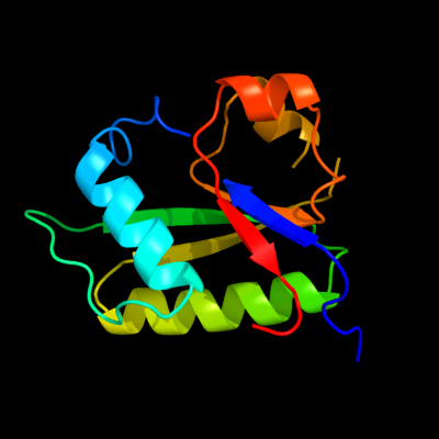
| c3nohA_ | PDB header: peptide binding protein | Chain: A: PDB Molecule: putative peptide binding protein; | PDBTitle: crystal structure of a putative peptide binding protein (rumgna_00914)2 from ruminococcus gnavus atcc 29149 at 1.60 a resolution |
| Added to library: Sat Aug 28 20:27:59 2010 | |
| Links to external resources | |
|---|---|
| 1 | . | . | . | . | . | . | . | . | 10 | . | . | . | . | . | . | . | . | . | 20 | . | . | . | . | . | . | . | . | . | 30 | . | . | . | . | . | . | . | . | . | 40 | . | . | . | . | . | . | . | . | . | 50 | . | . | . | . | . | . | . | . | . | 60 | . | . | . | . | . | . | . | . | . | 70 | |
| Sequence | S | A | Q | L | E | G | S | Y | I | F | C | X | N | P | L | L | D | K | L | S | D | E | D | I | R | E | Q | L | K | A | F | V | T | G | K | T | D | S | I | R | T | D | T | E | L | S | F | D | I | Y | V | S | E | T | D | Y | A | L | I | R | Y | A | D | S | L | C | E | R | L | N |
| Predicted secondary structure |  | | |  |  | | | | | | | | | | | | | | | | | | | | | | | | | | | | | | | | | | | | | |||||||||||||||||||||||||||||
| SS confidence | ||||||||||||||||||||||||||||||||||||||||||||||||||||||||||||||||||||||
| Known secondary structure (DSSP) | S | | | | | | | T | T | S | G | G | G | | | | | | | | | | | | T | T | S | S | S | S | | | | | | | | T | T | | | | | | | | | | | | | | | | . | . | . | . | . | . | . | . | . | 80 | . | . | . | . | . | . | . | . | . | 90 | . | . | . | . | . | . | . | . | . | 100 | . | . | . | . | . | . | . | . | . | 110 | . | . | . | . | . | . | . | . | . | 120 | . | . | . |
| Sequence | D | A | G | A | D | V | Q | I | K | Q | Y | S | G | T | X | L | R | S | R | A | V | S | G | K | Y | E | A | F | L | S | E | S | D | L | V | S | T | D | A | L | E | N | A | D | Y | I | I | L | D | S | A | E | X | |||||||||||||||||
| Predicted secondary structure | | | | | | | | | | | | | | | | | | | | | | | | | | | | | | | ||||||||||||||||||||||||||||||||||||||||
| SS confidence | ||||||||||||||||||||||||||||||||||||||||||||||||||||||||||||||||||||||
| Known secondary structure (DSSP) | | T | T | | | | | | | | | | | | | | | | | | T | S | | | | | | T | T | S | S | | | | | | T | S | | | | | | | G | G | G |
| Download: | PDB structure | FASTA sequence |
Phyre is now FREE for commercial users! All images and data generated by Phyre2 are free to use in any publication with acknowledgement Accessibility Statement
| ||||||||||||||||||||||||