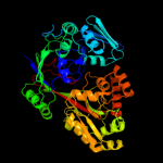
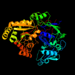
1 c3tpaA_
100.0
28
PDB header: heme binding proteinChain: A: PDB Molecule: heme-binding protein a;PDBTitle: structure of hbpa2 from haemophilus parasuis
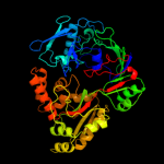
2 d1dpea_
100.0
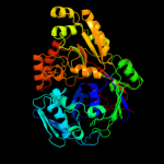
27
Fold: Periplasmic binding protein-like IISuperfamily: Periplasmic binding protein-like IIFamily: Phosphate binding protein-like3 c3m8uA_
100.0
26
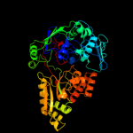
PDB header: transport proteinChain: A: PDB Molecule: heme-binding protein a;PDBTitle: crystal structure of glutathione-binding protein a (gbpa) from2 haemophilus parasuis sh0165 in complex with glutathione disulfide3 (gssg)
4 d1zlqa1
100.0
24
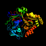
Fold: Periplasmic binding protein-like IISuperfamily: Periplasmic binding protein-like IIFamily: Phosphate binding protein-like5 c2wokA_
100.0
22
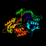
PDB header: peptide binding protein/peptideChain: A: PDB Molecule: clavulanic acid biosynthesis oligopeptidePDBTitle: clavulanic acid biosynthesis oligopeptide2 binding protein 2 complexed with bradykinin
6 d1jeta_
100.0
26
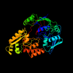
Fold: Periplasmic binding protein-like IISuperfamily: Periplasmic binding protein-like IIFamily: Phosphate binding protein-like7 c3o9pA_
100.0
28
PDB header: peptide binding protein/peptideChain: A: PDB Molecule: periplasmic murein peptide-binding protein;PDBTitle: the structure of the escherichia coli murein tripeptide binding2 protein mppa
8 d1xoca1
100.0
23
Fold: Periplasmic binding protein-like IISuperfamily: Periplasmic binding protein-like IIFamily: Phosphate binding protein-like9 d1uqwa_
100.0
24
Fold: Periplasmic binding protein-like IISuperfamily: Periplasmic binding protein-like IIFamily: Phosphate binding protein-like10 c3t66A_
100.0
23
PDB header: transport proteinChain: A: PDB Molecule: nickel abc transporter (nickel-binding protein);PDBTitle: crystal structure of nickel abc transporter from bacillus halodurans
11 c1ztyA_
100.0
21
PDB header: sugar binding protein, signaling proteinChain: A: PDB Molecule: chitin oligosaccharide binding protein;PDBTitle: crystal structure of the chitin oligasaccharide binding2 protein
12 c2grvC_
100.0
15
PDB header: biosynthetic proteinChain: C: PDB Molecule: lpqw;PDBTitle: crystal structure of lpqw
13 c3rqtA_
100.0
21
PDB header: unknown functionChain: A: PDB Molecule: putative uncharacterized protein;PDBTitle: 1.5 angstrom crystal structure of the complex of ligand binding2 component of abc-type import system from staphylococcus aureus with3 nickel and two histidines
14 c2o7jA_
100.0
22
PDB header: sugar binding proteinChain: A: PDB Molecule: oligopeptide abc transporter, periplasmicPDBTitle: the x-ray crystal structure of a thermophilic cellobiose2 binding protein bound with cellopentaose
15 d1vr5a1
100.0
20
Fold: Periplasmic binding protein-like IISuperfamily: Periplasmic binding protein-like IIFamily: Phosphate binding protein-like16 c3ftoA_
100.0
22
PDB header: peptide binding proteinChain: A: PDB Molecule: oligopeptide-binding protein oppa;PDBTitle: crystal structure of oppa in a open conformation
17 c3ry3B_
100.0
23
PDB header: transport proteinChain: B: PDB Molecule: putative solute-binding protein;PDBTitle: putative solute-binding protein from yersinia pestis.
18 c2d5wA_
100.0
19
PDB header: peptide binding proteinChain: A: PDB Molecule: peptide abc transporter, peptide-binding protein;PDBTitle: the crystal structure of oligopeptide binding protein from thermus2 thermophilus hb8 complexed with pentapeptide
19 c3lvuB_
100.0
17
PDB header: transport proteinChain: B: PDB Molecule: abc transporter, periplasmic substrate-binding protein;PDBTitle: crystal structure of abc transporter, periplasmic substrate-binding2 protein spo2066 from silicibacter pomeroyi
20 c3pamB_
100.0
18
PDB header: transport proteinChain: B: PDB Molecule: transmembrane protein;PDBTitle: crystal structure of a domain of transmembrane protein of abc-type2 oligopeptide transport system from bartonella henselae str. houston-1
21 c3o6pA_
not modelled
100.0
33
PDB header: protein bindingChain: A: PDB Molecule: peptide abc transporter, peptide-binding protein;PDBTitle: crystal structure of peptide abc transporter, peptide-binding protein
22 c3chgB_
not modelled
96.2
21
PDB header: ligand binding proteinChain: B: PDB Molecule: glycine betaine-binding protein;PDBTitle: the compatible solute-binding protein opuac from bacillus2 subtilis in complex with dmsa
23 c3l6gA_
not modelled
96.0
22
PDB header: glycine betaine-binding proteinChain: A: PDB Molecule: betaine abc transporter permease and substrate bindingPDBTitle: crystal structure of lactococcal opuac in its open conformation
24 c3tmgA_
not modelled
95.5
17
PDB header: transport proteinChain: A: PDB Molecule: glycine betaine, l-proline abc transporter,PDBTitle: crystal structure of glycine betaine, l-proline abc transporter,2 glycine/betaine/l-proline-binding protein (prox) from borrelia3 burgdorferi
25 c2rejA_
not modelled
94.5
17
PDB header: choline-binding proteinChain: A: PDB Molecule: putative glycine betaine abc transporter protein;PDBTitle: abc-transporter choline binding protein in unliganded semi-2 closed conformation
26 d1r9la_
not modelled
94.4
22
Fold: Periplasmic binding protein-like IISuperfamily: Periplasmic binding protein-like IIFamily: Phosphate binding protein-like27 c3nohA_
not modelled
93.6
20
PDB header: peptide binding proteinChain: A: PDB Molecule: putative peptide binding protein;PDBTitle: crystal structure of a putative peptide binding protein (rumgna_00914)2 from ruminococcus gnavus atcc 29149 at 1.60 a resolution
28 c3r6uA_
not modelled
90.9
20
PDB header: transport proteinChain: A: PDB Molecule: choline-binding protein;PDBTitle: crystal structure of choline binding protein opubc from bacillus2 subtilis
29 c3f6sI_
not modelled
88.6
18
PDB header: electron transportChain: I: PDB Molecule: flavodoxin;PDBTitle: desulfovibrio desulfuricans (atcc 29577) oxidized flavodoxin2 alternate conformers
30 d1sw5a_
not modelled
87.6
11
Fold: Periplasmic binding protein-like IISuperfamily: Periplasmic binding protein-like IIFamily: Phosphate binding protein-like31 c3pppA_
not modelled
82.3
16
PDB header: transport proteinChain: A: PDB Molecule: glycine betaine/carnitine/choline-binding protein;PDBTitle: structures of the substrate-binding protein provide insights into the2 multiple compatible solutes binding specificities of bacillus3 subtilis abc transporter opuc
32 d1e5da1
not modelled
80.9
14
Fold: Flavodoxin-likeSuperfamily: FlavoproteinsFamily: Flavodoxin-related33 d1ycga1
not modelled
80.1
19
Fold: Flavodoxin-likeSuperfamily: FlavoproteinsFamily: Flavodoxin-related34 c3hlyA_
not modelled
79.9
14
PDB header: flavoproteinChain: A: PDB Molecule: flavodoxin-like domain;PDBTitle: crystal structure of the flavodoxin-like domain from2 synechococcus sp q5mzp6_synp6 protein. northeast structural3 genomics consortium target snr135d.
35 d1f4pa_
not modelled
77.1
16
Fold: Flavodoxin-likeSuperfamily: FlavoproteinsFamily: Flavodoxin-related36 d2fz5a1
not modelled
72.3
11
Fold: Flavodoxin-likeSuperfamily: FlavoproteinsFamily: Flavodoxin-related37 d1ykga1
not modelled
63.3
12
Fold: Flavodoxin-likeSuperfamily: FlavoproteinsFamily: Cytochrome p450 reductase N-terminal domain-like38 d1tlla2
not modelled
63.1
8
Fold: Flavodoxin-likeSuperfamily: FlavoproteinsFamily: Cytochrome p450 reductase N-terminal domain-like39 d5nula_
not modelled
62.7
10
Fold: Flavodoxin-likeSuperfamily: FlavoproteinsFamily: Flavodoxin-related40 d1ja1a2
not modelled
61.8
11
Fold: Flavodoxin-likeSuperfamily: FlavoproteinsFamily: Cytochrome p450 reductase N-terminal domain-like41 c3ir1F_
not modelled
59.8
19
PDB header: protein bindingChain: F: PDB Molecule: outer membrane lipoprotein gna1946;PDBTitle: crystal structure of lipoprotein gna1946 from neisseria2 meningitidis
42 c3un6A_
not modelled
56.9
10
PDB header: unknown functionChain: A: PDB Molecule: hypothetical protein saouhsc_00137;PDBTitle: 2.0 angstrom crystal structure of ligand binding component of abc-type2 import system from staphylococcus aureus with zinc bound
43 c1bvyF_
not modelled
54.6
22
PDB header: oxidoreductaseChain: F: PDB Molecule: protein (cytochrome p450 bm-3);PDBTitle: complex of the heme and fmn-binding domains of the2 cytochrome p450(bm-3)
44 d1bvyf_
not modelled
54.6
22
Fold: Flavodoxin-likeSuperfamily: FlavoproteinsFamily: Flavodoxin-related45 c2ek8A_
not modelled
54.0
10
PDB header: hydrolaseChain: A: PDB Molecule: aminopeptidase;PDBTitle: aminopeptidase from aneurinibacillus sp. strain am-1
46 c3kzgB_
not modelled
53.3
11
PDB header: transport proteinChain: B: PDB Molecule: arginine 3rd transport system periplasmic bindingPDBTitle: crystal structure of an arginine 3rd transport system2 periplasmic binding protein from legionella pneumophila
47 c3kn3C_
not modelled
52.8
11
PDB header: transcriptionChain: C: PDB Molecule: putative periplasmic protein;PDBTitle: crystal structure of lysr substrate binding domain (25-263) of2 putative periplasmic protein from wolinella succinogenes
48 c3lr1A_
not modelled
51.3
11
PDB header: transport proteinChain: A: PDB Molecule: tungstate abc transporter, periplasmic tungstate-PDBTitle: the crystal structure of the tungstate abc transporter from2 geobacter sulfurreducens
49 c2o1mB_
not modelled
50.1
18
PDB header: structural genomics, unknown functionChain: B: PDB Molecule: probable amino-acid abc transporterPDBTitle: crystal structure of the probable amino-acid abc2 transporter extracellular-binding protein ytmk from3 bacillus subtilis. northeast structural genomics4 consortium target sr572
50 c2hnbA_
not modelled
49.6
14
PDB header: electron transportChain: A: PDB Molecule: protein mioc;PDBTitle: solution structure of a bacterial holo-flavodoxin
51 c3gxaA_
not modelled
47.2
19
PDB header: protein bindingChain: A: PDB Molecule: outer membrane lipoprotein gna1946;PDBTitle: crystal structure of gna1946
52 c3fniA_
not modelled
45.0
17
PDB header: oxidoreductaseChain: A: PDB Molecule: putative diflavin flavoprotein a 3;PDBTitle: crystal structure of a diflavin flavoprotein a3 (all3895) from nostoc2 sp., northeast structural genomics consortium target nsr431a
53 c3tqwA_
not modelled
44.2
15
PDB header: transport proteinChain: A: PDB Molecule: methionine-binding protein;PDBTitle: structure of a abc transporter, periplasmic substrate-binding protein2 from coxiella burnetii
54 c3hr4C_
not modelled
44.1
9
PDB header: oxidoreductase/metal binding proteinChain: C: PDB Molecule: nitric oxide synthase, inducible;PDBTitle: human inos reductase and calmodulin complex
55 d1ydga_
not modelled
41.7
18
Fold: Flavodoxin-likeSuperfamily: FlavoproteinsFamily: WrbA-like56 d2arka1
not modelled
39.2
17
Fold: Flavodoxin-likeSuperfamily: FlavoproteinsFamily: WrbA-like57 d1vmea1
not modelled
38.0
20
Fold: Flavodoxin-likeSuperfamily: FlavoproteinsFamily: Flavodoxin-related58 d1s8na_
not modelled
37.5
12
Fold: Flavodoxin-likeSuperfamily: CheY-likeFamily: CheY-related59 c2rc9A_
not modelled
35.6
12
PDB header: membrane proteinChain: A: PDB Molecule: glutamate [nmda] receptor subunit 3a;PDBTitle: crystal structure of the nr3a ligand binding core complex with acpc at2 1.96 angstrom resolution
60 c2ra9A_
not modelled
35.6
13
PDB header: unknown functionChain: A: PDB Molecule: uncharacterized protein duf1285;PDBTitle: crystal structure of a duf1285 family protein (sbal_2486) from2 shewanella baltica os155 at 1.40 a resolution
61 d1xs5a_
not modelled
34.9
12
Fold: Periplasmic binding protein-like IISuperfamily: Periplasmic binding protein-like IIFamily: Phosphate binding protein-like62 c3ix1B_
not modelled
33.7
10
PDB header: biosynthetic proteinChain: B: PDB Molecule: n-formyl-4-amino-5-aminomethyl-2-methylpyrimidine bindingPDBTitle: periplasmic n-formyl-4-amino-5-aminomethyl-2-methylpyrimidine binding2 protein from bacillus halodurans
63 c3ix1A_
not modelled
33.7
10
PDB header: biosynthetic proteinChain: A: PDB Molecule: n-formyl-4-amino-5-aminomethyl-2-methylpyrimidine bindingPDBTitle: periplasmic n-formyl-4-amino-5-aminomethyl-2-methylpyrimidine binding2 protein from bacillus halodurans
64 c3hn0A_
not modelled
33.6
23
PDB header: transport proteinChain: A: PDB Molecule: nitrate transport protein;PDBTitle: crystal structure of an abc transporter (bdi_1369) from2 parabacteroides distasonis at 1.75 a resolution
65 c2x26A_
not modelled
32.4
13
PDB header: transport proteinChain: A: PDB Molecule: periplasmic aliphatic sulphonates-binding protein;PDBTitle: crystal structure of the periplasmic aliphatic sulphonate2 binding protein ssua from escherichia coli
66 c3k2dA_
not modelled
31.7
14
PDB header: immune systemChain: A: PDB Molecule: abc-type metal ion transport system, periplasmic component;PDBTitle: crystal structure of immunogenic lipoprotein a from vibrio vulnificus
67 c1vmeB_
not modelled
31.5
18
PDB header: electron transportChain: B: PDB Molecule: flavoprotein;PDBTitle: crystal structure of flavoprotein (tm0755) from thermotoga maritima at2 1.80 a resolution
68 c2q9uB_
not modelled
31.0
9
PDB header: oxidoreductaseChain: B: PDB Molecule: a-type flavoprotein;PDBTitle: crystal structure of the flavodiiron protein from giardia2 intestinalis
69 d1b1ca_
not modelled
30.5
10
Fold: Flavodoxin-likeSuperfamily: FlavoproteinsFamily: Cytochrome p450 reductase N-terminal domain-like70 c1xt8B_
not modelled
28.1
10
PDB header: transport proteinChain: B: PDB Molecule: putative amino-acid transporter periplasmic solute-bindingPDBTitle: crystal structure of cysteine-binding protein from campylobacter2 jejuni at 2.0 a resolution
71 d1twya_
not modelled
27.8
15
Fold: Periplasmic binding protein-like IISuperfamily: Periplasmic binding protein-like IIFamily: Phosphate binding protein-like72 c1twyG_
not modelled
27.8
15
PDB header: structural genomics, unknown functionChain: G: PDB Molecule: abc transporter, periplasmic substrate-binding protein;PDBTitle: crystal structure of an abc-type phosphate transport receptor from2 vibrio cholerae
73 c3o66A_
not modelled
27.1
9
PDB header: transport proteinChain: A: PDB Molecule: glycine betaine/carnitine/choline abc transporter;PDBTitle: crystal structure of glycine betaine/carnitine/choline abc transporter
74 c2wc1A_
not modelled
26.0
8
PDB header: electron transportChain: A: PDB Molecule: flavodoxin;PDBTitle: three-dimensional structure of the nitrogen fixation2 flavodoxin (niff) from rhodobacter capsulatus at 2.2 a
75 d2a5la1
not modelled
25.9
11
Fold: Flavodoxin-likeSuperfamily: FlavoproteinsFamily: WrbA-like76 c3iibA_
not modelled
25.8
17
PDB header: hydrolaseChain: A: PDB Molecule: peptidase m28;PDBTitle: crystal structure of peptidase m28 precursor (yp_926796.1) from2 shewanella amazonensis sb2b at 1.70 a resolution
77 c2y7iB_
not modelled
25.2
4
PDB header: arginine-binding proteinChain: B: PDB Molecule: stm4351;PDBTitle: structural basis for high arginine specificity in salmonella2 typhimurium periplasmic binding protein stm4351.
78 c3e4rA_
not modelled
25.0
14
PDB header: transport proteinChain: A: PDB Molecule: nitrate transport protein;PDBTitle: crystal structure of the alkanesulfonate binding protein2 (ssua) from the phytopathogenic bacteria xanthomonas3 axonopodis pv. citri bound to hepes
79 c2zykA_
not modelled
24.5
20
PDB header: sugar binding proteinChain: A: PDB Molecule: solute-binding protein;PDBTitle: crystal structure of cyclo/maltodextrin-binding protein2 complexed with gamma-cyclodextrin
80 c2ylnA_
not modelled
24.1
18
PDB header: transport proteinChain: A: PDB Molecule: putative abc transporter, periplasmic binding protein,PDBTitle: crystal structure of the l-cystine solute receptor of2 neisseria gonorrhoeae in the closed conformation
81 d1mb3a_
not modelled
23.8
9
Fold: Flavodoxin-likeSuperfamily: CheY-likeFamily: CheY-related82 c3r39A_
not modelled
23.3
18
PDB header: transport proteinChain: A: PDB Molecule: putative periplasmic binding protein;PDBTitle: crystal structure of periplasmic d-alanine abc transporter from2 salmonella enterica
83 c3kbrA_
not modelled
22.4
7
PDB header: lyaseChain: A: PDB Molecule: cyclohexadienyl dehydratase;PDBTitle: the crystal structure of cyclohexadienyl dehydratase precursor from2 pseudomonas aeruginosa pa01
84 c3c97A_
not modelled
22.3
10
PDB header: signaling protein, transferaseChain: A: PDB Molecule: signal transduction histidine kinase;PDBTitle: crystal structure of the response regulator receiver domain2 of a signal transduction histidine kinase from aspergillus3 oryzae
85 c2bpoA_
not modelled
21.9
18
PDB header: reductaseChain: A: PDB Molecule: nadph-cytochrom p450 reductase;PDBTitle: crystal structure of the yeast cpr triple mutant: d74g,2 y75f, k78a.
86 d1wdna_
not modelled
21.7
9
Fold: Periplasmic binding protein-like IISuperfamily: Periplasmic binding protein-like IIFamily: Phosphate binding protein-like87 c1ychD_
not modelled
21.5
17
PDB header: oxidoreductaseChain: D: PDB Molecule: nitric oxide reductase;PDBTitle: x-ray crystal structures of moorella thermoacetica fpra.2 novel diiron site structure and mechanistic insights into3 a scavenging nitric oxide reductase
88 d1xhfa1
not modelled
21.5
12
Fold: Flavodoxin-likeSuperfamily: CheY-likeFamily: CheY-related89 c2q2aD_
not modelled
21.3
9
PDB header: transport proteinChain: D: PDB Molecule: artj;PDBTitle: crystal structures of the arginine-, lysine-, histidine-2 binding protein artj from the thermophilic bacterium3 geobacillus stearothermophilus
90 c1p99A_
not modelled
21.2
14
PDB header: structural genomics, unknown functionChain: A: PDB Molecule: hypothetical protein pg110;PDBTitle: 1.7a crystal structure of protein pg110 from staphylococcus2 aureus
91 d1p99a_
not modelled
21.2
14
Fold: Periplasmic binding protein-like IISuperfamily: Periplasmic binding protein-like IIFamily: Phosphate binding protein-like92 d1us5a_
not modelled
21.0
14
Fold: Periplasmic binding protein-like IISuperfamily: Periplasmic binding protein-like IIFamily: Phosphate binding protein-like93 d2a9pa1
not modelled
20.9
15
Fold: Flavodoxin-likeSuperfamily: CheY-likeFamily: CheY-related94 c2j48A_
not modelled
20.8
14
PDB header: transferaseChain: A: PDB Molecule: two-component sensor kinase;PDBTitle: nmr structure of the pseudo-receiver domain of the cika2 protein.
95 d1zgza1
not modelled
20.5
12
Fold: Flavodoxin-likeSuperfamily: CheY-likeFamily: CheY-related96 c3muqB_
not modelled
20.4
9
PDB header: structural genomics, unknown functionChain: B: PDB Molecule: uncharacterized conserved protein;PDBTitle: the crystal structure of a conserved functionally unknown protein from2 vibrio parahaemolyticus rimd 2210633
97 c3d7nA_
not modelled
20.2
9
PDB header: electron transportChain: A: PDB Molecule: flavodoxin, wrba-like protein;PDBTitle: the crystal structure of the flavodoxin, wrba-like protein from2 agrobacterium tumefaciens
98 d2a5sa1
not modelled
20.1
12
Fold: Periplasmic binding protein-like IISuperfamily: Periplasmic binding protein-like IIFamily: Phosphate binding protein-like99 c3f6cB_
not modelled
19.9
22
PDB header: dna binding proteinChain: B: PDB Molecule: positive transcription regulator evga;PDBTitle: crystal structure of n-terminal domain of positive transcription2 regulator evga from escherichia coli