1 c2yvaB_
100.0
100
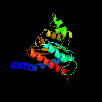
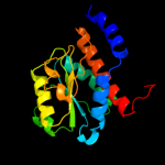
PDB header: dna binding proteinChain: B: PDB Molecule: dnaa initiator-associating protein diaa;PDBTitle: crystal structure of escherichia coli diaa
2 d1x92a_
100.0
58
Fold: SIS domainSuperfamily: SIS domainFamily: mono-SIS domain3 d1tk9a_
100.0
40
Fold: SIS domainSuperfamily: SIS domainFamily: mono-SIS domain4 c2x3yA_
100.0
40
PDB header: isomeraseChain: A: PDB Molecule: phosphoheptose isomerase;PDBTitle: crystal structure of gmha from burkholderia pseudomallei
5 c3trjC_
100.0
46
PDB header: isomeraseChain: C: PDB Molecule: phosphoheptose isomerase;PDBTitle: structure of a phosphoheptose isomerase from francisella tularensis
6 d1x94a_
100.0
42
Fold: SIS domainSuperfamily: SIS domainFamily: mono-SIS domain7 c3cvjB_
99.9
17
PDB header: isomeraseChain: B: PDB Molecule: putative phosphoheptose isomerase;PDBTitle: crystal structure of a putative phosphoheptose isomerase (bh3325) from2 bacillus halodurans c-125 at 2.00 a resolution
8 d1nria_
99.9
16
Fold: SIS domainSuperfamily: SIS domainFamily: mono-SIS domain9 c1nriA_
99.9
16
PDB header: structural genomics, unknown functionChain: A: PDB Molecule: hypothetical protein hi0754;PDBTitle: crystal structure of putative phosphosugar isomerase hi0754 from2 haemophilus influenzae
10 c3fxaA_
99.9
20
PDB header: sugar binding proteinChain: A: PDB Molecule: sis domain protein;PDBTitle: crystal structure of a putative sugar-phosphate isomerase2 (lmof2365_0531) from listeria monocytogenes str. 4b f2365 at 1.60 a3 resolution
11 c3etnD_
99.9
18
PDB header: isomeraseChain: D: PDB Molecule: putative phosphosugar isomerase involved in capsulePDBTitle: crystal structure of putative phosphosugar isomerase involved in2 capsule formation (yp_209877.1) from bacteroides fragilis nctc 93433 at 1.70 a resolution
12 c2xhzC_
99.8
14
PDB header: isomeraseChain: C: PDB Molecule: arabinose 5-phosphate isomerase;PDBTitle: probing the active site of the sugar isomerase domain from e. coli2 arabinose-5-phosphate isomerase via x-ray crystallography
13 d1m3sa_
99.8
17
Fold: SIS domainSuperfamily: SIS domainFamily: mono-SIS domain14 c3shoA_
99.8
22
PDB header: transcription regulatorChain: A: PDB Molecule: transcriptional regulator, rpir family;PDBTitle: crystal structure of rpir transcription factor from sphaerobacter2 thermophilus (sugar isomerase domain)
15 d1vima_
99.8
19
Fold: SIS domainSuperfamily: SIS domainFamily: mono-SIS domain16 d1jeoa_
99.7
16
Fold: SIS domainSuperfamily: SIS domainFamily: mono-SIS domain17 c3hbaA_
99.7
17
PDB header: isomeraseChain: A: PDB Molecule: putative phosphosugar isomerase;PDBTitle: crystal structure of a putative phosphosugar isomerase (sden_2705)2 from shewanella denitrificans os217 at 2.00 a resolution
18 c2a3nA_
99.7
15
PDB header: sugar binding proteinChain: A: PDB Molecule: putative glucosamine-fructose-6-phosphate aminotransferase;PDBTitle: crystal structure of a putative glucosamine-fructose-6-phosphate2 aminotransferase (stm4540.s) from salmonella typhimurium lt2 at 1.353 a resolution
19 c3fj1A_
99.7
19
PDB header: isomeraseChain: A: PDB Molecule: putative phosphosugar isomerase;PDBTitle: crystal structure of putative phosphosugar isomerase (yp_167080.1)2 from silicibacter pomeroyi dss-3 at 1.75 a resolution
20 c3knzA_
99.7
15
PDB header: sugar binding proteinChain: A: PDB Molecule: putative sugar binding protein;PDBTitle: crystal structure of putative sugar binding protein (np_459565.1) from2 salmonella typhimurium lt2 at 2.50 a resolution
21 c2puwA_
not modelled
99.6
13
PDB header: transferaseChain: A: PDB Molecule: isomerase domain of glutamine-fructose-6-phosphatePDBTitle: the crystal structure of isomerase domain of glucosamine-6-phosphate2 synthase from candida albicans
22 d1j5xa_
not modelled
99.6
19
Fold: SIS domainSuperfamily: SIS domainFamily: double-SIS domain23 c2amlB_
not modelled
99.6
19
PDB header: transferaseChain: B: PDB Molecule: sis domain protein;PDBTitle: crystal structure of lmo0035 protein (46906266) from listeria2 monocytogenes 4b f2365 at 1.50 a resolution
24 c2zj3A_
not modelled
99.6
14
PDB header: transferaseChain: A: PDB Molecule: glucosamine--fructose-6-phosphatePDBTitle: isomerase domain of human glucose:fructose-6-phosphate2 amidotransferase
25 c3g68A_
not modelled
99.6
14
PDB header: isomeraseChain: A: PDB Molecule: putative phosphosugar isomerase;PDBTitle: crystal structure of a putative phosphosugar isomerase (cd3275) from2 clostridium difficile 630 at 1.80 a resolution
26 c3euaD_
not modelled
99.6
14
PDB header: isomeraseChain: D: PDB Molecule: putative fructose-aminoacid-6-phosphate deglycase;PDBTitle: crystal structure of a putative phosphosugar isomerase (bsu32610) from2 bacillus subtilis at 1.90 a resolution
27 c3tbfA_
not modelled
99.6
12
PDB header: transferaseChain: A: PDB Molecule: glucosamine--fructose-6-phosphate aminotransferasePDBTitle: c-terminal domain of glucosamine-fructose-6-phosphate aminotransferase2 from francisella tularensis.
28 d1moqa_
not modelled
99.6
13
Fold: SIS domainSuperfamily: SIS domainFamily: double-SIS domain29 d1x9ia_
not modelled
99.5
24
Fold: SIS domainSuperfamily: SIS domainFamily: double-SIS domain30 c3fkjA_
not modelled
99.5
16
PDB header: isomeraseChain: A: PDB Molecule: putative phosphosugar isomerases;PDBTitle: crystal structure of a putative phosphosugar isomerase (stm_0572) from2 salmonella typhimurium lt2 at 2.12 a resolution
31 c2decA_
not modelled
99.4
15
PDB header: structural genomics, unknown functionChain: A: PDB Molecule: 325aa long hypothetical protein;PDBTitle: crystal structure of the ph0510 protein from pyrococcus horikoshii ot3
32 c3i0zB_
not modelled
99.4
22
PDB header: isomeraseChain: B: PDB Molecule: putative tagatose-6-phosphate ketose/aldose isomerase;PDBTitle: crystal structure of putative putative tagatose-6-phosphate2 ketose/aldose isomerase (np_344614.1) from streptococcus pneumoniae3 tigr4 at 1.70 a resolution
33 c3odpA_
not modelled
99.4
17
PDB header: isomeraseChain: A: PDB Molecule: putative tagatose-6-phosphate ketose/aldose isomerase;PDBTitle: crystal structure of a putative tagatose-6-phosphate ketose/aldose2 isomerase (nt01cx_0292) from clostridium novyi nt at 2.35 a3 resolution
34 c3c3jA_
not modelled
99.3
24
PDB header: isomeraseChain: A: PDB Molecule: putative tagatose-6-phosphate ketose/aldose isomerase;PDBTitle: crystal structure of tagatose-6-phosphate ketose/aldose isomerase from2 escherichia coli
35 c1jxaA_
not modelled
99.3
12
PDB header: transferaseChain: A: PDB Molecule: glucosamine 6-phosphate synthase;PDBTitle: glucosamine 6-phosphate synthase with glucose 6-phosphate
36 c2q8nB_
not modelled
98.4
14
PDB header: isomeraseChain: B: PDB Molecule: glucose-6-phosphate isomerase;PDBTitle: crystal structure of glucose-6-phosphate isomerase (ec2 5.3.1.9) (tm1385) from thermotoga maritima at 1.82 a3 resolution
37 c3ff1B_
not modelled
98.3
19
PDB header: isomeraseChain: B: PDB Molecule: glucose-6-phosphate isomerase;PDBTitle: structure of glucose 6-phosphate isomerase from staphylococcus aureus
38 d1c7qa_
not modelled
98.1
16
Fold: SIS domainSuperfamily: SIS domainFamily: Phosphoglucose isomerase, PGI39 c3jx9B_
not modelled
98.0
13
PDB header: isomeraseChain: B: PDB Molecule: putative phosphoheptose isomerase;PDBTitle: crystal structure of putative phosphoheptose isomerase2 (yp_001815198.1) from exiguobacterium sp. 255-15 at 1.95 a resolution
40 c1zzgB_
not modelled
97.9
16
PDB header: isomeraseChain: B: PDB Molecule: glucose-6-phosphate isomerase;PDBTitle: crystal structure of hypothetical protein tt0462 from thermus2 thermophilus hb8
41 c3ljkA_
not modelled
97.6
12
PDB header: isomeraseChain: A: PDB Molecule: glucose-6-phosphate isomerase;PDBTitle: glucose-6-phosphate isomerase from francisella tularensis.
42 d1gzda_
not modelled
97.4
15
Fold: SIS domainSuperfamily: SIS domainFamily: Phosphoglucose isomerase, PGI43 c2wu8A_
not modelled
97.4
15
PDB header: isomeraseChain: A: PDB Molecule: glucose-6-phosphate isomerase;PDBTitle: structural studies of phosphoglucose isomerase from2 mycobacterium tuberculosis h37rv
44 d1u0fa_
not modelled
97.4
16
Fold: SIS domainSuperfamily: SIS domainFamily: Phosphoglucose isomerase, PGI45 d1iata_
not modelled
97.4
14
Fold: SIS domainSuperfamily: SIS domainFamily: Phosphoglucose isomerase, PGI46 d1hm5a_
not modelled
97.3
14
Fold: SIS domainSuperfamily: SIS domainFamily: Phosphoglucose isomerase, PGI47 c3hjbA_
not modelled
97.3
15
PDB header: isomeraseChain: A: PDB Molecule: glucose-6-phosphate isomerase;PDBTitle: 1.5 angstrom crystal structure of glucose-6-phosphate isomerase from2 vibrio cholerae.
48 c1t10A_
not modelled
97.2
12
PDB header: isomeraseChain: A: PDB Molecule: glucose-6-phosphate isomerase;PDBTitle: phosphoglucose isomerase from leishmania mexicana in complex with2 substrate d-fructose-6-phosphate
49 c2o2cB_
not modelled
97.2
15
PDB header: isomeraseChain: B: PDB Molecule: glucose-6-phosphate isomerase, glycosomal;PDBTitle: crystal structure of phosphoglucose isomerase from t. brucei2 containing glucose-6-phosphate in the active site
50 d1q50a_
not modelled
97.1
14
Fold: SIS domainSuperfamily: SIS domainFamily: Phosphoglucose isomerase, PGI51 c3nbuC_
not modelled
97.1
12
PDB header: isomeraseChain: C: PDB Molecule: glucose-6-phosphate isomerase;PDBTitle: crystal structure of pgi glucosephosphate isomerase
52 c3ujhB_
not modelled
96.8
13
PDB header: isomeraseChain: B: PDB Molecule: glucose-6-phosphate isomerase;PDBTitle: crystal structure of substrate-bound glucose-6-phosphate isomerase2 from toxoplasma gondii
53 c3bbnB_
not modelled
96.3
17
PDB header: ribosomeChain: B: PDB Molecule: ribosomal protein s2;PDBTitle: homology model for the spinach chloroplast 30s subunit2 fitted to 9.4a cryo-em map of the 70s chlororibosome.
54 c3pr3B_
not modelled
96.3
19
PDB header: isomeraseChain: B: PDB Molecule: glucose-6-phosphate isomerase;PDBTitle: crystal structure of plasmodium falciparum glucose-6-phosphate2 isomerase (pf14_0341) in complex with fructose-6-phosphate
55 c2zkqb_
not modelled
96.2
20
PDB header: ribosomal protein/rnaChain: B: PDB Molecule: rna expansion segment es3;PDBTitle: structure of a mammalian ribosomal 40s subunit within an2 80s complex obtained by docking homology models of the rna3 and proteins into an 8.7 a cryo-em map
56 c3iz6A_
not modelled
96.1
15
PDB header: ribosomeChain: A: PDB Molecule: 40s ribosomal protein sa (s2p);PDBTitle: localization of the small subunit ribosomal proteins into a 5.5 a2 cryo-em map of triticum aestivum translating 80s ribosome
57 c3bchA_
not modelled
95.8
20
PDB header: cell adhesion, ribosomal proteinChain: A: PDB Molecule: 40s ribosomal protein sa;PDBTitle: crystal structure of the human laminin receptor precursor
58 d1y5ia2
not modelled
95.7
8
Fold: Formate dehydrogenase/DMSO reductase, domains 1-3Superfamily: Formate dehydrogenase/DMSO reductase, domains 1-3Family: Formate dehydrogenase/DMSO reductase, domains 1-359 c1h0hA_
not modelled
95.7
11
PDB header: dehydrogenaseChain: A: PDB Molecule: formate dehydrogenase (large subunit);PDBTitle: tungsten containing formate dehydrogenase from2 desulfovibrio gigas
60 d2jioa2
not modelled
95.7
13
Fold: Formate dehydrogenase/DMSO reductase, domains 1-3Superfamily: Formate dehydrogenase/DMSO reductase, domains 1-3Family: Formate dehydrogenase/DMSO reductase, domains 1-361 c3izbA_
not modelled
95.7
15
PDB header: ribosomeChain: A: PDB Molecule: 40s ribosomal protein rps0 (s2p);PDBTitle: localization of the small subunit ribosomal proteins into a 6.1 a2 cryo-em map of saccharomyces cerevisiae translating 80s ribosome
62 c1s1hB_
not modelled
95.6
14
PDB header: ribosomeChain: B: PDB Molecule: 40s ribosomal protein s0-a;PDBTitle: structure of the ribosomal 80s-eef2-sordarin complex from2 yeast obtained by docking atomic models for rna and protein3 components into a 11.7 a cryo-em map. this file, 1s1h,4 contains 40s subunit. the 60s ribosomal subunit is in file5 1s1i.
63 c1y5iA_
not modelled
95.5
8
PDB header: oxidoreductaseChain: A: PDB Molecule: respiratory nitrate reductase 1 alpha chain;PDBTitle: the crystal structure of the narghi mutant nari-k86a
64 c2v45A_
not modelled
95.5
14
PDB header: oxidoreductaseChain: A: PDB Molecule: periplasmic nitrate reductase;PDBTitle: a new catalytic mechanism of periplasmic nitrate reductase2 from desulfovibrio desulfuricans atcc 27774 from3 crystallographic and epr data and based on detailed4 analysis of the sixth ligand
65 d1ogya2
not modelled
95.4
10
Fold: Formate dehydrogenase/DMSO reductase, domains 1-3Superfamily: Formate dehydrogenase/DMSO reductase, domains 1-3Family: Formate dehydrogenase/DMSO reductase, domains 1-366 c2xznB_
not modelled
95.4
19
PDB header: ribosomeChain: B: PDB Molecule: rps0e;PDBTitle: crystal structure of the eukaryotic 40s ribosomal2 subunit in complex with initiation factor 1. this file3 contains the 40s subunit and initiation factor for4 molecule 2
67 d1h0ha2
not modelled
95.3
12
Fold: Formate dehydrogenase/DMSO reductase, domains 1-3Superfamily: Formate dehydrogenase/DMSO reductase, domains 1-3Family: Formate dehydrogenase/DMSO reductase, domains 1-368 c2ivfA_
not modelled
95.3
11
PDB header: oxidoreductaseChain: A: PDB Molecule: ethylbenzene dehydrogenase alpha-subunit;PDBTitle: ethylbenzene dehydrogenase from aromatoleum aromaticum
69 d2uubb1
not modelled
95.2
23
Fold: Flavodoxin-likeSuperfamily: Ribosomal protein S2Family: Ribosomal protein S270 d2iv2x2
not modelled
95.2
9
Fold: Formate dehydrogenase/DMSO reductase, domains 1-3Superfamily: Formate dehydrogenase/DMSO reductase, domains 1-3Family: Formate dehydrogenase/DMSO reductase, domains 1-371 c2e7zA_
not modelled
95.1
13
PDB header: lyaseChain: A: PDB Molecule: acetylene hydratase ahy;PDBTitle: acetylene hydratase from pelobacter acetylenicus
72 c1kqgA_
not modelled
95.0
9
PDB header: oxidoreductaseChain: A: PDB Molecule: formate dehydrogenase, nitrate-inducible, major subunit;PDBTitle: formate dehydrogenase n from e. coli
73 d2gy9b1
not modelled
94.9
20
Fold: Flavodoxin-likeSuperfamily: Ribosomal protein S2Family: Ribosomal protein S274 c2nyaF_
not modelled
94.8
9
PDB header: oxidoreductaseChain: F: PDB Molecule: periplasmic nitrate reductase;PDBTitle: crystal structure of the periplasmic nitrate reductase2 (nap) from escherichia coli
75 d1kqfa2
not modelled
94.7
9
Fold: Formate dehydrogenase/DMSO reductase, domains 1-3Superfamily: Formate dehydrogenase/DMSO reductase, domains 1-3Family: Formate dehydrogenase/DMSO reductase, domains 1-376 c1ogyA_
not modelled
94.7
10
PDB header: oxidoreductaseChain: A: PDB Molecule: periplasmic nitrate reductase;PDBTitle: crystal structure of the heterodimeric nitrate reductase2 from rhodobacter sphaeroides
77 c2vpyE_
not modelled
94.6
11
PDB header: oxidoreductaseChain: E: PDB Molecule: thiosulfate reductase;PDBTitle: polysulfide reductase with bound quinone inhibitor,2 pentachlorophenol (pcp)
78 c2iv2X_
not modelled
94.4
9
PDB header: oxidoreductaseChain: X: PDB Molecule: formate dehydrogenase h;PDBTitle: reinterpretation of reduced form of formate dehydrogenase h2 from e. coli
79 d1vi6a_
not modelled
93.8
21
Fold: Flavodoxin-likeSuperfamily: Ribosomal protein S2Family: Ribosomal protein S280 d1vlfm2
not modelled
92.8
5
Fold: Formate dehydrogenase/DMSO reductase, domains 1-3Superfamily: Formate dehydrogenase/DMSO reductase, domains 1-3Family: Formate dehydrogenase/DMSO reductase, domains 1-381 c1tmoA_
not modelled
92.1
9
PDB header: oxidoreductaseChain: A: PDB Molecule: trimethylamine n-oxide reductase;PDBTitle: trimethylamine n-oxide reductase from shewanella massilia
82 d1dmra2
not modelled
92.1
11
Fold: Formate dehydrogenase/DMSO reductase, domains 1-3Superfamily: Formate dehydrogenase/DMSO reductase, domains 1-3Family: Formate dehydrogenase/DMSO reductase, domains 1-383 d1p3da1
not modelled
91.5
11
Fold: MurCD N-terminal domainSuperfamily: MurCD N-terminal domainFamily: MurCD N-terminal domain84 c1vlfQ_
not modelled
91.5
5
PDB header: oxidoreductaseChain: Q: PDB Molecule: pyrogallol hydroxytransferase large subunit;PDBTitle: crystal structure of pyrogallol-phloroglucinol2 transhydroxylase from pelobacter acidigallici complexed3 with inhibitor 1,2,4,5-tetrahydroxy-benzene
85 c3jwpA_
not modelled
90.8
13
PDB header: transcriptionChain: A: PDB Molecule: transcriptional regulatory protein sir2 homologue;PDBTitle: crystal structure of plasmodium falciparum sir2a (pf13_0152) in2 complex with amp
86 c3uagA_
not modelled
90.3
16
PDB header: ligaseChain: A: PDB Molecule: protein (udp-n-acetylmuramoyl-l-alanine:d-PDBTitle: udp-n-acetylmuramoyl-l-alanine:d-glutamate ligase
87 c2axqA_
not modelled
90.2
17
PDB header: oxidoreductaseChain: A: PDB Molecule: saccharopine dehydrogenase;PDBTitle: apo histidine-tagged saccharopine dehydrogenase (l-glu2 forming) from saccharomyces cerevisiae
88 d1tmoa2
not modelled
89.7
9
Fold: Formate dehydrogenase/DMSO reductase, domains 1-3Superfamily: Formate dehydrogenase/DMSO reductase, domains 1-3Family: Formate dehydrogenase/DMSO reductase, domains 1-389 c1h5nC_
not modelled
89.5
9
PDB header: oxidoreductaseChain: C: PDB Molecule: dmso reductase;PDBTitle: dmso reductase modified by the presence of dms and air
90 d2ax3a2
not modelled
89.0
12
Fold: YjeF N-terminal domain-likeSuperfamily: YjeF N-terminal domain-likeFamily: YjeF N-terminal domain-like91 d1j6ua1
not modelled
88.6
17
Fold: MurCD N-terminal domainSuperfamily: MurCD N-terminal domainFamily: MurCD N-terminal domain92 c3lk7A_
not modelled
88.5
16
PDB header: ligaseChain: A: PDB Molecule: udp-n-acetylmuramoylalanine--d-glutamate ligase;PDBTitle: the crystal structure of udp-n-acetylmuramoylalanine-d-2 glutamate (murd) ligase from streptococcus agalactiae to3 1.5a
93 d2jfga1
not modelled
88.5
14
Fold: MurCD N-terminal domainSuperfamily: MurCD N-terminal domainFamily: MurCD N-terminal domain94 c2q1yB_
not modelled
88.2
22
PDB header: cell cycle, signaling proteinChain: B: PDB Molecule: cell division protein ftsz;PDBTitle: crystal structure of cell division protein ftsz from mycobacterium2 tuberculosis in complex with gtp-gamma-s
95 d1ma3a_
not modelled
88.0
7
Fold: DHS-like NAD/FAD-binding domainSuperfamily: DHS-like NAD/FAD-binding domainFamily: Sir2 family of transcriptional regulators96 c1j6uA_
not modelled
86.9
19
PDB header: ligaseChain: A: PDB Molecule: udp-n-acetylmuramate-alanine ligase murc;PDBTitle: crystal structure of udp-n-acetylmuramate-alanine ligase2 murc (tm0231) from thermotoga maritima at 2.3 a resolution
97 c2f00A_
not modelled
86.8
18
PDB header: ligaseChain: A: PDB Molecule: udp-n-acetylmuramate--l-alanine ligase;PDBTitle: escherichia coli murc
98 d2b4ya1
not modelled
86.7
9
Fold: DHS-like NAD/FAD-binding domainSuperfamily: DHS-like NAD/FAD-binding domainFamily: Sir2 family of transcriptional regulators99 c1w59B_
not modelled
86.2
19
PDB header: cell divisionChain: B: PDB Molecule: cell division protein ftsz homolog 1;PDBTitle: ftsz dimer, empty (m. jannaschii)
100 c3k35D_
not modelled
85.9
12
PDB header: hydrolaseChain: D: PDB Molecule: nad-dependent deacetylase sirtuin-6;PDBTitle: crystal structure of human sirt6
101 d1m2ka_
not modelled
85.8
11
Fold: DHS-like NAD/FAD-binding domainSuperfamily: DHS-like NAD/FAD-binding domainFamily: Sir2 family of transcriptional regulators102 d1yc5a1
not modelled
85.7
5
Fold: DHS-like NAD/FAD-binding domainSuperfamily: DHS-like NAD/FAD-binding domainFamily: Sir2 family of transcriptional regulators103 c3d3jA_
not modelled
85.3
11
PDB header: protein bindingChain: A: PDB Molecule: enhancer of mrna-decapping protein 3;PDBTitle: crystal structure of human edc3p
104 c3pkiF_
not modelled
85.0
12
PDB header: hydrolaseChain: F: PDB Molecule: nad-dependent deacetylase sirtuin-6;PDBTitle: human sirt6 crystal structure in complex with adp ribose
105 c2ax3A_
not modelled
84.2
12
PDB header: transferaseChain: A: PDB Molecule: hypothetical protein tm0922;PDBTitle: crystal structure of a putative carbohydrate kinase (tm0922) from2 thermotoga maritima msb8 at 2.25 a resolution
106 c2rhoB_
not modelled
83.2
22
PDB header: cell cycleChain: B: PDB Molecule: cell division protein ftsz;PDBTitle: synthetic gene encoded bacillus subtilis ftsz ncs dimer with2 bound gdp and gtp-gamma-s
107 c1ir6A_
not modelled
82.9
14
PDB header: hydrolaseChain: A: PDB Molecule: exonuclease recj;PDBTitle: crystal structure of exonuclease recj bound to manganese
108 d1ir6a_
not modelled
82.9
14
Fold: DHH phosphoesterasesSuperfamily: DHH phosphoesterasesFamily: Exonuclease RecJ109 d1pjqa1
not modelled
81.5
21
Fold: NAD(P)-binding Rossmann-fold domainsSuperfamily: NAD(P)-binding Rossmann-fold domainsFamily: Siroheme synthase N-terminal domain-like110 d1rq2a1
not modelled
80.4
25
Fold: Tubulin nucleotide-binding domain-likeSuperfamily: Tubulin nucleotide-binding domain-likeFamily: Tubulin, GTPase domain111 c1e5lA_
not modelled
78.5
15
PDB header: oxidoreductaseChain: A: PDB Molecule: saccharopine reductase;PDBTitle: apo saccharopine reductase from magnaporthe grisea
112 d1s5pa_
not modelled
78.4
21
Fold: DHS-like NAD/FAD-binding domainSuperfamily: DHS-like NAD/FAD-binding domainFamily: Sir2 family of transcriptional regulators113 c2dg2D_
not modelled
77.2
11
PDB header: protein bindingChain: D: PDB Molecule: apolipoprotein a-i binding protein;PDBTitle: crystal structure of mouse apolipoprotein a-i binding2 protein
114 c3eagA_
not modelled
77.2
23
PDB header: ligaseChain: A: PDB Molecule: udp-n-acetylmuramate:l-alanyl-gamma-d-glutamyl-meso-PDBTitle: the crystal structure of udp-n-acetylmuramate:l-alanyl-gamma-d-2 glutamyl-meso-diaminopimelate ligase (mpl) from neisseria3 meningitides
115 c3d3kD_
not modelled
77.1
12
PDB header: protein bindingChain: D: PDB Molecule: enhancer of mrna-decapping protein 3;PDBTitle: crystal structure of human edc3p
116 c2vawA_
not modelled
76.9
22
PDB header: cell cycleChain: A: PDB Molecule: cell division protein ftsz;PDBTitle: ftsz pseudomonas aeruginosa gdp
117 c3q2oB_
not modelled
76.8
21
PDB header: lyaseChain: B: PDB Molecule: phosphoribosylaminoimidazole carboxylase, atpase subunit;PDBTitle: crystal structure of purk: n5-carboxyaminoimidazole ribonucleotide2 synthetase
118 c3ijpA_
not modelled
76.7
14
PDB header: oxidoreductaseChain: A: PDB Molecule: dihydrodipicolinate reductase;PDBTitle: crystal structure of dihydrodipicolinate reductase from2 bartonella henselae at 2.0a resolution
119 d1ibja_
not modelled
74.2
17
Fold: PLP-dependent transferase-likeSuperfamily: PLP-dependent transferasesFamily: Cystathionine synthase-like120 d1kjqa2
not modelled
74.2
8
Fold: PreATP-grasp domainSuperfamily: PreATP-grasp domainFamily: BC N-terminal domain-like