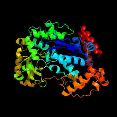
1 d2qxfa1
100.0
100
Fold: Ribonuclease H-like motifSuperfamily: Ribonuclease H-likeFamily: DnaQ-like 3'-5' exonuclease2 d1y97a1
100.0
15
Fold: Ribonuclease H-like motifSuperfamily: Ribonuclease H-likeFamily: DnaQ-like 3'-5' exonuclease3 d1j9aa_
100.0
18
Fold: Ribonuclease H-like motifSuperfamily: Ribonuclease H-likeFamily: DnaQ-like 3'-5' exonuclease4 d3b6oa1
100.0
18
Fold: Ribonuclease H-like motifSuperfamily: Ribonuclease H-likeFamily: DnaQ-like 3'-5' exonuclease5 d2f96a1
100.0
20
Fold: Ribonuclease H-like motifSuperfamily: Ribonuclease H-likeFamily: DnaQ-like 3'-5' exonuclease6 c3u6fA_
100.0
17
PDB header: hydrolase/dnaChain: A: PDB Molecule: three prime repair exonuclease 1;PDBTitle: mouse trex1 d200n mutant
7 c3tr8A_
100.0
17
PDB header: hydrolaseChain: A: PDB Molecule: oligoribonuclease;PDBTitle: structure of an oligoribonuclease (orn) from coxiella burnetii
8 d1w0ha_
100.0
11
Fold: Ribonuclease H-like motifSuperfamily: Ribonuclease H-likeFamily: DnaQ-like 3'-5' exonuclease9 d2igia1
100.0
16
Fold: Ribonuclease H-like motifSuperfamily: Ribonuclease H-likeFamily: DnaQ-like 3'-5' exonuclease10 c2gbzA_
100.0
17
PDB header: hydrolaseChain: A: PDB Molecule: oligoribonuclease;PDBTitle: the crystal structure of xc847 from xanthomonas campestris: a 3-52 oligoribonuclease of dnaq fold family with a novel opposingly-shifted3 helix
11 c3cm6A_
100.0
15
PDB header: hydrolase, apoptosisChain: A: PDB Molecule: cell death-related nuclease 4;PDBTitle: crystal structure of cell-death related nuclease 4 (crn-4)2 bound with er
12 c2p1jB_
100.0
20
PDB header: transferaseChain: B: PDB Molecule: dna polymerase iii polc-type;PDBTitle: crystal structure of a polc-type dna polymerase iii2 exonuclease domain from thermotoga maritima
13 c2xriA_
100.0
14
PDB header: hydrolaseChain: A: PDB Molecule: eri1 exoribonuclease 3;PDBTitle: crystal structure of human eri1 exoribonuclease 3
14 d2guia1
100.0
20
Fold: Ribonuclease H-like motifSuperfamily: Ribonuclease H-likeFamily: DnaQ-like 3'-5' exonuclease15 c1zbhA_
100.0
13
PDB header: hydrolase/rnaChain: A: PDB Molecule: 3'-5' exonuclease eri1;PDBTitle: 3'-end specific recognition of histone mrna stem-loop by 3'-2 exonuclease
16 c1zbuB_
99.9
14
PDB header: hydrolaseChain: B: PDB Molecule: 3'-5' exonuclease eri1;PDBTitle: crystal structure of full-length 3'-exonuclease
17 c2is3B_
99.9
15
PDB header: hydrolaseChain: B: PDB Molecule: ribonuclease t;PDBTitle: crystal structure of escherichia coli rnase t
18 d1wlja_
99.9
16
Fold: Ribonuclease H-like motifSuperfamily: Ribonuclease H-likeFamily: DnaQ-like 3'-5' exonuclease19 d1uoca_
99.7
15
Fold: Ribonuclease H-like motifSuperfamily: Ribonuclease H-likeFamily: CAF1-like ribonuclease20 d2d5ra1
99.4
13
Fold: Ribonuclease H-like motifSuperfamily: Ribonuclease H-likeFamily: CAF1-like ribonuclease21 c2p51A_
not modelled
99.4
16
PDB header: hydrolase, gene regulationChain: A: PDB Molecule: spcc18.06c protein;PDBTitle: crystal structure of the s. pombe pop2p deadenylation2 subunit
22 d1x9ma1
not modelled
99.3
16
Fold: Ribonuclease H-like motifSuperfamily: Ribonuclease H-likeFamily: DnaQ-like 3'-5' exonuclease23 d1kfsa1
not modelled
99.2
18
Fold: Ribonuclease H-like motifSuperfamily: Ribonuclease H-likeFamily: DnaQ-like 3'-5' exonuclease24 c2kzzA_
not modelled
99.1
14
PDB header: transferase/dnaChain: A: PDB Molecule: protein (dna polymerase i);PDBTitle: klenow fragment with normal substrate and zinc only
25 c1tk0A_
not modelled
99.0
14
PDB header: transferase/electron transport/dnaChain: A: PDB Molecule: dna polymerase;PDBTitle: t7 dna polymerase ternary complex with 8 oxo guanosine and2 ddctp at the insertion site
26 c2gv9B_
not modelled
99.0
15
PDB header: transferaseChain: B: PDB Molecule: dna polymerase;PDBTitle: crystal structure of the herpes simplex virus type 1 dna polymerase
27 d1qhta1
not modelled
99.0
15
Fold: Ribonuclease H-like motifSuperfamily: Ribonuclease H-likeFamily: DnaQ-like 3'-5' exonuclease28 d1wn7a1
not modelled
98.9
17
Fold: Ribonuclease H-like motifSuperfamily: Ribonuclease H-likeFamily: DnaQ-like 3'-5' exonuclease29 d1tgoa1
not modelled
98.9
18
Fold: Ribonuclease H-like motifSuperfamily: Ribonuclease H-likeFamily: DnaQ-like 3'-5' exonuclease30 c1njzA_
not modelled
98.8
19
PDB header: transferase/dnaChain: A: PDB Molecule: dna polymerase i;PDBTitle: cytosine-thymine mismatch at the polymerase active site
31 d2hhva1
not modelled
98.7
16
Fold: Ribonuclease H-like motifSuperfamily: Ribonuclease H-likeFamily: DnaQ-like 3'-5' exonuclease32 d1d5aa1
not modelled
98.7
17
Fold: Ribonuclease H-like motifSuperfamily: Ribonuclease H-likeFamily: DnaQ-like 3'-5' exonuclease33 c3d45B_
not modelled
98.7
20
PDB header: hydrolaseChain: B: PDB Molecule: poly(a)-specific ribonuclease parn;PDBTitle: crystal structure of mouse parn in complex with m7gpppg
34 c3iayA_
not modelled
98.7
18
PDB header: transferase/dnaChain: A: PDB Molecule: dna polymerase delta catalytic subunit;PDBTitle: ternary complex of dna polymerase delta
35 c2a1sC_
not modelled
98.6
23
PDB header: hydrolaseChain: C: PDB Molecule: poly(a)-specific ribonuclease parn;PDBTitle: crystal structure of native parn nuclease domain
36 c2vwkA_
not modelled
98.5
16
PDB header: dna replicationChain: A: PDB Molecule: dna polymerase;PDBTitle: uracil recognition in archaeal dna polymerases captured by2 x-ray crystallography. v93q polymerase variant
37 c1d5aA_
not modelled
98.4
19
PDB header: transferaseChain: A: PDB Molecule: protein (dna polymerase);PDBTitle: crystal structure of an archaebacterial dna polymerase2 d.tok. deposition of second native structure at 2.43 angstrom
38 d1ih7a1
not modelled
98.4
16
Fold: Ribonuclease H-like motifSuperfamily: Ribonuclease H-likeFamily: DnaQ-like 3'-5' exonuclease39 d1s5ja1
not modelled
98.4
12
Fold: Ribonuclease H-like motifSuperfamily: Ribonuclease H-likeFamily: DnaQ-like 3'-5' exonuclease40 c4ktqA_
not modelled
98.2
15
PDB header: transferase/dnaChain: A: PDB Molecule: protein (large fragment of dna polymerase i);PDBTitle: binary complex of the large fragment of dna polymerase i2 from t. aquaticus bound to a primer/template dna
41 c1s5jA_
not modelled
98.2
13
PDB header: transferaseChain: A: PDB Molecule: dna polymerase i;PDBTitle: insight in dna replication: the crystal structure of dna2 polymerase b1 from the archaeon sulfolobus solfataricus
42 d1q8ia1
not modelled
98.2
16
Fold: Ribonuclease H-like motifSuperfamily: Ribonuclease H-likeFamily: DnaQ-like 3'-5' exonuclease43 c2dtuA_
not modelled
98.1
13
PDB header: transferase/dnaChain: A: PDB Molecule: dna polymerase;PDBTitle: crystal structure of the beta hairpin loop deletion variant2 of rb69 gp43 in complex with dna containing an abasic site3 analog
44 d1noya_
not modelled
98.1
18
Fold: Ribonuclease H-like motifSuperfamily: Ribonuclease H-likeFamily: DnaQ-like 3'-5' exonuclease45 c1q8iA_
not modelled
97.9
16
PDB header: transferaseChain: A: PDB Molecule: dna polymerase ii;PDBTitle: crystal structure of escherichia coli dna polymerase ii
46 c1yt3A_
not modelled
97.7
18
PDB header: hydrolase,translationChain: A: PDB Molecule: ribonuclease d;PDBTitle: crystal structure of escherichia coli rnase d, an2 exoribonuclease involved in structured rna processing
47 d1yt3a3
not modelled
97.7
16
Fold: Ribonuclease H-like motifSuperfamily: Ribonuclease H-likeFamily: DnaQ-like 3'-5' exonuclease48 d2hbka2
not modelled
97.5
18
Fold: Ribonuclease H-like motifSuperfamily: Ribonuclease H-likeFamily: DnaQ-like 3'-5' exonuclease49 c2e6mA_
not modelled
97.4
13
PDB header: hydrolaseChain: A: PDB Molecule: werner syndrome atp-dependent helicase homolog;PDBTitle: structure of mouse werner exonuclease domain
50 c2hbkA_
not modelled
97.2
18
PDB header: hydrolase, gene regulationChain: A: PDB Molecule: exosome complex exonuclease rrp6;PDBTitle: structure of the yeast nuclear exosome component, rrp6p,2 reveals an interplay between the active site and the hrdc3 domain; protein in complex with mn
51 c3cymA_
not modelled
95.6
24
PDB header: structural genomics, unknown functionChain: A: PDB Molecule: uncharacterized protein bad_0989;PDBTitle: crystal structure of protein bad_0989 from bifidobacterium2 adolescentis
52 c3f2cA_
not modelled
95.3
12
PDB header: transferase/dnaChain: A: PDB Molecule: geobacillus kaustophilus dna polc;PDBTitle: dna polymerase polc from geobacillus kaustophilus complex with dna,2 dgtp and mn
53 c3sahA_
not modelled
94.4
20
PDB header: hydrolaseChain: A: PDB Molecule: exosome component 10;PDBTitle: crystal structure of the human rrp6 catalytic domain with y436a2 mutation in the catalytic site
54 c1cmwA_
not modelled
84.3
14
PDB header: transferaseChain: A: PDB Molecule: protein (dna polymerase i);PDBTitle: crystal structure of taq dna-polymerase shows a new2 orientation for the structure-specific nuclease domain
55 d1vk0a_
not modelled
82.8
11
Fold: Ribonuclease H-like motifSuperfamily: Ribonuclease H-likeFamily: DnaQ-like 3'-5' exonuclease56 c3ikmD_
not modelled
79.4
34
PDB header: transferaseChain: D: PDB Molecule: dna polymerase subunit gamma-1;PDBTitle: crystal structure of human mitochondrial dna polymerase2 holoenzyme
57 d2py5a1
not modelled
56.5
17
Fold: Ribonuclease H-like motifSuperfamily: Ribonuclease H-likeFamily: DnaQ-like 3'-5' exonuclease58 c3iykA_
not modelled
32.9
24
PDB header: virusChain: A: PDB Molecule: vp5;PDBTitle: bluetongue virus structure reveals a sialic acid binding domain,2 amphipathic helices and a central coiled coil in the outer capsid3 proteins
59 d1m0da_
not modelled
26.8
26
Fold: Restriction endonuclease-likeSuperfamily: Restriction endonuclease-likeFamily: Endonuclease I (Holliday junction resolvase)60 c2ex3I_
not modelled
19.5
17
PDB header: transferase/replicationChain: I: PDB Molecule: dna polymerase;PDBTitle: bacteriophage phi29 dna polymerase bound to terminal protein
61 c2r6cG_
not modelled
18.2
13
PDB header: replicationChain: G: PDB Molecule: dnag primase, helicase binding domain;PDBTitle: crystal form bh2
62 c3lp5A_
not modelled
13.0
19
PDB header: hydrolaseChain: A: PDB Molecule: putative cell surface hydrolase;PDBTitle: the crystal structure of the putative cell surface hydrolase from2 lactobacillus plantarum wcfs1
63 c3hhjA_
not modelled
10.6
11
PDB header: hydrolaseChain: A: PDB Molecule: mutator mutt protein;PDBTitle: crystal structure of mutator mutt from bartonella henselae
64 c3a7mA_
not modelled
9.9
18
PDB header: gene regulation, chaperoneChain: A: PDB Molecule: flagellar protein flit;PDBTitle: structure of flit, the flagellar type iii chaperone for flid
65 c2d3wB_
not modelled
9.7
13
PDB header: biosynthetic proteinChain: B: PDB Molecule: probable atp-dependent transporter sufc;PDBTitle: crystal structure of escherichia coli sufc, an atpase2 compenent of the suf iron-sulfur cluster assembly machinery
66 c2jo8B_
not modelled
9.5
19
PDB header: transferaseChain: B: PDB Molecule: serine/threonine-protein kinase 4;PDBTitle: solution structure of c-terminal domain of human mammalian2 sterile 20-like kinase 1 (mst1)
67 d1f0la3
not modelled
8.7
19
Fold: Toxins' membrane translocation domainsSuperfamily: Diphtheria toxin, middle domainFamily: Diphtheria toxin, middle domain68 d2fmme1
not modelled
8.3
28
Fold: ENT-likeSuperfamily: ENT-likeFamily: Emsy N terminal (ENT) domain-like69 d1j6ua3
not modelled
8.2
9
Fold: Ribokinase-likeSuperfamily: MurD-like peptide ligases, catalytic domainFamily: MurCDEF70 c2fmmE_
not modelled
8.0
28
PDB header: transcriptionChain: E: PDB Molecule: protein emsy;PDBTitle: crystal structure of emsy-hp1 complex
71 d1z67a1
not modelled
7.8
15
Fold: YidB-likeSuperfamily: YidB-likeFamily: YidB-like72 c2r6fA_
not modelled
7.6
23
PDB header: hydrolaseChain: A: PDB Molecule: excinuclease abc subunit a;PDBTitle: crystal structure of bacillus stearothermophilus uvra
73 c3r03B_
not modelled
6.6
8
PDB header: hydrolaseChain: B: PDB Molecule: nudix hydrolase;PDBTitle: the crystal structure of nudix hydrolase from rhodospirillum rubrum
74 c2fpgA_
not modelled
6.3
13
PDB header: ligaseChain: A: PDB Molecule: succinyl-coa ligase [gdp-forming] alpha-chain,PDBTitle: crystal structure of pig gtp-specific succinyl-coa2 synthetase in complex with gdp
75 c3nrwA_
not modelled
6.3
19
PDB header: recombinationChain: A: PDB Molecule: phage integrase/site-specific recombinase;PDBTitle: crystal structure of the n-terminal domain of phage integrase/site-2 specific recombinase (tnp) from haloarcula marismortui, northeast3 structural genomics consortium target hmr208a
76 c3fleB_
not modelled
6.0
19
PDB header: structural genomics, unknown functionChain: B: PDB Molecule: se_1780 protein;PDBTitle: se_1780 protein of unknown function from staphylococcus epidermidis.
77 d1bh9b_
not modelled
5.9
14
Fold: Histone-foldSuperfamily: Histone-foldFamily: TBP-associated factors, TAFs78 c3tovB_
not modelled
5.8
17
PDB header: transferaseChain: B: PDB Molecule: glycosyl transferase family 9;PDBTitle: the crystal structure of the glycosyl transferase family 9 from2 veillonella parvula dsm 2008
79 c3uo9B_
not modelled
5.6
14
PDB header: hydrolase/hydrolase inhibitorChain: B: PDB Molecule: glutaminase kidney isoform, mitochondrial;PDBTitle: crystal structure of human gac in complex with glutamate and bptes
80 c2h1fB_
not modelled
5.5
21
PDB header: transferaseChain: B: PDB Molecule: lipopolysaccharide heptosyltransferase-1;PDBTitle: e. coli heptosyltransferase waac with adp
81 c3fp5A_
not modelled
5.5
19
PDB header: lipid binding proteinChain: A: PDB Molecule: acyl-coa binding protein;PDBTitle: crystal structure of acbp from moniliophthora perniciosa
82 c2eqxA_
not modelled
5.3
9
PDB header: structural genomics, unknown functionChain: A: PDB Molecule: kelch repeat and btb domain-containing protein 4;PDBTitle: solution structure of the back domain of kelch repeat and2 btb domain-containing protein 4