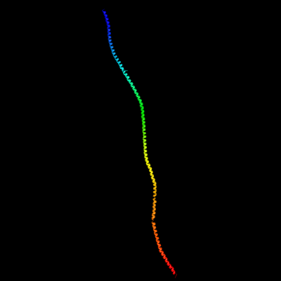
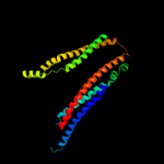
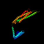
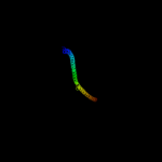
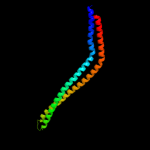
1 c1c1gA_
99.0
6
PDB header: contractile proteinChain: A: PDB Molecule: tropomyosin;PDBTitle: crystal structure of tropomyosin at 7 angstroms resolution2 in the spermine-induced crystal form
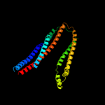
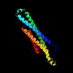
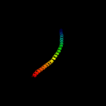
2 c2ch7A_
98.4
10
PDB header: chemotaxisChain: A: PDB Molecule: methyl-accepting chemotaxis protein;PDBTitle: crystal structure of the cytoplasmic domain of a bacterial2 chemoreceptor from thermotoga maritima
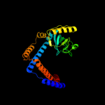
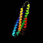
3 c2oevA_
97.6
7
PDB header: protein transportChain: A: PDB Molecule: programmed cell death 6-interacting protein;PDBTitle: crystal structure of alix/aip1
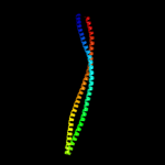
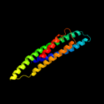
4 c2efrB_
97.3
12
PDB header: contractile proteinChain: B: PDB Molecule: general control protein gcn4 and tropomyosin 1 alpha chain;PDBTitle: crystal structure of the c-terminal tropomyosin fragment with n- and2 c-terminal extensions of the leucine zipper at 1.8 angstroms3 resolution
5 c2oexB_
97.1
8
PDB header: protein transportChain: B: PDB Molecule: programmed cell death 6-interacting protein;PDBTitle: structure of alix/aip1 v domain
6 c1ciiA_
96.9
12
PDB header: transmembrane proteinChain: A: PDB Molecule: colicin ia;PDBTitle: colicin ia
7 c3ojaB_
96.3
10
PDB header: protein bindingChain: B: PDB Molecule: anopheles plasmodium-responsive leucine-rich repeat proteinPDBTitle: crystal structure of lrim1/apl1c complex
8 c1yvlB_
95.3
9
PDB header: signaling proteinChain: B: PDB Molecule: signal transducer and activator of transcriptionPDBTitle: structure of unphosphorylated stat1
9 c2d3eD_
95.3
13
PDB header: contractile proteinChain: D: PDB Molecule: general control protein gcn4 and tropomyosin 1PDBTitle: crystal structure of the c-terminal fragment of rabbit2 skeletal alpha-tropomyosin
10 c3u59C_
94.9
6
PDB header: contractile proteinChain: C: PDB Molecule: tropomyosin beta chain;PDBTitle: n-terminal 98-aa fragment of smooth muscle tropomyosin beta
11 c2fxmB_
94.6
8
PDB header: contractile proteinChain: B: PDB Molecule: myosin heavy chain, cardiac muscle beta isoform;PDBTitle: structure of the human beta-myosin s2 fragment
12 c2b9cA_
94.5
9
PDB header: contractile proteinChain: A: PDB Molecule: striated-muscle alpha tropomyosin;PDBTitle: structure of tropomyosin's mid-region: bending and binding2 sites for actin
13 c1bg1A_
94.5
13
PDB header: transcription/dnaChain: A: PDB Molecule: protein (transcription factor stat3b);PDBTitle: transcription factor stat3b/dna complex
14 c3l9oA_
94.2
8
PDB header: hydrolaseChain: A: PDB Molecule: atp-dependent rna helicase dob1;PDBTitle: crystal structure of mtr4, a co-factor of the nuclear exosome
15 c3g67A_
94.0
13
PDB header: signaling proteinChain: A: PDB Molecule: methyl-accepting chemotaxis protein;PDBTitle: crystal structure of a soluble chemoreceptor from thermotoga2 maritima
16 c3na7A_
93.7
10
PDB header: gene regulation, chaperoneChain: A: PDB Molecule: hp0958;PDBTitle: 2.2 angstrom structure of the hp0958 protein from helicobacter pylori2 ccug 17874
17 c1bf5A_
93.7
8
PDB header: gene regulation/dnaChain: A: PDB Molecule: signal transducer and activator of transcriptionPDBTitle: tyrosine phosphorylated stat-1/dna complex
18 c3ol1A_
93.7
8
PDB header: structural proteinChain: A: PDB Molecule: vimentin;PDBTitle: crystal structure of vimentin (fragment 144-251) from homo sapiens,2 northeast structural genomics consortium target hr4796b
19 c3o0zD_
93.7
11
PDB header: transferaseChain: D: PDB Molecule: rho-associated protein kinase 1;PDBTitle: crystal structure of a coiled-coil domain from human rock i
20 c3cwgA_
93.5
11
PDB header: transcriptionChain: A: PDB Molecule: signal transducer and activator of transcriptionPDBTitle: unphosphorylated mouse stat3 core fragment
21 c3ghgK_
not modelled
93.4
10
PDB header: blood clottingChain: K: PDB Molecule: fibrinogen beta chain;PDBTitle: crystal structure of human fibrinogen
22 c4a55B_
not modelled
93.0
5
PDB header: transferaseChain: B: PDB Molecule: phosphatidylinositol 3-kinase regulatory subunit alpha;PDBTitle: crystal structure of p110alpha in complex with ish2 of p85alpha and2 the inhibitor pik-108
23 c2gl2B_
not modelled
92.7
13
PDB header: cell adhesionChain: B: PDB Molecule: adhesion a;PDBTitle: crystal structure of the tetra muntant (t66g,r67g,f68g,2 y69g) of bacterial adhesin fada
24 c1deqO_
not modelled
92.2
12
PDB header: PDB COMPND: 25 c1qu7A_
not modelled
90.9
12
PDB header: signaling proteinChain: A: PDB Molecule: methyl-accepting chemotaxis protein i;PDBTitle: four helical-bundle structure of the cytoplasmic domain of a serine2 chemotaxis receptor
26 c2y3aB_
not modelled
89.5
8
PDB header: transferaseChain: B: PDB Molecule: phosphatidylinositol 3-kinase regulatory subunit beta;PDBTitle: crystal structure of p110beta in complex with icsh2 of p85beta and2 the drug gdc-0941
27 c1y4cA_
not modelled
89.1
13
PDB header: de novo proteinChain: A: PDB Molecule: maltose binding protein fused with designedPDBTitle: designed helical protein fusion mbp
28 c1ei3E_
not modelled
88.6
6
PDB header: PDB COMPND: 29 c2v66C_
not modelled
87.9
9
PDB header: structural proteinChain: C: PDB Molecule: nuclear distribution protein nude-like 1;PDBTitle: crystal structure of the coiled-coil domain of ndel1 (a.a.2 58 to 169)c
30 c2wpqA_
not modelled
86.8
8
PDB header: membrane proteinChain: A: PDB Molecule: trimeric autotransporter adhesin fragment;PDBTitle: salmonella enterica sada 479-519 fused to gcn4 adaptors (2 sadak3, in-register fusion)
31 c1deqF_
not modelled
85.9
11
PDB header: PDB COMPND: 32 c2dq3A_
not modelled
83.9
16
PDB header: ligaseChain: A: PDB Molecule: seryl-trna synthetase;PDBTitle: crystal structure of aq_298
33 c1deqD_
not modelled
83.1
8
PDB header: PDB COMPND: 34 c2v1yB_
not modelled
80.9
8
PDB header: transferaseChain: B: PDB Molecule: phosphatidylinositol 3-kinase regulatory subunit alpha;PDBTitle: structure of a phosphoinositide 3-kinase alpha adaptor-2 binding domain (abd) in a complex with the ish2 domain3 from p85 alpha
35 c2v71A_
not modelled
80.2
13
PDB header: nuclear proteinChain: A: PDB Molecule: nuclear distribution protein nude-like 1;PDBTitle: coiled-coil region of nudel
36 c1hciB_
not modelled
80.0
8
PDB header: triple-helix coiled coilChain: B: PDB Molecule: alpha-actinin 2;PDBTitle: crystal structure of the rod domain of alpha-actinin
37 c1ei3C_
not modelled
79.0
8
PDB header: PDB COMPND: 38 c3dtpA_
not modelled
78.9
10
PDB header: contractile proteinChain: A: PDB Molecule: myosin 2 heavy chain chimera of smooth andPDBTitle: tarantula heavy meromyosin obtained by flexible docking to2 tarantula muscle thick filament cryo-em 3d-map
39 c3u1aC_
not modelled
77.0
6
PDB header: contractile proteinChain: C: PDB Molecule: smooth muscle tropomyosin alpha;PDBTitle: n-terminal 81-aa fragment of smooth muscle tropomyosin alpha
40 c2e7sM_
not modelled
75.8
14
PDB header: endocytosis/exocytosisChain: M: PDB Molecule: rab guanine nucleotide exchange factor sec2;PDBTitle: crystal structure of the yeast sec2p gef domain
41 c3ojaA_
not modelled
73.8
10
PDB header: protein bindingChain: A: PDB Molecule: leucine-rich immune molecule 1;PDBTitle: crystal structure of lrim1/apl1c complex
42 c1jchC_
not modelled
73.6
9
PDB header: ribosome inhibitor, hydrolaseChain: C: PDB Molecule: colicin e3;PDBTitle: crystal structure of colicin e3 in complex with its immunity protein
43 c3hnwB_
not modelled
72.6
7
PDB header: structural genomics, unknown functionChain: B: PDB Molecule: uncharacterized protein;PDBTitle: crystal structure of a basic coiled-coil protein of unknown function2 from eubacterium eligens atcc 27750
44 c2i1jA_
not modelled
69.9
9
PDB header: cell adhesion, membrane proteinChain: A: PDB Molecule: moesin;PDBTitle: moesin from spodoptera frugiperda at 2.1 angstroms resolution
45 c1l8dB_
not modelled
69.4
13
PDB header: replicationChain: B: PDB Molecule: dna double-strand break repair rad50 atpase;PDBTitle: rad50 coiled-coil zn hook
46 c3a7pB_
not modelled
69.4
15
PDB header: protein transportChain: B: PDB Molecule: autophagy protein 16;PDBTitle: the crystal structure of saccharomyces cerevisiae atg16
47 c2qihA_
not modelled
66.3
13
PDB header: cell adhesionChain: A: PDB Molecule: protein uspa1;PDBTitle: crystal structure of 527-665 fragment of uspa1 protein from2 moraxella catarrhalis
48 c3hizB_
not modelled
64.6
8
PDB header: transferase/oncoproteinChain: B: PDB Molecule: phosphatidylinositol 3-kinase regulatory subunitPDBTitle: crystal structure of p110alpha h1047r mutant in complex with2 nish2 of p85alpha
49 c3iv1F_
not modelled
56.7
11
PDB header: hydrolaseChain: F: PDB Molecule: tumor susceptibility gene 101 protein;PDBTitle: coiled-coil domain of tumor susceptibility gene 101
50 c3r6nA_
not modelled
56.2
7
PDB header: cell adhesionChain: A: PDB Molecule: desmoplakin;PDBTitle: crystal structure of a rigid four spectrin repeat fragment of the2 human desmoplakin plakin domain
51 c2rd0B_
not modelled
53.3
9
PDB header: transferase/oncoproteinChain: B: PDB Molecule: phosphatidylinositol 3-kinase regulatory subunit alpha;PDBTitle: structure of a human p110alpha/p85alpha complex
52 c3qh9A_
not modelled
49.8
16
PDB header: structural proteinChain: A: PDB Molecule: liprin-beta-2;PDBTitle: human liprin-beta2 coiled-coil
53 c2eqbC_
not modelled
43.9
12
PDB header: endocytosis/exocytosisChain: C: PDB Molecule: rab guanine nucleotide exchange factor sec2;PDBTitle: crystal structure of the rab gtpase sec4p, the sec2p gef2 domain, and phosphate complex
54 c3layF_
not modelled
42.0
14
PDB header: metal binding proteinChain: F: PDB Molecule: zinc resistance-associated protein;PDBTitle: alpha-helical barrel formed by the decamer of the zinc resistance-2 associated protein (stm4172) from salmonella enterica subsp. enterica3 serovar typhimurium str. lt2
55 c2jeeA_
not modelled
38.6
12
PDB header: cell cycleChain: A: PDB Molecule: yiiu;PDBTitle: xray structure of e. coli yiiu
56 c1ic2B_
not modelled
34.7
9
PDB header: contractile proteinChain: B: PDB Molecule: tropomyosin alpha chain, skeletal muscle;PDBTitle: deciphering the design of the tropomyosin molecule
57 c2v4hA_
not modelled
32.9
18
PDB header: transcriptionChain: A: PDB Molecule: nf-kappa-b essential modulator;PDBTitle: nemo cc2-lz domain - 1d5 darpin complex
58 c2xdjF_
not modelled
32.8
10
PDB header: unknown functionChain: F: PDB Molecule: uncharacterized protein ybgf;PDBTitle: crystal structure of the n-terminal domain of e.coli ybgf
59 c3ipkA_
not modelled
30.1
11
PDB header: cell adhesionChain: A: PDB Molecule: agi/ii;PDBTitle: crystal structure of a3vp1 of agi/ii of streptococcus mutans
60 c1sjjB_
not modelled
29.7
10
PDB header: contractile proteinChain: B: PDB Molecule: actinin;PDBTitle: cryo-em structure of chicken gizzard smooth muscle alpha-2 actinin
61 c2no2A_
not modelled
29.4
10
PDB header: cell adhesionChain: A: PDB Molecule: huntingtin-interacting protein 1;PDBTitle: crystal structure of the dllrkn-containing coiled-coil2 domain of huntingtin-interacting protein 1
62 d1seta1
not modelled
28.2
11
Fold: Long alpha-hairpinSuperfamily: tRNA-binding armFamily: Seryl-tRNA synthetase (SerRS)63 c3m9bK_
not modelled
24.8
5
PDB header: chaperoneChain: K: PDB Molecule: proteasome-associated atpase;PDBTitle: crystal structure of the amino terminal coiled coil domain and the2 inter domain of the mycobacterium tuberculosis proteasomal atpase mpa
64 c3n4xB_
not modelled
22.4
8
PDB header: replicationChain: B: PDB Molecule: monopolin complex subunit csm1;PDBTitle: structure of csm1 full-length
65 c1ci6A_
not modelled
22.0
14
PDB header: transcriptionChain: A: PDB Molecule: transcription factor atf-4;PDBTitle: transcription factor atf4-c/ebp beta bzip heterodimer
66 c3qo8A_
not modelled
21.7
14
PDB header: ligaseChain: A: PDB Molecule: seryl-trna synthetase, cytoplasmic;PDBTitle: crystal structure of seryl-trna synthetase from candida albicans
67 c1gd2G_
not modelled
21.3
10
PDB header: transcription/dnaChain: G: PDB Molecule: transcription factor pap1;PDBTitle: crystal structure of bzip transcription factor pap1 bound2 to dna
68 c1gk4A_
not modelled
20.4
8
PDB header: vimentinChain: A: PDB Molecule: vimentin;PDBTitle: human vimentin coil 2b fragment (cys2)
69 c3q0xA_
not modelled
18.8
13
PDB header: structural proteinChain: A: PDB Molecule: centriole protein;PDBTitle: n-terminal coiled-coil dimer domain of c. reinhardtii sas-6 homolog2 bld12p
70 d2ap3a1
not modelled
17.9
7
Fold: Four-helical up-and-down bundleSuperfamily: MW0975(SA0943)-likeFamily: MW0975(SA0943)-like71 c1x8yA_
not modelled
17.6
11
PDB header: structural proteinChain: A: PDB Molecule: lamin a/c;PDBTitle: human lamin coil 2b
72 c2wt7B_
not modelled
16.7
14
PDB header: transcriptionChain: B: PDB Molecule: transcription factor mafb;PDBTitle: crystal structure of the bzip heterodimeric complex2 mafb:cfos bound to dna
73 c1wt6B_
not modelled
16.5
20
PDB header: transferaseChain: B: PDB Molecule: myotonin-protein kinase;PDBTitle: coiled-coil domain of dmpk
74 c2zvnF_
not modelled
15.8
17
PDB header: signaling protein/transcriptionChain: F: PDB Molecule: nf-kappa-b essential modulator;PDBTitle: nemo cozi domain incomplex with diubiquitin in p2121212 space group
75 c2x7aB_
not modelled
15.5
5
PDB header: immune systemChain: B: PDB Molecule: bone marrow stromal antigen 2;PDBTitle: structural basis of hiv-1 tethering to membranes by the2 bst2-tetherin ectodomain
76 c2bsgA_
not modelled
15.5
9
PDB header: viral proteinChain: A: PDB Molecule: fibritin;PDBTitle: the modeled structure of fibritin (gpwac) of bacteriophage2 t4 based on cryo-em reconstruction of the extended tail of3 bacteriophage t4
77 c2pmsD_
not modelled
15.5
11
PDB header: metal transport, hydrolaseChain: D: PDB Molecule: pneumococcal surface protein a (pspa);PDBTitle: crystal structure of the complex of human lactoferrin n-lobe and2 lactoferrin-binding domain of pneumococcal surface protein a
78 c1fosF_
not modelled
15.4
10
PDB header: transcription/dnaChain: F: PDB Molecule: c-jun proto-oncogene protein;PDBTitle: two human c-fos:c-jun:dna complexes
79 c2ke4A_
not modelled
15.4
12
PDB header: membrane proteinChain: A: PDB Molecule: cdc42-interacting protein 4;PDBTitle: the nmr structure of the tc10 and cdc42 interacting domain2 of cip4
80 c3batB_
not modelled
15.2
14
PDB header: contractile proteinChain: B: PDB Molecule: myosin heavy chain, striated muscle/generalPDBTitle: crystal structure of the n-terminal region of the scallop2 myosin rod, monoclinic (p21) form
81 c3cvfA_
not modelled
14.8
18
PDB header: signaling proteinChain: A: PDB Molecule: homer protein homolog 3;PDBTitle: crystal structure of the carboxy terminus of homer3
82 c1n73C_
not modelled
14.6
6
PDB header: blood clottingChain: C: PDB Molecule: fibrin gamma chain;PDBTitle: fibrin d-dimer, lamprey complexed with the peptide ligand: gly-his-2 arg-pro-amide
83 c2hpcF_
not modelled
13.3
9
PDB header: blood clottingChain: F: PDB Molecule: fibrinogen, gamma polypeptide;PDBTitle: crystal structure of fragment d from human fibrinogen complexed with2 gly-pro-arg-pro-amide.
84 c1t2kD_
not modelled
11.4
10
PDB header: transcription/dnaChain: D: PDB Molecule: cyclic-amp-dependent transcription factor atf-2;PDBTitle: structure of the dna binding domains of irf3, atf-2 and jun2 bound to dna
85 c2p22C_
not modelled
11.2
15
PDB header: transport proteinChain: C: PDB Molecule: protein srn2;PDBTitle: structure of the yeast escrt-i heterotetramer core
86 c1g8xB_
not modelled
10.9
8
PDB header: structural proteinChain: B: PDB Molecule: myosin ii heavy chain fused to alpha-actinin 3;PDBTitle: structure of a genetically engineered molecular motor
87 c3cveC_
not modelled
10.7
17
PDB header: signaling proteinChain: C: PDB Molecule: homer protein homolog 1;PDBTitle: crystal structure of the carboxy terminus of homer1
88 c1yv0I_
not modelled
10.4
8
PDB header: contractile proteinChain: I: PDB Molecule: troponin i, fast skeletal muscle;PDBTitle: crystal structure of skeletal muscle troponin in the ca2+-2 free state
89 c1junB_
not modelled
10.4
11
PDB header: transcription regulationChain: B: PDB Molecule: c-jun homodimer;PDBTitle: nmr study of c-jun homodimer
90 c3mkxC_
not modelled
9.9
13
PDB header: antiviral proteinChain: C: PDB Molecule: bone marrow stromal antigen 2;PDBTitle: crystal structure of bst2/tetherin
91 c3m06F_
not modelled
9.6
10
PDB header: protein bindingChain: F: PDB Molecule: tnf receptor-associated factor 2;PDBTitle: crystal structure of traf2
92 c1fosE_
not modelled
9.2
13
PDB header: transcription/dnaChain: E: PDB Molecule: p55-c-fos proto-oncogene protein;PDBTitle: two human c-fos:c-jun:dna complexes
93 c1u4qB_
not modelled
9.1
12
PDB header: structural proteinChain: B: PDB Molecule: spectrin alpha chain, brain;PDBTitle: crystal structure of repeats 15, 16 and 17 of chicken brain2 alpha spectrin
94 c1ru7B_
not modelled
9.0
15
PDB header: viral proteinChain: B: PDB Molecule: hemagglutinin;PDBTitle: 1934 human h1 hemagglutinin
95 c2xgjA_
not modelled
8.8
18
PDB header: hydrolase/rnaChain: A: PDB Molecule: atp-dependent rna helicase dob1;PDBTitle: structure of mtr4, a dexh helicase involved in nuclear rna2 processing and surveillance
96 c2e43A_
not modelled
8.7
11
PDB header: transcription/dnaChain: A: PDB Molecule: ccaat/enhancer-binding protein beta;PDBTitle: crystal structure of c/ebpbeta bzip homodimer k269a mutant2 bound to a high affinity dna fragment
97 c3lssA_
not modelled
8.6
9
PDB header: ligaseChain: A: PDB Molecule: seryl-trna synthetase;PDBTitle: trypanosoma brucei seryl-trna synthetase in complex with atp
98 c1ik9B_
not modelled
8.0
8
PDB header: gene regulation/ligaseChain: B: PDB Molecule: dna repair protein xrcc4;PDBTitle: crystal structure of a xrcc4-dna ligase iv complex
99 c2w6aB_
not modelled
7.9
11
PDB header: signaling proteinChain: B: PDB Molecule: arf gtpase-activating protein git1;PDBTitle: x-ray structure of the dimeric git1 coiled-coil domain