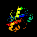
1 d1y97a1
100.0
20
Fold: Ribonuclease H-like motifSuperfamily: Ribonuclease H-likeFamily: DnaQ-like 3'-5' exonuclease2 d2qxfa1
100.0
20
Fold: Ribonuclease H-like motifSuperfamily: Ribonuclease H-likeFamily: DnaQ-like 3'-5' exonuclease3 c3u6fA_
100.0
18
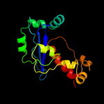
PDB header: hydrolase/dnaChain: A: PDB Molecule: three prime repair exonuclease 1;PDBTitle: mouse trex1 d200n mutant
4 c1zbhA_
100.0
17
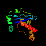
PDB header: hydrolase/rnaChain: A: PDB Molecule: 3'-5' exonuclease eri1;PDBTitle: 3'-end specific recognition of histone mrna stem-loop by 3'-2 exonuclease
5 c1zbuB_
100.0
18
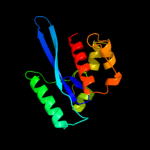
PDB header: hydrolaseChain: B: PDB Molecule: 3'-5' exonuclease eri1;PDBTitle: crystal structure of full-length 3'-exonuclease
6 d2guia1
100.0
30
Fold: Ribonuclease H-like motifSuperfamily: Ribonuclease H-likeFamily: DnaQ-like 3'-5' exonuclease7 c2p1jB_
100.0
34
PDB header: transferaseChain: B: PDB Molecule: dna polymerase iii polc-type;PDBTitle: crystal structure of a polc-type dna polymerase iii2 exonuclease domain from thermotoga maritima
8 c3cm6A_
100.0
17
PDB header: hydrolase, apoptosisChain: A: PDB Molecule: cell death-related nuclease 4;PDBTitle: crystal structure of cell-death related nuclease 4 (crn-4)2 bound with er
9 d3b6oa1
100.0
16
Fold: Ribonuclease H-like motifSuperfamily: Ribonuclease H-likeFamily: DnaQ-like 3'-5' exonuclease10 d1wlja_
100.0
20
Fold: Ribonuclease H-like motifSuperfamily: Ribonuclease H-likeFamily: DnaQ-like 3'-5' exonuclease11 d1j9aa_
100.0
17
Fold: Ribonuclease H-like motifSuperfamily: Ribonuclease H-likeFamily: DnaQ-like 3'-5' exonuclease12 c3tr8A_
100.0
17
PDB header: hydrolaseChain: A: PDB Molecule: oligoribonuclease;PDBTitle: structure of an oligoribonuclease (orn) from coxiella burnetii
13 d1w0ha_
100.0
16
Fold: Ribonuclease H-like motifSuperfamily: Ribonuclease H-likeFamily: DnaQ-like 3'-5' exonuclease14 c2xriA_
100.0
17
PDB header: hydrolaseChain: A: PDB Molecule: eri1 exoribonuclease 3;PDBTitle: crystal structure of human eri1 exoribonuclease 3
15 d2f96a1
100.0
22
Fold: Ribonuclease H-like motifSuperfamily: Ribonuclease H-likeFamily: DnaQ-like 3'-5' exonuclease16 d2igia1
100.0
19
Fold: Ribonuclease H-like motifSuperfamily: Ribonuclease H-likeFamily: DnaQ-like 3'-5' exonuclease17 c2gbzA_
99.9
16
PDB header: hydrolaseChain: A: PDB Molecule: oligoribonuclease;PDBTitle: the crystal structure of xc847 from xanthomonas campestris: a 3-52 oligoribonuclease of dnaq fold family with a novel opposingly-shifted3 helix
18 c2is3B_
99.9
21
PDB header: hydrolaseChain: B: PDB Molecule: ribonuclease t;PDBTitle: crystal structure of escherichia coli rnase t
19 d1uoca_
99.4
9
Fold: Ribonuclease H-like motifSuperfamily: Ribonuclease H-likeFamily: CAF1-like ribonuclease20 d1kfsa1
99.1
19
Fold: Ribonuclease H-like motifSuperfamily: Ribonuclease H-likeFamily: DnaQ-like 3'-5' exonuclease21 d2d5ra1
not modelled
99.1
12
Fold: Ribonuclease H-like motifSuperfamily: Ribonuclease H-likeFamily: CAF1-like ribonuclease22 d1x9ma1
not modelled
99.1
20
Fold: Ribonuclease H-like motifSuperfamily: Ribonuclease H-likeFamily: DnaQ-like 3'-5' exonuclease23 c2p51A_
not modelled
99.0
15
PDB header: hydrolase, gene regulationChain: A: PDB Molecule: spcc18.06c protein;PDBTitle: crystal structure of the s. pombe pop2p deadenylation2 subunit
24 c2kzzA_
not modelled
99.0
18
PDB header: transferase/dnaChain: A: PDB Molecule: protein (dna polymerase i);PDBTitle: klenow fragment with normal substrate and zinc only
25 c1tk0A_
not modelled
98.8
20
PDB header: transferase/electron transport/dnaChain: A: PDB Molecule: dna polymerase;PDBTitle: t7 dna polymerase ternary complex with 8 oxo guanosine and2 ddctp at the insertion site
26 c1njzA_
not modelled
98.8
14
PDB header: transferase/dnaChain: A: PDB Molecule: dna polymerase i;PDBTitle: cytosine-thymine mismatch at the polymerase active site
27 c3d45B_
not modelled
98.6
23
PDB header: hydrolaseChain: B: PDB Molecule: poly(a)-specific ribonuclease parn;PDBTitle: crystal structure of mouse parn in complex with m7gpppg
28 d2hhva1
not modelled
98.5
17
Fold: Ribonuclease H-like motifSuperfamily: Ribonuclease H-likeFamily: DnaQ-like 3'-5' exonuclease29 c2a1sC_
not modelled
98.5
23
PDB header: hydrolaseChain: C: PDB Molecule: poly(a)-specific ribonuclease parn;PDBTitle: crystal structure of native parn nuclease domain
30 d1d5aa1
not modelled
98.0
18
Fold: Ribonuclease H-like motifSuperfamily: Ribonuclease H-likeFamily: DnaQ-like 3'-5' exonuclease31 d1wn7a1
not modelled
97.9
16
Fold: Ribonuclease H-like motifSuperfamily: Ribonuclease H-likeFamily: DnaQ-like 3'-5' exonuclease32 c2gv9B_
not modelled
97.9
12
PDB header: transferaseChain: B: PDB Molecule: dna polymerase;PDBTitle: crystal structure of the herpes simplex virus type 1 dna polymerase
33 d1qhta1
not modelled
97.9
17
Fold: Ribonuclease H-like motifSuperfamily: Ribonuclease H-likeFamily: DnaQ-like 3'-5' exonuclease34 c1yt3A_
not modelled
97.7
22
PDB header: hydrolase,translationChain: A: PDB Molecule: ribonuclease d;PDBTitle: crystal structure of escherichia coli rnase d, an2 exoribonuclease involved in structured rna processing
35 d1tgoa1
not modelled
97.7
16
Fold: Ribonuclease H-like motifSuperfamily: Ribonuclease H-likeFamily: DnaQ-like 3'-5' exonuclease36 d1noya_
not modelled
97.6
16
Fold: Ribonuclease H-like motifSuperfamily: Ribonuclease H-likeFamily: DnaQ-like 3'-5' exonuclease37 d1q8ia1
not modelled
97.5
18
Fold: Ribonuclease H-like motifSuperfamily: Ribonuclease H-likeFamily: DnaQ-like 3'-5' exonuclease38 c3iayA_
not modelled
97.5
21
PDB header: transferase/dnaChain: A: PDB Molecule: dna polymerase delta catalytic subunit;PDBTitle: ternary complex of dna polymerase delta
39 d1ih7a1
not modelled
97.3
20
Fold: Ribonuclease H-like motifSuperfamily: Ribonuclease H-likeFamily: DnaQ-like 3'-5' exonuclease40 c2e6mA_
not modelled
97.3
12
PDB header: hydrolaseChain: A: PDB Molecule: werner syndrome atp-dependent helicase homolog;PDBTitle: structure of mouse werner exonuclease domain
41 c2dtuA_
not modelled
97.2
18
PDB header: transferase/dnaChain: A: PDB Molecule: dna polymerase;PDBTitle: crystal structure of the beta hairpin loop deletion variant2 of rb69 gp43 in complex with dna containing an abasic site3 analog
42 c1d5aA_
not modelled
97.1
16
PDB header: transferaseChain: A: PDB Molecule: protein (dna polymerase);PDBTitle: crystal structure of an archaebacterial dna polymerase2 d.tok. deposition of second native structure at 2.43 angstrom
43 c3cymA_
not modelled
97.1
18
PDB header: structural genomics, unknown functionChain: A: PDB Molecule: uncharacterized protein bad_0989;PDBTitle: crystal structure of protein bad_0989 from bifidobacterium2 adolescentis
44 c2vwkA_
not modelled
97.0
18
PDB header: dna replicationChain: A: PDB Molecule: dna polymerase;PDBTitle: uracil recognition in archaeal dna polymerases captured by2 x-ray crystallography. v93q polymerase variant
45 d1yt3a3
not modelled
96.9
21
Fold: Ribonuclease H-like motifSuperfamily: Ribonuclease H-likeFamily: DnaQ-like 3'-5' exonuclease46 c1q8iA_
not modelled
96.6
20
PDB header: transferaseChain: A: PDB Molecule: dna polymerase ii;PDBTitle: crystal structure of escherichia coli dna polymerase ii
47 d1s5ja1
not modelled
96.4
15
Fold: Ribonuclease H-like motifSuperfamily: Ribonuclease H-likeFamily: DnaQ-like 3'-5' exonuclease48 c4ktqA_
not modelled
96.4
16
PDB header: transferase/dnaChain: A: PDB Molecule: protein (large fragment of dna polymerase i);PDBTitle: binary complex of the large fragment of dna polymerase i2 from t. aquaticus bound to a primer/template dna
49 d2hbka2
not modelled
94.6
15
Fold: Ribonuclease H-like motifSuperfamily: Ribonuclease H-likeFamily: DnaQ-like 3'-5' exonuclease50 d1vk0a_
not modelled
93.0
17
Fold: Ribonuclease H-like motifSuperfamily: Ribonuclease H-likeFamily: DnaQ-like 3'-5' exonuclease51 c1s5jA_
not modelled
92.1
16
PDB header: transferaseChain: A: PDB Molecule: dna polymerase i;PDBTitle: insight in dna replication: the crystal structure of dna2 polymerase b1 from the archaeon sulfolobus solfataricus
52 c3ikmD_
not modelled
91.9
18
PDB header: transferaseChain: D: PDB Molecule: dna polymerase subunit gamma-1;PDBTitle: crystal structure of human mitochondrial dna polymerase2 holoenzyme
53 c2hbkA_
not modelled
85.4
20
PDB header: hydrolase, gene regulationChain: A: PDB Molecule: exosome complex exonuclease rrp6;PDBTitle: structure of the yeast nuclear exosome component, rrp6p,2 reveals an interplay between the active site and the hrdc3 domain; protein in complex with mn
54 c3mx2A_
not modelled
68.9
20
PDB header: nuclear proteinChain: A: PDB Molecule: nucleoprotein;PDBTitle: lassa fever virus nucleoprotein complexed with dttp
55 c3q7cA_
not modelled
68.4
20
PDB header: hydrolaseChain: A: PDB Molecule: nucleoprotein;PDBTitle: exonuclease domain of lassa virus nucleoprotein bound to manganese
56 c1cmwA_
not modelled
64.2
18
PDB header: transferaseChain: A: PDB Molecule: protein (dna polymerase i);PDBTitle: crystal structure of taq dna-polymerase shows a new2 orientation for the structure-specific nuclease domain
57 c3sahA_
not modelled
49.5
21
PDB header: hydrolaseChain: A: PDB Molecule: exosome component 10;PDBTitle: crystal structure of the human rrp6 catalytic domain with y436a2 mutation in the catalytic site
58 d2py5a1
not modelled
17.8
19
Fold: Ribonuclease H-like motifSuperfamily: Ribonuclease H-likeFamily: DnaQ-like 3'-5' exonuclease59 c1lj2B_
not modelled
9.4
13
PDB header: viral protein/ translationChain: B: PDB Molecule: nonstructural rna-binding protein 34;PDBTitle: recognition of eif4g by rotavirus nsp3 reveals a basis for2 mrna circularization
60 c3eukC_
not modelled
5.4
14
PDB header: cell cycleChain: C: PDB Molecule: chromosome partition protein mukb, linker;PDBTitle: crystal structure of muke-mukf(residues 292-443)-mukb(head2 domain)-atpgammas complex, asymmetric dimer
61 c3g6iA_
not modelled
5.3
18
PDB header: unknown functionChain: A: PDB Molecule: putative outer membrane protein, part of carbohydratePDBTitle: crystal structure of an outer membrane protein, part of a putative2 carbohydrate binding complex (bt_1022) from bacteroides3 thetaiotaomicron vpi-5482 at 1.93 a resolution