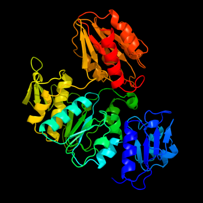
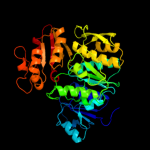
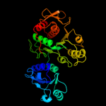
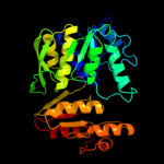
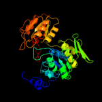
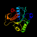
| 1 | c3uagA_
|
|
 |
100.0 |
98 |
PDB header:ligase
Chain: A: PDB Molecule:protein (udp-n-acetylmuramoyl-l-alanine:d-
PDBTitle: udp-n-acetylmuramoyl-l-alanine:d-glutamate ligase
|
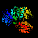
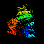
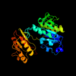
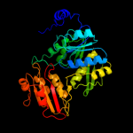
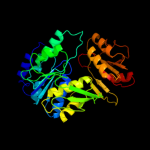
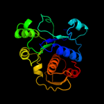
| 2 | c3lk7A_
|
|
 |
100.0 |
29 |
PDB header:ligase
Chain: A: PDB Molecule:udp-n-acetylmuramoylalanine--d-glutamate ligase;
PDBTitle: the crystal structure of udp-n-acetylmuramoylalanine-d-2 glutamate (murd) ligase from streptococcus agalactiae to3 1.5a
|
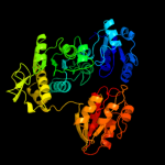
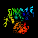
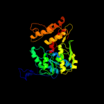
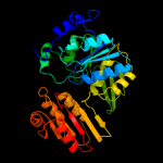
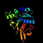
| 3 | c2f00A_
|
|
 |
100.0 |
18 |
PDB header:ligase
Chain: A: PDB Molecule:udp-n-acetylmuramate--l-alanine ligase;
PDBTitle: escherichia coli murc
|
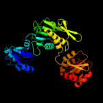
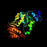
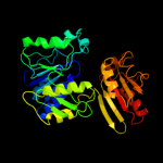
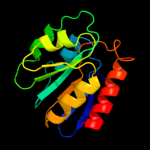
| 4 | c3hn7A_
|
|
 |
100.0 |
18 |
PDB header:ligase
Chain: A: PDB Molecule:udp-n-acetylmuramate-l-alanine ligase;
PDBTitle: crystal structure of a murein peptide ligase mpl (psyc_0032) from2 psychrobacter arcticus 273-4 at 1.65 a resolution
|
| 5 | c1j6uA_
|
|
 |
100.0 |
22 |
PDB header:ligase
Chain: A: PDB Molecule:udp-n-acetylmuramate-alanine ligase murc;
PDBTitle: crystal structure of udp-n-acetylmuramate-alanine ligase2 murc (tm0231) from thermotoga maritima at 2.3 a resolution
|
| 6 | c1gqqA_
|
|
 |
100.0 |
19 |
PDB header:cell wall biosynthesis
Chain: A: PDB Molecule:udp-n-acetylmuramate-l-alanine ligase;
PDBTitle: murc - crystal structure of the apo-enzyme from haemophilus2 influenzae
|
| 7 | c3eagA_
|
|
 |
100.0 |
20 |
PDB header:ligase
Chain: A: PDB Molecule:udp-n-acetylmuramate:l-alanyl-gamma-d-glutamyl-meso-
PDBTitle: the crystal structure of udp-n-acetylmuramate:l-alanyl-gamma-d-2 glutamyl-meso-diaminopimelate ligase (mpl) from neisseria3 meningitides
|
| 8 | c2am1A_
|
|
 |
100.0 |
22 |
PDB header:ligase
Chain: A: PDB Molecule:udp-n-acetylmuramoylalanine-d-glutamyl-lysine-d-alanyl-d-
PDBTitle: sp protein ligand 1
|
| 9 | c1e8cB_
|
|
 |
100.0 |
22 |
PDB header:ligase
Chain: B: PDB Molecule:udp-n-acetylmuramoylalanyl-d-glutamate--2,6-
PDBTitle: structure of mure the udp-n-acetylmuramyl tripeptide2 synthetase from e. coli
|
| 10 | c1gg4A_
|
|
 |
100.0 |
21 |
PDB header:ligase
Chain: A: PDB Molecule:udp-n-acetylmuramoylalanyl-d-glutamyl-2,6-
PDBTitle: crystal structure of escherichia coli udpmurnac-tripeptide2 d-alanyl-d-alanine-adding enzyme (murf) at 2.3 angstrom3 resolution
|
| 11 | c2wtzC_
|
|
 |
100.0 |
24 |
PDB header:ligase
Chain: C: PDB Molecule:udp-n-acetylmuramoyl-l-alanyl-d-glutamate-
PDBTitle: mure ligase of mycobacterium tuberculosis
|
| 12 | c2vosA_
|
|
 |
100.0 |
22 |
PDB header:ligase
Chain: A: PDB Molecule:folylpolyglutamate synthase protein folc;
PDBTitle: mycobacterium tuberculosis folylpolyglutamate synthase2 complexed with adp
|
| 13 | c1w78A_
|
|
 |
100.0 |
22 |
PDB header:synthase
Chain: A: PDB Molecule:folc bifunctional protein;
PDBTitle: e.coli folc in complex with dhpp and adp
|
| 14 | c2gc6A_
|
|
 |
100.0 |
16 |
PDB header:ligase
Chain: A: PDB Molecule:folylpolyglutamate synthase;
PDBTitle: s73a mutant of l. casei fpgs
|
| 15 | c3n2aA_
|
|
 |
100.0 |
22 |
PDB header:ligase
Chain: A: PDB Molecule:bifunctional folylpolyglutamate synthase/dihydrofolate
PDBTitle: crystal structure of bifunctional folylpolyglutamate2 synthase/dihydrofolate synthase from yersinia pestis co92
|
| 16 | c1o5zA_
|
|
 |
100.0 |
22 |
PDB header:ligase
Chain: A: PDB Molecule:folylpolyglutamate synthase/dihydrofolate synthase;
PDBTitle: crystal structure of folylpolyglutamate synthase (tm0166) from2 thermotoga maritima at 2.10 a resolution
|
| 17 | d2jfga3
|
|
 |
100.0 |
97 |
Fold:Ribokinase-like
Superfamily:MurD-like peptide ligases, catalytic domain
Family:MurCDEF |
| 18 | d2jfga2
|
|
 |
100.0 |
100 |
Fold:MurD-like peptide ligases, peptide-binding domain
Superfamily:MurD-like peptide ligases, peptide-binding domain
Family:MurCDEF C-terminal domain |
| 19 | d1p3da3
|
|
 |
100.0 |
19 |
Fold:Ribokinase-like
Superfamily:MurD-like peptide ligases, catalytic domain
Family:MurCDEF |
| 20 | d1e8ca3
|
|
 |
100.0 |
27 |
Fold:Ribokinase-like
Superfamily:MurD-like peptide ligases, catalytic domain
Family:MurCDEF |
| 21 | d1j6ua3 |
|
not modelled |
100.0 |
32 |
Fold:Ribokinase-like
Superfamily:MurD-like peptide ligases, catalytic domain
Family:MurCDEF |
| 22 | d2gc6a2 |
|
not modelled |
100.0 |
15 |
Fold:Ribokinase-like
Superfamily:MurD-like peptide ligases, catalytic domain
Family:Folylpolyglutamate synthetase |
| 23 | d1gg4a4 |
|
not modelled |
100.0 |
22 |
Fold:Ribokinase-like
Superfamily:MurD-like peptide ligases, catalytic domain
Family:MurCDEF |
| 24 | d1o5za2 |
|
not modelled |
100.0 |
20 |
Fold:Ribokinase-like
Superfamily:MurD-like peptide ligases, catalytic domain
Family:Folylpolyglutamate synthetase |
| 25 | d1gg4a1 |
|
not modelled |
99.7 |
18 |
Fold:MurD-like peptide ligases, peptide-binding domain
Superfamily:MurD-like peptide ligases, peptide-binding domain
Family:MurCDEF C-terminal domain |
| 26 | d2jfga1 |
|
not modelled |
99.7 |
100 |
Fold:MurCD N-terminal domain
Superfamily:MurCD N-terminal domain
Family:MurCD N-terminal domain |
| 27 | d1j6ua1 |
|
not modelled |
99.7 |
14 |
Fold:MurCD N-terminal domain
Superfamily:MurCD N-terminal domain
Family:MurCD N-terminal domain |
| 28 | d1p3da1 |
|
not modelled |
99.7 |
19 |
Fold:MurCD N-terminal domain
Superfamily:MurCD N-terminal domain
Family:MurCD N-terminal domain |
| 29 | d1e8ca2 |
|
not modelled |
99.6 |
18 |
Fold:MurD-like peptide ligases, peptide-binding domain
Superfamily:MurD-like peptide ligases, peptide-binding domain
Family:MurCDEF C-terminal domain |
| 30 | c3mvnA_ |
|
not modelled |
99.5 |
16 |
PDB header:ligase
Chain: A: PDB Molecule:udp-n-acetylmuramate:l-alanyl-gamma-d-glutamayl-medo-
PDBTitle: crystal structure of a domain from a putative udp-n-acetylmuramate:l-2 alanyl-gamma-d-glutamayl-medo-diaminopimelate ligase from haemophilus3 ducreyi 35000hp
|
| 31 | d1p3da2 |
|
not modelled |
99.5 |
17 |
Fold:MurD-like peptide ligases, peptide-binding domain
Superfamily:MurD-like peptide ligases, peptide-binding domain
Family:MurCDEF C-terminal domain |
| 32 | d1j6ua2 |
|
not modelled |
99.1 |
11 |
Fold:MurD-like peptide ligases, peptide-binding domain
Superfamily:MurD-like peptide ligases, peptide-binding domain
Family:MurCDEF C-terminal domain |
| 33 | d1o5za1 |
|
not modelled |
98.9 |
25 |
Fold:MurD-like peptide ligases, peptide-binding domain
Superfamily:MurD-like peptide ligases, peptide-binding domain
Family:Folylpolyglutamate synthetase, C-terminal domain |
| 34 | d2gc6a1 |
|
not modelled |
98.7 |
20 |
Fold:MurD-like peptide ligases, peptide-binding domain
Superfamily:MurD-like peptide ligases, peptide-binding domain
Family:Folylpolyglutamate synthetase, C-terminal domain |
| 35 | d1pjqa1 |
|
not modelled |
98.6 |
16 |
Fold:NAD(P)-binding Rossmann-fold domains
Superfamily:NAD(P)-binding Rossmann-fold domains
Family:Siroheme synthase N-terminal domain-like |
| 36 | c3d4oA_ |
|
not modelled |
98.2 |
17 |
PDB header:oxidoreductase
Chain: A: PDB Molecule:dipicolinate synthase subunit a;
PDBTitle: crystal structure of dipicolinate synthase subunit a (np_243269.1)2 from bacillus halodurans at 2.10 a resolution
|
| 37 | c3gg2B_ |
|
not modelled |
97.9 |
17 |
PDB header:oxidoreductase
Chain: B: PDB Molecule:sugar dehydrogenase, udp-glucose/gdp-mannose
PDBTitle: crystal structure of udp-glucose 6-dehydrogenase from2 porphyromonas gingivalis bound to product udp-glucuronate
|
| 38 | c2y0dB_ |
|
not modelled |
97.9 |
15 |
PDB header:oxidoreductase
Chain: B: PDB Molecule:udp-glucose dehydrogenase;
PDBTitle: bcec mutation y10k
|
| 39 | c3cumA_ |
|
not modelled |
97.9 |
17 |
PDB header:oxidoreductase
Chain: A: PDB Molecule:probable 3-hydroxyisobutyrate dehydrogenase;
PDBTitle: crystal structure of a possible 3-hydroxyisobutyrate dehydrogenase2 from pseudomonas aeruginosa pao1
|
| 40 | c1mv8A_ |
|
not modelled |
97.8 |
23 |
PDB header:oxidoreductase
Chain: A: PDB Molecule:gdp-mannose 6-dehydrogenase;
PDBTitle: 1.55 a crystal structure of a ternary complex of gdp-mannose2 dehydrogenase from psuedomonas aeruginosa
|
| 41 | c2rirA_ |
|
not modelled |
97.8 |
20 |
PDB header:oxidoreductase
Chain: A: PDB Molecule:dipicolinate synthase, a chain;
PDBTitle: crystal structure of dipicolinate synthase, a chain, from bacillus2 subtilis
|
| 42 | c1pjtB_ |
|
not modelled |
97.8 |
17 |
PDB header:transferase/oxidoreductase/lyase
Chain: B: PDB Molecule:siroheme synthase;
PDBTitle: the structure of the ser128ala point-mutant variant of cysg,2 the multifunctional3 methyltransferase/dehydrogenase/ferrochelatase for4 siroheme synthesis
|
| 43 | c1gdhA_ |
|
not modelled |
97.8 |
14 |
PDB header:oxidoreductase(choh (d)-nad(p)+ (a))
Chain: A: PDB Molecule:d-glycerate dehydrogenase;
PDBTitle: crystal structure of a nad-dependent d-glycerate2 dehydrogenase at 2.4 angstroms resolution
|
| 44 | d3cuma2 |
|
not modelled |
97.8 |
17 |
Fold:NAD(P)-binding Rossmann-fold domains
Superfamily:NAD(P)-binding Rossmann-fold domains
Family:6-phosphogluconate dehydrogenase-like, N-terminal domain |
| 45 | c3dhyC_ |
|
not modelled |
97.8 |
21 |
PDB header:hydrolase
Chain: C: PDB Molecule:adenosylhomocysteinase;
PDBTitle: crystal structures of mycobacterium tuberculosis s-adenosyl-l-2 homocysteine hydrolase in ternary complex with substrate and3 inhibitors
|
| 46 | c3dfzB_ |
|
not modelled |
97.8 |
16 |
PDB header:oxidoreductase
Chain: B: PDB Molecule:precorrin-2 dehydrogenase;
PDBTitle: sirc, precorrin-2 dehydrogenase
|
| 47 | d1nyta1 |
|
not modelled |
97.8 |
15 |
Fold:NAD(P)-binding Rossmann-fold domains
Superfamily:NAD(P)-binding Rossmann-fold domains
Family:Aminoacid dehydrogenase-like, C-terminal domain |
| 48 | c3gvpB_ |
|
not modelled |
97.8 |
19 |
PDB header:hydrolase
Chain: B: PDB Molecule:adenosylhomocysteinase 3;
PDBTitle: human sahh-like domain of human adenosylhomocysteinase 3
|
| 49 | c2g5cD_ |
|
not modelled |
97.8 |
19 |
PDB header:oxidoreductase
Chain: D: PDB Molecule:prephenate dehydrogenase;
PDBTitle: crystal structure of prephenate dehydrogenase from aquifex aeolicus
|
| 50 | c1d4fD_ |
|
not modelled |
97.7 |
20 |
PDB header:hydrolase
Chain: D: PDB Molecule:s-adenosylhomocysteine hydrolase;
PDBTitle: crystal structure of recombinant rat-liver d244e mutant s-2 adenosylhomocysteine hydrolase
|
| 51 | d1mx3a1 |
|
not modelled |
97.7 |
21 |
Fold:NAD(P)-binding Rossmann-fold domains
Superfamily:NAD(P)-binding Rossmann-fold domains
Family:Formate/glycerate dehydrogenases, NAD-domain |
| 52 | d2naca1 |
|
not modelled |
97.7 |
14 |
Fold:NAD(P)-binding Rossmann-fold domains
Superfamily:NAD(P)-binding Rossmann-fold domains
Family:Formate/glycerate dehydrogenases, NAD-domain |
| 53 | c3g79A_ |
|
not modelled |
97.7 |
11 |
PDB header:oxidoreductase
Chain: A: PDB Molecule:ndp-n-acetyl-d-galactosaminuronic acid dehydrogenase;
PDBTitle: crystal structure of ndp-n-acetyl-d-galactosaminuronic acid2 dehydrogenase from methanosarcina mazei go1
|
| 54 | c2hk8B_ |
|
not modelled |
97.7 |
17 |
PDB header:oxidoreductase
Chain: B: PDB Molecule:shikimate dehydrogenase;
PDBTitle: crystal structure of shikimate dehydrogenase from aquifex2 aeolicus at 2.35 angstrom resolution
|
| 55 | c1np3B_ |
|
not modelled |
97.7 |
23 |
PDB header:oxidoreductase
Chain: B: PDB Molecule:ketol-acid reductoisomerase;
PDBTitle: crystal structure of class i acetohydroxy acid isomeroreductase from2 pseudomonas aeruginosa
|
| 56 | c3oneA_ |
|
not modelled |
97.7 |
19 |
PDB header:hydrolase/hydrolase substrate
Chain: A: PDB Molecule:adenosylhomocysteinase;
PDBTitle: crystal structure of lupinus luteus s-adenosyl-l-homocysteine2 hydrolase in complex with adenine
|
| 57 | d1li4a1 |
|
not modelled |
97.7 |
22 |
Fold:NAD(P)-binding Rossmann-fold domains
Superfamily:NAD(P)-binding Rossmann-fold domains
Family:Formate/glycerate dehydrogenases, NAD-domain |
| 58 | c1nytC_ |
|
not modelled |
97.7 |
15 |
PDB header:oxidoreductase
Chain: C: PDB Molecule:shikimate 5-dehydrogenase;
PDBTitle: shikimate dehydrogenase aroe complexed with nadp+
|
| 59 | d1gdha1 |
|
not modelled |
97.7 |
15 |
Fold:NAD(P)-binding Rossmann-fold domains
Superfamily:NAD(P)-binding Rossmann-fold domains
Family:Formate/glycerate dehydrogenases, NAD-domain |
| 60 | c3n7uD_ |
|
not modelled |
97.7 |
22 |
PDB header:oxidoreductase
Chain: D: PDB Molecule:formate dehydrogenase;
PDBTitle: nad-dependent formate dehydrogenase from higher-plant arabidopsis2 thaliana in complex with nad and azide
|
| 61 | d1np3a2 |
|
not modelled |
97.7 |
23 |
Fold:NAD(P)-binding Rossmann-fold domains
Superfamily:NAD(P)-binding Rossmann-fold domains
Family:6-phosphogluconate dehydrogenase-like, N-terminal domain |
| 62 | c2o3jC_ |
|
not modelled |
97.7 |
18 |
PDB header:oxidoreductase
Chain: C: PDB Molecule:udp-glucose 6-dehydrogenase;
PDBTitle: structure of caenorhabditis elegans udp-glucose dehydrogenase
|
| 63 | c2j6iC_ |
|
not modelled |
97.7 |
16 |
PDB header:oxidoreductase
Chain: C: PDB Molecule:formate dehydrogenase;
PDBTitle: candida boidinii formate dehydrogenase (fdh) c-terminal2 mutant
|
| 64 | c2dbqA_ |
|
not modelled |
97.7 |
19 |
PDB header:oxidoreductase
Chain: A: PDB Molecule:glyoxylate reductase;
PDBTitle: crystal structure of glyoxylate reductase (ph0597) from pyrococcus2 horikoshii ot3, complexed with nadp (i41)
|
| 65 | d1vpda2 |
|
not modelled |
97.6 |
23 |
Fold:NAD(P)-binding Rossmann-fold domains
Superfamily:NAD(P)-binding Rossmann-fold domains
Family:6-phosphogluconate dehydrogenase-like, N-terminal domain |
| 66 | d1a4ia1 |
|
not modelled |
97.6 |
19 |
Fold:NAD(P)-binding Rossmann-fold domains
Superfamily:NAD(P)-binding Rossmann-fold domains
Family:Aminoacid dehydrogenase-like, C-terminal domain |
| 67 | c3ojlA_ |
|
not modelled |
97.6 |
19 |
PDB header:oxidoreductase
Chain: A: PDB Molecule:cap5o;
PDBTitle: native structure of the udp-n-acetyl-mannosamine dehydrogenase cap5o2 from staphylococcus aureus
|
| 68 | c2g76A_ |
|
not modelled |
97.6 |
21 |
PDB header:oxidoreductase
Chain: A: PDB Molecule:d-3-phosphoglycerate dehydrogenase;
PDBTitle: crystal structure of human 3-phosphoglycerate dehydrogenase
|
| 69 | c3n58D_ |
|
not modelled |
97.6 |
20 |
PDB header:hydrolase
Chain: D: PDB Molecule:adenosylhomocysteinase;
PDBTitle: crystal structure of s-adenosyl-l-homocysteine hydrolase from brucella2 melitensis in ternary complex with nad and adenosine, orthorhombic3 form
|
| 70 | d2dlda1 |
|
not modelled |
97.6 |
19 |
Fold:NAD(P)-binding Rossmann-fold domains
Superfamily:NAD(P)-binding Rossmann-fold domains
Family:Formate/glycerate dehydrogenases, NAD-domain |
| 71 | d1v8ba1 |
|
not modelled |
97.6 |
22 |
Fold:NAD(P)-binding Rossmann-fold domains
Superfamily:NAD(P)-binding Rossmann-fold domains
Family:Formate/glycerate dehydrogenases, NAD-domain |
| 72 | d1leha1 |
|
not modelled |
97.6 |
15 |
Fold:NAD(P)-binding Rossmann-fold domains
Superfamily:NAD(P)-binding Rossmann-fold domains
Family:Aminoacid dehydrogenase-like, C-terminal domain |
| 73 | c1wwkA_ |
|
not modelled |
97.6 |
18 |
PDB header:oxidoreductase
Chain: A: PDB Molecule:phosphoglycerate dehydrogenase;
PDBTitle: crystal structure of phosphoglycerate dehydrogenase from pyrococcus2 horikoshii ot3
|
| 74 | c2eklA_ |
|
not modelled |
97.6 |
18 |
PDB header:oxidoreductase
Chain: A: PDB Molecule:d-3-phosphoglycerate dehydrogenase;
PDBTitle: structure of st1218 protein from sulfolobus tokodaii
|
| 75 | d1j4aa1 |
|
not modelled |
97.6 |
20 |
Fold:NAD(P)-binding Rossmann-fold domains
Superfamily:NAD(P)-binding Rossmann-fold domains
Family:Formate/glycerate dehydrogenases, NAD-domain |
| 76 | c2omeA_ |
|
not modelled |
97.5 |
17 |
PDB header:oxidoreductase
Chain: A: PDB Molecule:c-terminal-binding protein 2;
PDBTitle: crystal structure of human ctbp2 dehydrogenase complexed with nad(h)
|
| 77 | c2nacA_ |
|
not modelled |
97.5 |
14 |
PDB header:oxidoreductase(aldehyde(d),nad+(a))
Chain: A: PDB Molecule:nad-dependent formate dehydrogenase;
PDBTitle: high resolution structures of holo and apo formate dehydrogenase
|
| 78 | d1l7da1 |
|
not modelled |
97.5 |
16 |
Fold:NAD(P)-binding Rossmann-fold domains
Superfamily:NAD(P)-binding Rossmann-fold domains
Family:Formate/glycerate dehydrogenases, NAD-domain |
| 79 | c2gcgB_ |
|
not modelled |
97.5 |
23 |
PDB header:oxidoreductase
Chain: B: PDB Molecule:glyoxylate reductase/hydroxypyruvate reductase;
PDBTitle: ternary crystal structure of human glyoxylate2 reductase/hydroxypyruvate reductase
|
| 80 | d1c1da1 |
|
not modelled |
97.5 |
21 |
Fold:NAD(P)-binding Rossmann-fold domains
Superfamily:NAD(P)-binding Rossmann-fold domains
Family:Aminoacid dehydrogenase-like, C-terminal domain |
| 81 | d2cvza2 |
|
not modelled |
97.5 |
18 |
Fold:NAD(P)-binding Rossmann-fold domains
Superfamily:NAD(P)-binding Rossmann-fold domains
Family:6-phosphogluconate dehydrogenase-like, N-terminal domain |
| 82 | d1bg6a2 |
|
not modelled |
97.5 |
14 |
Fold:NAD(P)-binding Rossmann-fold domains
Superfamily:NAD(P)-binding Rossmann-fold domains
Family:6-phosphogluconate dehydrogenase-like, N-terminal domain |
| 83 | c1yb4A_ |
|
not modelled |
97.5 |
13 |
PDB header:oxidoreductase
Chain: A: PDB Molecule:tartronic semialdehyde reductase;
PDBTitle: crystal structure of the tartronic semialdehyde reductase from2 salmonella typhimurium lt2
|
| 84 | d1mv8a2 |
|
not modelled |
97.5 |
18 |
Fold:NAD(P)-binding Rossmann-fold domains
Superfamily:NAD(P)-binding Rossmann-fold domains
Family:6-phosphogluconate dehydrogenase-like, N-terminal domain |
| 85 | d1p77a1 |
|
not modelled |
97.5 |
14 |
Fold:NAD(P)-binding Rossmann-fold domains
Superfamily:NAD(P)-binding Rossmann-fold domains
Family:Aminoacid dehydrogenase-like, C-terminal domain |
| 86 | c1v8bA_ |
|
not modelled |
97.5 |
20 |
PDB header:hydrolase
Chain: A: PDB Molecule:adenosylhomocysteinase;
PDBTitle: crystal structure of a hydrolase
|
| 87 | c3g0oA_ |
|
not modelled |
97.5 |
18 |
PDB header:oxidoreductase
Chain: A: PDB Molecule:3-hydroxyisobutyrate dehydrogenase;
PDBTitle: crystal structure of 3-hydroxyisobutyrate dehydrogenase2 (ygbj) from salmonella typhimurium
|
| 88 | c1vpdA_ |
|
not modelled |
97.5 |
18 |
PDB header:oxidoreductase
Chain: A: PDB Molecule:tartronate semialdehyde reductase;
PDBTitle: x-ray crystal structure of tartronate semialdehyde reductase2 [salmonella typhimurium lt2]
|
| 89 | d1ygya1 |
|
not modelled |
97.5 |
22 |
Fold:NAD(P)-binding Rossmann-fold domains
Superfamily:NAD(P)-binding Rossmann-fold domains
Family:Formate/glycerate dehydrogenases, NAD-domain |
| 90 | c1ygyA_ |
|
not modelled |
97.5 |
26 |
PDB header:oxidoreductase
Chain: A: PDB Molecule:d-3-phosphoglycerate dehydrogenase;
PDBTitle: crystal structure of d-3-phosphoglycerate dehydrogenase from2 mycobacterium tuberculosis
|
| 91 | c3prjB_ |
|
not modelled |
97.5 |
17 |
PDB header:oxidoreductase
Chain: B: PDB Molecule:udp-glucose 6-dehydrogenase;
PDBTitle: role of packing defects in the evolution of allostery and induced fit2 in human udp-glucose dehydrogenase.
|
| 92 | d1b0aa1 |
|
not modelled |
97.5 |
23 |
Fold:NAD(P)-binding Rossmann-fold domains
Superfamily:NAD(P)-binding Rossmann-fold domains
Family:Aminoacid dehydrogenase-like, C-terminal domain |
| 93 | c3pefA_ |
|
not modelled |
97.4 |
14 |
PDB header:oxidoreductase
Chain: A: PDB Molecule:6-phosphogluconate dehydrogenase, nad-binding;
PDBTitle: crystal structure of gamma-hydroxybutyrate dehydrogenase from2 geobacter metallireducens in complex with nadp+
|
| 94 | c1bg6A_ |
|
not modelled |
97.4 |
15 |
PDB header:oxidoreductase
Chain: A: PDB Molecule:n-(1-d-carboxylethyl)-l-norvaline dehydrogenase;
PDBTitle: crystal structure of the n-(1-d-carboxylethyl)-l-norvaline2 dehydrogenase from arthrobacter sp. strain 1c
|
| 95 | c3d64A_ |
|
not modelled |
97.4 |
22 |
PDB header:hydrolase
Chain: A: PDB Molecule:adenosylhomocysteinase;
PDBTitle: crystal structure of s-adenosyl-l-homocysteine hydrolase from2 burkholderia pseudomallei
|
| 96 | d1f0ya2 |
|
not modelled |
97.4 |
18 |
Fold:NAD(P)-binding Rossmann-fold domains
Superfamily:NAD(P)-binding Rossmann-fold domains
Family:6-phosphogluconate dehydrogenase-like, N-terminal domain |
| 97 | c2f1kD_ |
|
not modelled |
97.4 |
20 |
PDB header:oxidoreductase
Chain: D: PDB Molecule:prephenate dehydrogenase;
PDBTitle: crystal structure of synechocystis arogenate dehydrogenase
|
| 98 | d1uxja1 |
|
not modelled |
97.4 |
23 |
Fold:NAD(P)-binding Rossmann-fold domains
Superfamily:NAD(P)-binding Rossmann-fold domains
Family:LDH N-terminal domain-like |
| 99 | d1e5qa1 |
|
not modelled |
97.4 |
16 |
Fold:NAD(P)-binding Rossmann-fold domains
Superfamily:NAD(P)-binding Rossmann-fold domains
Family:Glyceraldehyde-3-phosphate dehydrogenase-like, N-terminal domain |
| 100 | c1j4aA_ |
|
not modelled |
97.4 |
17 |
PDB header:oxidoreductase
Chain: A: PDB Molecule:d-lactate dehydrogenase;
PDBTitle: insights into domain closure, substrate specificity and2 catalysis of d-lactate dehydrogenase from lactobacillus3 bulgaricus
|
| 101 | c2uyyD_ |
|
not modelled |
97.4 |
21 |
PDB header:cytokine
Chain: D: PDB Molecule:n-pac protein;
PDBTitle: structure of the cytokine-like nuclear factor n-pac
|
| 102 | d1qp8a1 |
|
not modelled |
97.4 |
21 |
Fold:NAD(P)-binding Rossmann-fold domains
Superfamily:NAD(P)-binding Rossmann-fold domains
Family:Formate/glycerate dehydrogenases, NAD-domain |
| 103 | c2pi1C_ |
|
not modelled |
97.4 |
19 |
PDB header:oxidoreductase
Chain: C: PDB Molecule:d-lactate dehydrogenase;
PDBTitle: crystal structure of d-lactate dehydrogenase from aquifex2 aeolicus complexed with nad and lactic acid
|
| 104 | c3hn2A_ |
|
not modelled |
97.4 |
17 |
PDB header:oxidoreductase
Chain: A: PDB Molecule:2-dehydropantoate 2-reductase;
PDBTitle: crystal structure of 2-dehydropantoate 2-reductase from geobacter2 metallireducens gs-15
|
| 105 | c3gg9C_ |
|
not modelled |
97.4 |
13 |
PDB header:oxidoreductase
Chain: C: PDB Molecule:d-3-phosphoglycerate dehydrogenase oxidoreductase protein;
PDBTitle: crystal structure of putative d-3-phosphoglycerate dehydrogenase2 oxidoreductase from ralstonia solanacearum
|
| 106 | c3evtA_ |
|
not modelled |
97.4 |
24 |
PDB header:oxidoreductase
Chain: A: PDB Molecule:phosphoglycerate dehydrogenase;
PDBTitle: crystal structure of phosphoglycerate dehydrogenase from2 lactobacillus plantarum
|
| 107 | c1e5lA_ |
|
not modelled |
97.4 |
17 |
PDB header:oxidoreductase
Chain: A: PDB Molecule:saccharopine reductase;
PDBTitle: apo saccharopine reductase from magnaporthe grisea
|
| 108 | c3hg7A_ |
|
not modelled |
97.4 |
15 |
PDB header:oxidoreductase
Chain: A: PDB Molecule:d-isomer specific 2-hydroxyacid dehydrogenase family
PDBTitle: crystal structure of d-isomer specific 2-hydroxyacid dehydrogenase2 family protein from aeromonas salmonicida subsp. salmonicida a449
|
| 109 | c2eggA_ |
|
not modelled |
97.3 |
15 |
PDB header:oxidoreductase
Chain: A: PDB Molecule:shikimate 5-dehydrogenase;
PDBTitle: crystal structure of shikimate 5-dehydrogenase (aroe) from2 geobacillus kaustophilus
|
| 110 | d1pjca1 |
|
not modelled |
97.3 |
17 |
Fold:NAD(P)-binding Rossmann-fold domains
Superfamily:NAD(P)-binding Rossmann-fold domains
Family:Formate/glycerate dehydrogenases, NAD-domain |
| 111 | c3o8qB_ |
|
not modelled |
97.3 |
14 |
PDB header:oxidoreductase
Chain: B: PDB Molecule:shikimate 5-dehydrogenase i alpha;
PDBTitle: 1.45 angstrom resolution crystal structure of shikimate 5-2 dehydrogenase (aroe) from vibrio cholerae
|
| 112 | c3bazA_ |
|
not modelled |
97.3 |
22 |
PDB header:oxidoreductase
Chain: A: PDB Molecule:hydroxyphenylpyruvate reductase;
PDBTitle: structure of hydroxyphenylpyruvate reductase from coleus blumei in2 complex with nadp+
|
| 113 | c2q3eH_ |
|
not modelled |
97.3 |
15 |
PDB header:oxidoreductase
Chain: H: PDB Molecule:udp-glucose 6-dehydrogenase;
PDBTitle: structure of human udp-glucose dehydrogenase complexed with nadh and2 udp-glucose
|
| 114 | c2axqA_ |
|
not modelled |
97.3 |
19 |
PDB header:oxidoreductase
Chain: A: PDB Molecule:saccharopine dehydrogenase;
PDBTitle: apo histidine-tagged saccharopine dehydrogenase (l-glu2 forming) from saccharomyces cerevisiae
|
| 115 | c2d0iC_ |
|
not modelled |
97.3 |
19 |
PDB header:oxidoreductase
Chain: C: PDB Molecule:dehydrogenase;
PDBTitle: crystal structure ph0520 protein from pyrococcus horikoshii ot3
|
| 116 | d2f1ka2 |
|
not modelled |
97.3 |
19 |
Fold:NAD(P)-binding Rossmann-fold domains
Superfamily:NAD(P)-binding Rossmann-fold domains
Family:6-phosphogluconate dehydrogenase-like, N-terminal domain |
| 117 | c2w2kB_ |
|
not modelled |
97.3 |
15 |
PDB header:oxidoreductase
Chain: B: PDB Molecule:d-mandelate dehydrogenase;
PDBTitle: crystal structure of the apo forms of rhodotorula graminis2 d-mandelate dehydrogenase at 1.8a.
|
| 118 | c3l6dB_ |
|
not modelled |
97.3 |
16 |
PDB header:oxidoreductase
Chain: B: PDB Molecule:putative oxidoreductase;
PDBTitle: crystal structure of putative oxidoreductase from pseudomonas putida2 kt2440
|
| 119 | c3gvxA_ |
|
not modelled |
97.3 |
14 |
PDB header:oxidoreductase
Chain: A: PDB Molecule:glycerate dehydrogenase related protein;
PDBTitle: crystal structure of glycerate dehydrogenase related2 protein from thermoplasma acidophilum
|
| 120 | c3pgjB_ |
|
not modelled |
97.3 |
13 |
PDB header:oxidoreductase
Chain: B: PDB Molecule:shikimate dehydrogenase;
PDBTitle: 2.49 angstrom resolution crystal structure of shikimate 5-2 dehydrogenase (aroe) from vibrio cholerae o1 biovar eltor str. n169613 in complex with shikimate
|