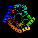
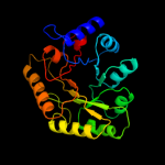
1 c1w8gA_
100.0
100
PDB header: plp-binding proteinChain: A: PDB Molecule: hypothetical upf0001 protein yggs;PDBTitle: crystal structure of e. coli k-12 yggs
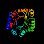
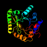
2 c3cpgA_
100.0
33
PDB header: structural genomics, unknown functionChain: A: PDB Molecule: uncharacterized protein;PDBTitle: crystal structure of an unknown protein from bifidobacterium2 adolescentis
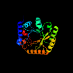
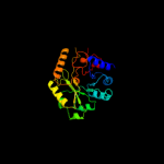
3 c3r79B_
100.0
38
PDB header: structure genomics, unknown functionChain: B: PDB Molecule: uncharacterized protein;PDBTitle: crystal structure of an uncharactertized protein from agrobacterium2 tumefaciens
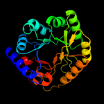
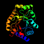
4 d1ct5a_
100.0
36
Fold: TIM beta/alpha-barrelSuperfamily: PLP-binding barrelFamily: "Hypothetical" protein ybl036c5 c1xfcB_
100.0
15
PDB header: isomeraseChain: B: PDB Molecule: alanine racemase;PDBTitle: the 1.9 a crystal structure of alanine racemase from mycobacterium2 tuberculosis contains a conserved entryway into the active site
6 d1bd0a2
100.0
16
Fold: TIM beta/alpha-barrelSuperfamily: PLP-binding barrelFamily: Alanine racemase-like, N-terminal domain7 d1vfsa2
100.0
18
Fold: TIM beta/alpha-barrelSuperfamily: PLP-binding barrelFamily: Alanine racemase-like, N-terminal domain8 c3e6eC_
100.0
16
PDB header: isomeraseChain: C: PDB Molecule: alanine racemase;PDBTitle: crystal structure of alanine racemase from e.faecalis2 complex with cycloserine
9 c3oo2A_
100.0
13
PDB header: isomeraseChain: A: PDB Molecule: alanine racemase 1;PDBTitle: 2.37 angstrom resolution crystal structure of an alanine racemase2 (alr) from staphylococcus aureus subsp. aureus col
10 c2dy3B_
100.0
17
PDB header: isomeraseChain: B: PDB Molecule: alanine racemase;PDBTitle: crystal structure of alanine racemase from corynebacterium glutamicum
11 c3mubB_
100.0
18
PDB header: isomeraseChain: B: PDB Molecule: alanine racemase;PDBTitle: the crystal structure of alanine racemase from streptococcus2 pneumoniae
12 d1rcqa2
100.0
19
Fold: TIM beta/alpha-barrelSuperfamily: PLP-binding barrelFamily: Alanine racemase-like, N-terminal domain13 c1niuA_
100.0
17
PDB header: isomeraseChain: A: PDB Molecule: alanine racemase;PDBTitle: alanine racemase with bound inhibitor derived from l-2 cycloserine
14 c3kw3B_
100.0
13
PDB header: isomeraseChain: B: PDB Molecule: alanine racemase;PDBTitle: crystal structure of alanine racemase from bartonella henselae with2 covalently bound pyridoxal phosphate
15 c1vftA_
100.0
17
PDB header: isomeraseChain: A: PDB Molecule: alanine racemase;PDBTitle: crystal structure of l-cycloserine-bound form of alanine2 racemase from d-cycloserine-producing streptomyces3 lavendulae
16 c3oo2B_
100.0
13
PDB header: isomeraseChain: B: PDB Molecule: alanine racemase 1;PDBTitle: 2.37 angstrom resolution crystal structure of an alanine racemase2 (alr) from staphylococcus aureus subsp. aureus col
17 c3co8B_
100.0
13
PDB header: isomeraseChain: B: PDB Molecule: alanine racemase;PDBTitle: crystal structure of alanine racemase from oenococcus oeni
18 c3hurA_
100.0
19
PDB header: isomeraseChain: A: PDB Molecule: alanine racemase;PDBTitle: crystal structure of alanine racemase from oenococcus oeni
19 c2vd9A_
100.0
18
PDB header: isomeraseChain: A: PDB Molecule: alanine racemase;PDBTitle: the crystal structure of alanine racemase from bacillus2 anthracis (ba0252) with bound l-ala-p
20 c2rjgC_
100.0
11
PDB header: isomeraseChain: C: PDB Molecule: alanine racemase;PDBTitle: crystal structure of biosynthetic alaine racemase from escherichia2 coli
21 c2odoC_
not modelled
100.0
17
PDB header: isomeraseChain: C: PDB Molecule: alanine racemase;PDBTitle: crystal structure of pseudomonas fluorescens alanine racemase
22 c3anuA_
not modelled
100.0
13
PDB header: lyaseChain: A: PDB Molecule: d-serine dehydratase;PDBTitle: crystal structure of d-serine dehydratase from chicken kidney
23 c3llxA_
not modelled
100.0
16
PDB header: isomeraseChain: A: PDB Molecule: predicted amino acid aldolase or racemase;PDBTitle: crystal structure of an ala racemase-like protein (il1761) from2 idiomarina loihiensis at 1.50 a resolution
24 c3gwqB_
not modelled
100.0
12
PDB header: lyaseChain: B: PDB Molecule: d-serine deaminase;PDBTitle: crystal structure of a putative d-serine deaminase (bxe_a4060) from2 burkholderia xenovorans lb400 at 2.00 a resolution
25 c2qghA_
not modelled
100.0
9
PDB header: lyaseChain: A: PDB Molecule: diaminopimelate decarboxylase;PDBTitle: crystal structure of diaminopimelate decarboxylase from helicobacter2 pylori complexed with l-lysine
26 c2j66A_
not modelled
100.0
12
PDB header: lyaseChain: A: PDB Molecule: btrk;PDBTitle: structural characterisation of btrk decarboxylase from2 butirosin biosynthesis
27 c2p3eA_
not modelled
100.0
11
PDB header: lyaseChain: A: PDB Molecule: diaminopimelate decarboxylase;PDBTitle: crystal structure of aq1208 from aquifex aeolicus
28 c3n2bD_
not modelled
100.0
16
PDB header: lyaseChain: D: PDB Molecule: diaminopimelate decarboxylase;PDBTitle: 1.8 angstrom resolution crystal structure of diaminopimelate2 decarboxylase (lysa) from vibrio cholerae.
29 c1tufA_
not modelled
99.9
13
PDB header: lyaseChain: A: PDB Molecule: diaminopimelate decarboxylase;PDBTitle: crystal structure of diaminopimelate decarboxylase from m.2 jannaschi
30 c1njjC_
not modelled
99.9
11
PDB header: lyaseChain: C: PDB Molecule: ornithine decarboxylase;PDBTitle: crystal structure determination of t. brucei ornithine2 decarboxylase bound to d-ornithine and to g418
31 d1twia2
not modelled
99.9
13
Fold: TIM beta/alpha-barrelSuperfamily: PLP-binding barrelFamily: Alanine racemase-like, N-terminal domain32 c2nvaH_
not modelled
99.9
12
PDB header: lyaseChain: H: PDB Molecule: arginine decarboxylase, a207r protein;PDBTitle: the x-ray crystal structure of the paramecium bursaria2 chlorella virus arginine decarboxylase bound to agmatine
33 d1hkva2
not modelled
99.9
15
Fold: TIM beta/alpha-barrelSuperfamily: PLP-binding barrelFamily: Alanine racemase-like, N-terminal domain34 d7odca2
not modelled
99.9
13
Fold: TIM beta/alpha-barrelSuperfamily: PLP-binding barrelFamily: Alanine racemase-like, N-terminal domain35 d1f3ta2
not modelled
99.9
12
Fold: TIM beta/alpha-barrelSuperfamily: PLP-binding barrelFamily: Alanine racemase-like, N-terminal domain36 c2pljA_
not modelled
99.9
15
PDB header: lyaseChain: A: PDB Molecule: lysine/ornithine decarboxylase;PDBTitle: crystal structure of lysine/ornithine decarboxylase complexed with2 putrescine from vibrio vulnificus
37 c2o0tB_
not modelled
99.8
15
PDB header: lyaseChain: B: PDB Molecule: diaminopimelate decarboxylase;PDBTitle: the three dimensional structure of diaminopimelate decarboxylase from2 mycobacterium tuberculosis reveals a tetrameric enzyme organisation
38 c1knwA_
not modelled
99.8
13
PDB header: lyaseChain: A: PDB Molecule: diaminopimelate decarboxylase;PDBTitle: crystal structure of diaminopimelate decarboxylase
39 d1d7ka2
not modelled
99.8
11
Fold: TIM beta/alpha-barrelSuperfamily: PLP-binding barrelFamily: Alanine racemase-like, N-terminal domain40 c2yxxA_
not modelled
99.8
13
PDB header: lyaseChain: A: PDB Molecule: diaminopimelate decarboxylase;PDBTitle: crystal structure analysis of diaminopimelate decarboxylate (lysa)
41 c2on3A_
not modelled
99.8
13
PDB header: lyaseChain: A: PDB Molecule: ornithine decarboxylase;PDBTitle: a structural insight into the inhibition of human and2 leishmania donovani ornithine decarboxylases by 3-aminooxy-3 1-aminopropane
42 c3nzqB_
not modelled
99.7
15
PDB header: lyaseChain: B: PDB Molecule: biosynthetic arginine decarboxylase;PDBTitle: crystal structure of biosynthetic arginine decarboxylase adc (spea)2 from escherichia coli, northeast structural genomics consortium3 target er600
43 c3btnA_
not modelled
99.7
15
PDB header: biosynthetic proteinChain: A: PDB Molecule: antizyme inhibitor 1;PDBTitle: crystal structure of antizyme inhibitor, an ornithine2 decarboxylase homologous protein
44 c3n2oA_
not modelled
99.7
11
PDB header: lyaseChain: A: PDB Molecule: biosynthetic arginine decarboxylase;PDBTitle: x-ray crystal structure of arginine decarboxylase complexed with2 arginine from vibrio vulnificus
45 c1d7kB_
not modelled
99.7
12
PDB header: lyaseChain: B: PDB Molecule: human ornithine decarboxylase;PDBTitle: crystal structure of human ornithine decarboxylase at 2.12 angstroms resolution
46 c3nzpA_
not modelled
99.6
13
PDB header: lyaseChain: A: PDB Molecule: arginine decarboxylase;PDBTitle: crystal structure of the biosynthetic arginine decarboxylase spea from2 campylobacter jejuni, northeast structural genomics consortium target3 br53
47 d1knwa2
not modelled
99.6
13
Fold: TIM beta/alpha-barrelSuperfamily: PLP-binding barrelFamily: Alanine racemase-like, N-terminal domain48 c3n29A_
not modelled
99.5
11
PDB header: lyaseChain: A: PDB Molecule: carboxynorspermidine decarboxylase;PDBTitle: crystal structure of carboxynorspermidine decarboxylase complexed with2 norspermidine from campylobacter jejuni
49 c3mt1B_
not modelled
99.1
13
PDB header: lyaseChain: B: PDB Molecule: putative carboxynorspermidine decarboxylase protein;PDBTitle: crystal structure of putative carboxynorspermidine decarboxylase2 protein from sinorhizobium meliloti
50 d1rpxa_
not modelled
95.4
13
Fold: TIM beta/alpha-barrelSuperfamily: Ribulose-phoshate binding barrelFamily: D-ribulose-5-phosphate 3-epimerase51 d2flia1
not modelled
94.6
17
Fold: TIM beta/alpha-barrelSuperfamily: Ribulose-phoshate binding barrelFamily: D-ribulose-5-phosphate 3-epimerase52 c2h90A_
not modelled
94.4
17
PDB header: oxidoreductaseChain: A: PDB Molecule: xenobiotic reductase a;PDBTitle: xenobiotic reductase a in complex with coumarin
53 d1h1ya_
not modelled
93.6
17
Fold: TIM beta/alpha-barrelSuperfamily: Ribulose-phoshate binding barrelFamily: D-ribulose-5-phosphate 3-epimerase54 c3hf3A_
not modelled
92.1
16
PDB header: oxidoreductaseChain: A: PDB Molecule: chromate reductase;PDBTitle: old yellow enzyme from thermus scotoductus sa-01
55 d1tqja_
not modelled
92.0
15
Fold: TIM beta/alpha-barrelSuperfamily: Ribulose-phoshate binding barrelFamily: D-ribulose-5-phosphate 3-epimerase56 c3ct7E_
not modelled
91.5
16
PDB header: isomeraseChain: E: PDB Molecule: d-allulose-6-phosphate 3-epimerase;PDBTitle: crystal structure of d-allulose 6-phosphate 3-epimerase2 from escherichia coli k-12
57 d1djqa1
not modelled
91.2
10
Fold: TIM beta/alpha-barrelSuperfamily: FMN-linked oxidoreductasesFamily: FMN-linked oxidoreductases58 d1ps9a1
not modelled
91.2
14
Fold: TIM beta/alpha-barrelSuperfamily: FMN-linked oxidoreductasesFamily: FMN-linked oxidoreductases59 c2y85D_
not modelled
91.0
12
PDB header: isomeraseChain: D: PDB Molecule: phosphoribosyl isomerase a;PDBTitle: crystal structure of mycobacterium tuberculosis phosphoribosyl2 isomerase with bound rcdrp
60 d1tqxa_
not modelled
87.9
12
Fold: TIM beta/alpha-barrelSuperfamily: Ribulose-phoshate binding barrelFamily: D-ribulose-5-phosphate 3-epimerase61 c1djnB_
not modelled
87.6
10
PDB header: oxidoreductaseChain: B: PDB Molecule: trimethylamine dehydrogenase;PDBTitle: structural and biochemical characterization of recombinant wild type2 trimethylamine dehydrogenase from methylophilus methylotrophus (sp.3 w3a1)
62 c3inpA_
not modelled
86.7
10
PDB header: isomeraseChain: A: PDB Molecule: d-ribulose-phosphate 3-epimerase;PDBTitle: 2.05 angstrom resolution crystal structure of d-ribulose-phosphate 3-2 epimerase from francisella tularensis.
63 c3k30B_
not modelled
86.3
17
PDB header: oxidoreductaseChain: B: PDB Molecule: histamine dehydrogenase;PDBTitle: histamine dehydrogenase from nocardiodes simplex
64 c2vwtA_
not modelled
85.8
14
PDB header: lyaseChain: A: PDB Molecule: yfau, 2-keto-3-deoxy sugar aldolase;PDBTitle: crystal structure of yfau, a metal ion dependent class ii2 aldolase from escherichia coli k12 - mg-pyruvate product3 complex
65 c1ps9A_
not modelled
82.9
13
PDB header: oxidoreductaseChain: A: PDB Molecule: 2,4-dienoyl-coa reductase;PDBTitle: the crystal structure and reaction mechanism of e. coli 2,4-2 dienoyl coa reductase
66 c3qz6A_
not modelled
80.7
10
PDB header: lyaseChain: A: PDB Molecule: hpch/hpai aldolase;PDBTitle: the crystal structure of hpch/hpai aldolase from desulfitobacterium2 hafniense dcb-2
67 d1dxea_
not modelled
79.7
13
Fold: TIM beta/alpha-barrelSuperfamily: Phosphoenolpyruvate/pyruvate domainFamily: HpcH/HpaI aldolase68 c2v5jB_
not modelled
79.4
12
PDB header: lyaseChain: B: PDB Molecule: 2,4-dihydroxyhept-2-ene-1,7-dioic acid aldolase;PDBTitle: apo class ii aldolase hpch
69 c1izcA_
not modelled
79.1
14
PDB header: lyaseChain: A: PDB Molecule: macrophomate synthase intermolecular diels-alderase;PDBTitle: crystal structure analysis of macrophomate synthase
70 d1izca_
not modelled
79.1
14
Fold: TIM beta/alpha-barrelSuperfamily: Phosphoenolpyruvate/pyruvate domainFamily: HpcH/HpaI aldolase71 d1qapa1
not modelled
76.1
13
Fold: TIM beta/alpha-barrelSuperfamily: Nicotinate/Quinolinate PRTase C-terminal domain-likeFamily: NadC C-terminal domain-like72 d1v6ta_
not modelled
75.6
14
Fold: 7-stranded beta/alpha barrelSuperfamily: Glycoside hydrolase/deacetylaseFamily: LamB/YcsF-like73 d2dfaa1
not modelled
72.3
19
Fold: 7-stranded beta/alpha barrelSuperfamily: Glycoside hydrolase/deacetylaseFamily: LamB/YcsF-like74 d1o4ua1
not modelled
70.7
12
Fold: TIM beta/alpha-barrelSuperfamily: Nicotinate/Quinolinate PRTase C-terminal domain-likeFamily: NadC C-terminal domain-like75 c3cu2A_
not modelled
67.5
6
PDB header: isomeraseChain: A: PDB Molecule: ribulose-5-phosphate 3-epimerase;PDBTitle: crystal structure of ribulose-5-phosphate 3-epimerase (yp_718263.1)2 from haemophilus somnus 129pt at 1.91 a resolution
76 c3gr7A_
not modelled
64.9
14
PDB header: oxidoreductaseChain: A: PDB Molecule: nadph dehydrogenase;PDBTitle: structure of oye from geobacillus kaustophilus, hexagonal2 crystal form
77 c3l5aA_
not modelled
62.5
9
PDB header: oxidoreductaseChain: A: PDB Molecule: nadh/flavin oxidoreductase/nadh oxidase;PDBTitle: crystal structure of a probable nadh-dependent flavin oxidoreductase2 from staphylococcus aureus
78 c3kruC_
not modelled
61.7
13
PDB header: oxidoreductaseChain: C: PDB Molecule: nadh:flavin oxidoreductase/nadh oxidase;PDBTitle: crystal structure of the thermostable old yellow enzyme from2 thermoanaerobacter pseudethanolicus e39
79 d1vyra_
not modelled
58.3
11
Fold: TIM beta/alpha-barrelSuperfamily: FMN-linked oxidoreductasesFamily: FMN-linked oxidoreductases80 c2gq8A_
not modelled
57.6
15
PDB header: oxidoreductaseChain: A: PDB Molecule: oxidoreductase, fmn-binding;PDBTitle: structure of sye1, an oye homologue from s. ondeidensis, in complex2 with p-hydroxyacetophenone
81 d1qo2a_
not modelled
56.9
12
Fold: TIM beta/alpha-barrelSuperfamily: Ribulose-phoshate binding barrelFamily: Histidine biosynthesis enzymes82 c3qc3B_
not modelled
56.2
16
PDB header: isomeraseChain: B: PDB Molecule: d-ribulose-5-phosphate-3-epimerase;PDBTitle: crystal structure of a d-ribulose-5-phosphate-3-epimerase (np_954699)2 from homo sapiens at 2.20 a resolution
83 c2yciX_
not modelled
55.7
9
PDB header: transferaseChain: X: PDB Molecule: 5-methyltetrahydrofolate corrinoid/iron sulfur proteinPDBTitle: methyltransferase native
84 c2c3zA_
not modelled
52.5
12
PDB header: lyaseChain: A: PDB Molecule: indole-3-glycerol phosphate synthase;PDBTitle: crystal structure of a truncated variant of indole-3-2 glycerol phosphate synthase from sulfolobus solfataricus
85 d1y0ea_
not modelled
51.0
11
Fold: TIM beta/alpha-barrelSuperfamily: Ribulose-phoshate binding barrelFamily: NanE-like86 c2x5eA_
not modelled
48.9
15
PDB header: unknown functionChain: A: PDB Molecule: upf0271 protein pa4511;PDBTitle: crystal structure of the hypothetical protein pa4511 from2 pseudomonas aeruginosa
87 d1qpoa1
not modelled
47.1
8
Fold: TIM beta/alpha-barrelSuperfamily: Nicotinate/Quinolinate PRTase C-terminal domain-likeFamily: NadC C-terminal domain-like88 d1gwja_
not modelled
46.8
14
Fold: TIM beta/alpha-barrelSuperfamily: FMN-linked oxidoreductasesFamily: FMN-linked oxidoreductases89 d1gvfa_
not modelled
45.2
12
Fold: TIM beta/alpha-barrelSuperfamily: AldolaseFamily: Class II FBP aldolase90 d1z41a1
not modelled
44.6
12
Fold: TIM beta/alpha-barrelSuperfamily: FMN-linked oxidoreductasesFamily: FMN-linked oxidoreductases91 d1vc4a_
not modelled
42.8
13
Fold: TIM beta/alpha-barrelSuperfamily: Ribulose-phoshate binding barrelFamily: Tryptophan biosynthesis enzymes92 c2zviB_
not modelled
42.8
15
PDB header: isomeraseChain: B: PDB Molecule: 2,3-diketo-5-methylthiopentyl-1-phosphatePDBTitle: crystal structure of 2,3-diketo-5-methylthiopentyl-1-2 phosphate enolase from bacillus subtilis
93 c2vkzH_
not modelled
40.7
12
PDB header: transferaseChain: H: PDB Molecule: fatty acid synthase subunit beta;PDBTitle: structure of the cerulenin-inhibited fungal fatty acid2 synthase type i multienzyme complex
94 d1vzwa1
not modelled
39.7
13
Fold: TIM beta/alpha-barrelSuperfamily: Ribulose-phoshate binding barrelFamily: Histidine biosynthesis enzymes95 c2v82A_
not modelled
39.0
13
PDB header: lyaseChain: A: PDB Molecule: 2-dehydro-3-deoxy-6-phosphogalactonate aldolase;PDBTitle: kdpgal complexed to kdpgal
96 c1yadD_
not modelled
38.4
12
PDB header: transcriptionChain: D: PDB Molecule: regulatory protein teni;PDBTitle: structure of teni from bacillus subtilis
97 c2qv5A_
not modelled
36.4
16
PDB header: structural genomics, unknown functionChain: A: PDB Molecule: uncharacterized protein atu2773;PDBTitle: crystal structure of uncharacterized protein atu2773 from2 agrobacterium tumefaciens c58
98 d1wbha1
not modelled
36.4
15
Fold: TIM beta/alpha-barrelSuperfamily: AldolaseFamily: Class I aldolase99 c2dzaA_
not modelled
35.3
14
PDB header: transferaseChain: A: PDB Molecule: dihydropteroate synthase;PDBTitle: crystal structure of dihydropteroate synthase from thermus2 thermophilus hb8 in complex with 4-aminobenzoate
100 c2oemA_
not modelled
34.5
15
PDB header: isomeraseChain: A: PDB Molecule: 2,3-diketo-5-methylthiopentyl-1-phosphate enolase;PDBTitle: crystal structure of a rubisco-like protein from geobacillus2 kaustophilus liganded with mg2+ and 2,3-diketohexane 1-phosphate
101 d1ka9f_
not modelled
34.4
15
Fold: TIM beta/alpha-barrelSuperfamily: Ribulose-phoshate binding barrelFamily: Histidine biosynthesis enzymes102 c1rr2A_
not modelled
33.0
12
PDB header: transferaseChain: A: PDB Molecule: transcarboxylase 5s subunit;PDBTitle: propionibacterium shermanii transcarboxylase 5s subunit bound to 2-2 ketobutyric acid
103 d1ajza_
not modelled
32.8
17
Fold: TIM beta/alpha-barrelSuperfamily: Dihydropteroate synthetase-likeFamily: Dihydropteroate synthetase104 c3atyA_
not modelled
31.1
13
PDB header: oxidoreductaseChain: A: PDB Molecule: prostaglandin f2a synthase;PDBTitle: crystal structure of tcoye
105 c2jfqA_
not modelled
26.9
10
PDB header: isomeraseChain: A: PDB Molecule: glutamate racemase;PDBTitle: crystal structure of staphylococcus aureus glutamate2 racemase in complex with d-glutamate
106 c3gkaB_
not modelled
26.6
15
PDB header: oxidoreductaseChain: B: PDB Molecule: n-ethylmaleimide reductase;PDBTitle: crystal structure of n-ethylmaleimidine reductase from2 burkholderia pseudomallei
107 d1thfd_
not modelled
23.0
10
Fold: TIM beta/alpha-barrelSuperfamily: Ribulose-phoshate binding barrelFamily: Histidine biosynthesis enzymes108 d2zdra2
not modelled
21.6
13
Fold: TIM beta/alpha-barrelSuperfamily: AldolaseFamily: NeuB-like109 c3ju3A_
not modelled
21.6
12
PDB header: oxidoreductaseChain: A: PDB Molecule: probable 2-oxoacid ferredoxin oxidoreductase, alpha chain;PDBTitle: crystal structure of alpha chain of probable 2-oxoacid ferredoxin2 oxidoreductase from thermoplasma acidophilum
110 c2x7vA_
not modelled
20.1
7
PDB header: hydrolaseChain: A: PDB Molecule: probable endonuclease 4;PDBTitle: crystal structure of thermotoga maritima endonuclease iv in2 the presence of zinc