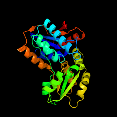
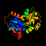
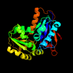
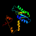
| 1 | c2jfnA_
|
|
 |
100.0 |
100 |
PDB header:isomerase
Chain: A: PDB Molecule:glutamate racemase;
PDBTitle: crystal structure of escherichia coli glutamate racemase2 in complex with l-glutamate and activator udp-murnac-ala
|
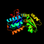
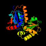
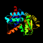
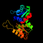
| 2 | c3hfrA_
|
|
 |
100.0 |
27 |
PDB header:isomerase
Chain: A: PDB Molecule:glutamate racemase;
PDBTitle: crystal structure of glutamate racemase from listeria monocytogenes
|
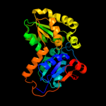
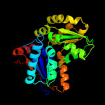
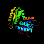
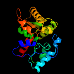
| 3 | c2jfoB_
|
|
 |
100.0 |
26 |
PDB header:isomerase
Chain: B: PDB Molecule:glutamate racemase;
PDBTitle: crystal structure of enterococcus faecalis glutamate2 racemase in complex with d- and l-glutamate
|
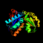
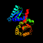
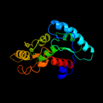
| 4 | c2jfqA_
|
|
 |
100.0 |
28 |
PDB header:isomerase
Chain: A: PDB Molecule:glutamate racemase;
PDBTitle: crystal structure of staphylococcus aureus glutamate2 racemase in complex with d-glutamate
|
| 5 | c2dwuA_
|
|
 |
100.0 |
29 |
PDB header:isomerase
Chain: A: PDB Molecule:glutamate racemase;
PDBTitle: crystal structure of glutamate racemase isoform race1 from bacillus2 anthracis
|
| 6 | c2gzmB_
|
|
 |
100.0 |
28 |
PDB header:isomerase
Chain: B: PDB Molecule:glutamate racemase;
PDBTitle: crystal structure of the glutamate racemase from bacillus2 anthracis
|
| 7 | c1zuwA_
|
|
 |
100.0 |
27 |
PDB header:isomerase
Chain: A: PDB Molecule:glutamate racemase 1;
PDBTitle: crystal structure of b.subtilis glutamate racemase (race) with d-glu
|
| 8 | c2ohoA_
|
|
 |
100.0 |
29 |
PDB header:isomerase
Chain: A: PDB Molecule:glutamate racemase;
PDBTitle: structural basis for glutamate racemase inhibitor
|
| 9 | c3outC_
|
|
 |
100.0 |
22 |
PDB header:isomerase
Chain: C: PDB Molecule:glutamate racemase;
PDBTitle: crystal structure of glutamate racemase from francisella tularensis2 subsp. tularensis schu s4 in complex with d-glutamate.
|
| 10 | c2jfzB_
|
|
 |
100.0 |
26 |
PDB header:isomerase
Chain: B: PDB Molecule:glutamate racemase;
PDBTitle: crystal structure of helicobacter pylori glutamate racemase2 in complex with d-glutamate and an inhibitor
|
| 11 | c1b74A_
|
|
 |
100.0 |
27 |
PDB header:isomerase
Chain: A: PDB Molecule:glutamate racemase;
PDBTitle: glutamate racemase from aquifex pyrophilus
|
| 12 | c2zskA_
|
|
 |
100.0 |
18 |
PDB header:unknown function
Chain: A: PDB Molecule:226aa long hypothetical aspartate racemase;
PDBTitle: crystal structure of ph1733, an aspartate racemase2 homologue, from pyrococcus horikoshii ot3
|
| 13 | d1b74a1
|
|
 |
100.0 |
34 |
Fold:ATC-like
Superfamily:Aspartate/glutamate racemase
Family:Aspartate/glutamate racemase |
| 14 | c3ojcD_
|
|
 |
100.0 |
18 |
PDB header:isomerase
Chain: D: PDB Molecule:putative aspartate/glutamate racemase;
PDBTitle: crystal structure of a putative asp/glu racemase from yersinia pestis
|
| 15 | c3s81A_
|
|
 |
100.0 |
17 |
PDB header:isomerase
Chain: A: PDB Molecule:putative aspartate racemase;
PDBTitle: crystal structure of putative aspartate racemase from salmonella2 typhimurium
|
| 16 | c2dx7B_
|
|
 |
100.0 |
14 |
PDB header:isomerase
Chain: B: PDB Molecule:aspartate racemase;
PDBTitle: crystal structure of pyrococcus horikoshii ot3 aspartate racemase2 complex with citric acid
|
| 17 | d1b74a2
|
|
 |
100.0 |
23 |
Fold:ATC-like
Superfamily:Aspartate/glutamate racemase
Family:Aspartate/glutamate racemase |
| 18 | c2vlbC_
|
|
 |
100.0 |
17 |
PDB header:lyase
Chain: C: PDB Molecule:arylmalonate decarboxylase;
PDBTitle: structure of unliganded arylmalonate decarboxylase
|
| 19 | c3qvjB_
|
|
 |
99.9 |
18 |
PDB header:isomerase
Chain: B: PDB Molecule:putative hydantoin racemase;
PDBTitle: allantoin racemase from klebsiella pneumoniae
|
| 20 | c2eq5D_
|
|
 |
99.9 |
18 |
PDB header:isomerase
Chain: D: PDB Molecule:228aa long hypothetical hydantoin racemase;
PDBTitle: crystal structure of hydantoin racemase from pyrococcus horikoshii ot3
|
| 21 | c2dgdD_ |
|
not modelled |
99.8 |
11 |
PDB header:lyase
Chain: D: PDB Molecule:223aa long hypothetical arylmalonate decarboxylase;
PDBTitle: crystal structure of st0656, a function unknown protein from2 sulfolobus tokodaii
|
| 22 | d1jfla2 |
|
not modelled |
99.8 |
16 |
Fold:ATC-like
Superfamily:Aspartate/glutamate racemase
Family:Aspartate/glutamate racemase |
| 23 | d2dx7a1 |
|
not modelled |
99.8 |
14 |
Fold:ATC-like
Superfamily:Aspartate/glutamate racemase
Family:Aspartate/glutamate racemase |
| 24 | d1jfla1 |
|
not modelled |
99.1 |
15 |
Fold:ATC-like
Superfamily:Aspartate/glutamate racemase
Family:Aspartate/glutamate racemase |
| 25 | c2xecD_ |
|
not modelled |
98.6 |
16 |
PDB header:isomerase
Chain: D: PDB Molecule:putative maleate isomerase;
PDBTitle: nocardia farcinica maleate cis-trans isomerase bound to2 tris
|
| 26 | c3dahB_ |
|
not modelled |
94.4 |
15 |
PDB header:transferase
Chain: B: PDB Molecule:ribose-phosphate pyrophosphokinase;
PDBTitle: 2.3 a crystal structure of ribose-phosphate pyrophosphokinase from2 burkholderia pseudomallei
|
| 27 | c1dkrB_ |
|
not modelled |
94.0 |
15 |
PDB header:transferase
Chain: B: PDB Molecule:phosphoribosyl pyrophosphate synthetase;
PDBTitle: crystal structures of bacillus subtilis phosphoribosylpyrophosphate2 synthetase: molecular basis of allosteric inhibition and activation.
|
| 28 | c2dpyA_ |
|
not modelled |
93.6 |
18 |
PDB header:hydrolase
Chain: A: PDB Molecule:flagellum-specific atp synthase;
PDBTitle: crystal structure of the flagellar type iii atpase flii
|
| 29 | d1m3ua_ |
|
not modelled |
93.4 |
17 |
Fold:TIM beta/alpha-barrel
Superfamily:Phosphoenolpyruvate/pyruvate domain
Family:Ketopantoate hydroxymethyltransferase PanB |
| 30 | c3a5dM_ |
|
not modelled |
93.4 |
15 |
PDB header:hydrolase
Chain: M: PDB Molecule:v-type atp synthase beta chain;
PDBTitle: inter-subunit interaction and quaternary rearrangement2 defined by the central stalk of prokaryotic v1-atpase
|
| 31 | d1oy0a_ |
|
not modelled |
91.1 |
14 |
Fold:TIM beta/alpha-barrel
Superfamily:Phosphoenolpyruvate/pyruvate domain
Family:Ketopantoate hydroxymethyltransferase PanB |
| 32 | d1skye3 |
|
not modelled |
91.1 |
13 |
Fold:P-loop containing nucleoside triphosphate hydrolases
Superfamily:P-loop containing nucleoside triphosphate hydrolases
Family:RecA protein-like (ATPase-domain) |
| 33 | c3lp6D_ |
|
not modelled |
90.7 |
20 |
PDB header:lyase
Chain: D: PDB Molecule:phosphoribosylaminoimidazole carboxylase catalytic subunit;
PDBTitle: crystal structure of rv3275c-e60a from mycobacterium tuberculosis at2 1.7a resolution
|
| 34 | c3ez4B_ |
|
not modelled |
87.5 |
19 |
PDB header:transferase
Chain: B: PDB Molecule:3-methyl-2-oxobutanoate hydroxymethyltransferase;
PDBTitle: crystal structure of 3-methyl-2-oxobutanoate2 hydroxymethyltransferase from burkholderia pseudomallei
|
| 35 | c2pjuD_ |
|
not modelled |
87.1 |
14 |
PDB header:transcription
Chain: D: PDB Molecule:propionate catabolism operon regulatory protein;
PDBTitle: crystal structure of propionate catabolism operon2 regulatory protein prpr
|
| 36 | d1wbha1 |
|
not modelled |
86.5 |
13 |
Fold:TIM beta/alpha-barrel
Superfamily:Aldolase
Family:Class I aldolase |
| 37 | c1kjjA_ |
|
not modelled |
85.7 |
17 |
PDB header:transferase
Chain: A: PDB Molecule:phosphoribosylglycinamide formyltransferase 2;
PDBTitle: crystal structure of glycniamide ribonucleotide2 transformylase in complex with mg-atp-gamma-s
|
| 38 | d1o5oa_ |
|
not modelled |
84.7 |
20 |
Fold:PRTase-like
Superfamily:PRTase-like
Family:Phosphoribosyltransferases (PRTases) |
| 39 | c2q5cA_ |
|
not modelled |
84.7 |
16 |
PDB header:transcription
Chain: A: PDB Molecule:ntrc family transcriptional regulator;
PDBTitle: crystal structure of ntrc family transcriptional regulator from2 clostridium acetobutylicum
|
| 40 | c2xd4A_ |
|
not modelled |
84.6 |
11 |
PDB header:ligase
Chain: A: PDB Molecule:phosphoribosylamine--glycine ligase;
PDBTitle: nucleotide-bound structures of bacillus subtilis glycinamide2 ribonucleotide synthetase
|
| 41 | d2pjua1 |
|
not modelled |
82.8 |
14 |
Fold:Chelatase-like
Superfamily:PrpR receptor domain-like
Family:PrpR receptor domain-like |
| 42 | c2c61A_ |
|
not modelled |
82.4 |
13 |
PDB header:hydrolase
Chain: A: PDB Molecule:a-type atp synthase non-catalytic subunit b;
PDBTitle: crystal structure of the non-catalytic b subunit of a-type2 atpase from m. mazei go1
|
| 43 | c3ouzA_ |
|
not modelled |
81.9 |
12 |
PDB header:ligase
Chain: A: PDB Molecule:biotin carboxylase;
PDBTitle: crystal structure of biotin carboxylase-adp complex from campylobacter2 jejuni
|
| 44 | c1fx0B_ |
|
not modelled |
80.8 |
14 |
PDB header:hydrolase
Chain: B: PDB Molecule:atp synthase beta chain;
PDBTitle: crystal structure of the chloroplast f1-atpase from spinach
|
| 45 | c2axqA_ |
|
not modelled |
80.7 |
16 |
PDB header:oxidoreductase
Chain: A: PDB Molecule:saccharopine dehydrogenase;
PDBTitle: apo histidine-tagged saccharopine dehydrogenase (l-glu2 forming) from saccharomyces cerevisiae
|
| 46 | d1o66a_ |
|
not modelled |
80.6 |
14 |
Fold:TIM beta/alpha-barrel
Superfamily:Phosphoenolpyruvate/pyruvate domain
Family:Ketopantoate hydroxymethyltransferase PanB |
| 47 | d1skyb3 |
|
not modelled |
79.4 |
16 |
Fold:P-loop containing nucleoside triphosphate hydrolases
Superfamily:P-loop containing nucleoside triphosphate hydrolases
Family:RecA protein-like (ATPase-domain) |
| 48 | c2gr2A_ |
|
not modelled |
78.2 |
21 |
PDB header:oxidoreductase
Chain: A: PDB Molecule:ferredoxin reductase;
PDBTitle: crystal structure of ferredoxin reductase, bpha4 (oxidized form)
|
| 49 | c3gkaB_ |
|
not modelled |
76.7 |
9 |
PDB header:oxidoreductase
Chain: B: PDB Molecule:n-ethylmaleimide reductase;
PDBTitle: crystal structure of n-ethylmaleimidine reductase from2 burkholderia pseudomallei
|
| 50 | d1cjca2 |
|
not modelled |
76.6 |
16 |
Fold:Nucleotide-binding domain
Superfamily:Nucleotide-binding domain
Family:N-terminal domain of adrenodoxin reductase-like |
| 51 | c3h5lB_ |
|
not modelled |
76.2 |
12 |
PDB header:transport protein
Chain: B: PDB Molecule:putative branched-chain amino acid abc
PDBTitle: crystal structure of a putative branched-chain amino acid2 abc transporter from silicibacter pomeroyi
|
| 52 | c3bolB_ |
|
not modelled |
75.3 |
22 |
PDB header:transferase
Chain: B: PDB Molecule:5-methyltetrahydrofolate s-homocysteine
PDBTitle: cobalamin-dependent methionine synthase (1-566) from2 thermotoga maritima complexed with zn2+
|
| 53 | c2jizD_ |
|
not modelled |
75.3 |
15 |
PDB header:hydrolase
Chain: D: PDB Molecule:atp synthase subunit beta;
PDBTitle: the structure of f1-atpase inhibited by resveratrol.
|
| 54 | c3noyA_ |
|
not modelled |
75.0 |
21 |
PDB header:oxidoreductase
Chain: A: PDB Molecule:4-hydroxy-3-methylbut-2-en-1-yl diphosphate synthase;
PDBTitle: crystal structure of ispg (gcpe)
|
| 55 | c3hf3A_ |
|
not modelled |
74.6 |
13 |
PDB header:oxidoreductase
Chain: A: PDB Molecule:chromate reductase;
PDBTitle: old yellow enzyme from thermus scotoductus sa-01
|
| 56 | c3d2fC_ |
|
not modelled |
74.3 |
19 |
PDB header:chaperone
Chain: C: PDB Molecule:heat shock protein homolog sse1;
PDBTitle: crystal structure of a complex of sse1p and hsp70
|
| 57 | c2v7yA_ |
|
not modelled |
74.2 |
21 |
PDB header:chaperone
Chain: A: PDB Molecule:chaperone protein dnak;
PDBTitle: crystal structure of the molecular chaperone dnak from2 geobacillus kaustophilus hta426 in post-atp hydrolysis3 state
|
| 58 | d1v9sa1 |
|
not modelled |
73.7 |
16 |
Fold:PRTase-like
Superfamily:PRTase-like
Family:Phosphoribosyltransferases (PRTases) |
| 59 | c2c4kD_ |
|
not modelled |
73.4 |
14 |
PDB header:regulatory protein
Chain: D: PDB Molecule:phosphoribosyl pyrophosphate synthetase-
PDBTitle: crystal structure of human phosphoribosylpyrophosphate2 synthetase-associated protein 39 (pap39)
|
| 60 | c1dkgD_ |
|
not modelled |
72.8 |
26 |
PDB header:complex (hsp24/hsp70)
Chain: D: PDB Molecule:molecular chaperone dnak;
PDBTitle: crystal structure of the nucleotide exchange factor grpe2 bound to the atpase domain of the molecular chaperone dnak
|
| 61 | c2oblA_ |
|
not modelled |
72.2 |
16 |
PDB header:hydrolase
Chain: A: PDB Molecule:escn;
PDBTitle: structural and biochemical analysis of a prototypical atpase from the2 type iii secretion system of pathogenic bacteria
|
| 62 | c3gr7A_ |
|
not modelled |
72.2 |
13 |
PDB header:oxidoreductase
Chain: A: PDB Molecule:nadph dehydrogenase;
PDBTitle: structure of oye from geobacillus kaustophilus, hexagonal2 crystal form
|
| 63 | c2ys6A_ |
|
not modelled |
72.1 |
12 |
PDB header:ligase
Chain: A: PDB Molecule:phosphoribosylglycinamide synthetase;
PDBTitle: crystal structure of gar synthetase from geobacillus kaustophilus
|
| 64 | c3a5dB_ |
|
not modelled |
71.8 |
19 |
PDB header:hydrolase
Chain: B: PDB Molecule:v-type atp synthase alpha chain;
PDBTitle: inter-subunit interaction and quaternary rearrangement2 defined by the central stalk of prokaryotic v1-atpase
|
| 65 | c2w6jD_ |
|
not modelled |
71.5 |
15 |
PDB header:hydrolase
Chain: D: PDB Molecule:atp synthase subunit beta, mitochondrial;
PDBTitle: low resolution structures of bovine mitochondrial f1-atpase2 during controlled dehydration: hydration state 5.
|
| 66 | c1skyE_ |
|
not modelled |
70.0 |
11 |
PDB header:atp synthase
Chain: E: PDB Molecule:f1-atpase;
PDBTitle: crystal structure of the nucleotide free alpha3beta3 sub-complex of2 f1-atpase from the thermophilic bacillus ps3
|
| 67 | c1kmhA_ |
|
not modelled |
69.9 |
17 |
PDB header:hydrolase
Chain: A: PDB Molecule:atpase alpha subunit;
PDBTitle: crystal structure of spinach chloroplast f1-atpase2 complexed with tentoxin
|
| 68 | c2o14A_ |
|
not modelled |
67.5 |
20 |
PDB header:structural genomics, unknown function
Chain: A: PDB Molecule:hypothetical protein yxim;
PDBTitle: x-ray crystal structure of protein yxim_bacsu from bacillus2 subtilis. northeast structural genomics consortium target3 sr595
|
| 69 | d1fx0b3 |
|
not modelled |
67.4 |
12 |
Fold:P-loop containing nucleoside triphosphate hydrolases
Superfamily:P-loop containing nucleoside triphosphate hydrolases
Family:RecA protein-like (ATPase-domain) |
| 70 | c2h31A_ |
|
not modelled |
65.4 |
17 |
PDB header:ligase, lyase
Chain: A: PDB Molecule:multifunctional protein ade2;
PDBTitle: crystal structure of human paics, a bifunctional carboxylase and2 synthetase in purine biosynthesis
|
| 71 | c1m6iA_ |
|
not modelled |
64.9 |
16 |
PDB header:oxidoreductase
Chain: A: PDB Molecule:programmed cell death protein 8;
PDBTitle: crystal structure of apoptosis inducing factor (aif)
|
| 72 | c3zu0A_ |
|
not modelled |
64.1 |
15 |
PDB header:hydrolase
Chain: A: PDB Molecule:nad nucleotidase;
PDBTitle: structure of haemophilus influenzae nad nucleotidase (nadn)
|
| 73 | d1vhca_ |
|
not modelled |
64.0 |
16 |
Fold:TIM beta/alpha-barrel
Superfamily:Aldolase
Family:Class I aldolase |
| 74 | d1gv4a1 |
|
not modelled |
63.9 |
18 |
Fold:FAD/NAD(P)-binding domain
Superfamily:FAD/NAD(P)-binding domain
Family:FAD/NAD-linked reductases, N-terminal and central domains |
| 75 | c1ulzA_ |
|
not modelled |
63.9 |
15 |
PDB header:ligase
Chain: A: PDB Molecule:pyruvate carboxylase n-terminal domain;
PDBTitle: crystal structure of the biotin carboxylase subunit of pyruvate2 carboxylase
|
| 76 | c2v3aA_ |
|
not modelled |
63.8 |
17 |
PDB header:oxidoreductase
Chain: A: PDB Molecule:rubredoxin reductase;
PDBTitle: crystal structure of rubredoxin reductase from pseudomonas2 aeruginosa.
|
| 77 | c3sg0A_ |
|
not modelled |
63.5 |
18 |
PDB header:signaling protein
Chain: A: PDB Molecule:extracellular ligand-binding receptor;
PDBTitle: the crystal structure of an extracellular ligand-binding receptor from2 rhodopseudomonas palustris haa2
|
| 78 | c2h90A_ |
|
not modelled |
63.3 |
14 |
PDB header:oxidoreductase
Chain: A: PDB Molecule:xenobiotic reductase a;
PDBTitle: xenobiotic reductase a in complex with coumarin
|
| 79 | c3jz3B_ |
|
not modelled |
63.1 |
43 |
PDB header:transferase
Chain: B: PDB Molecule:sensor protein qsec;
PDBTitle: structure of the cytoplasmic segment of histidine kinase qsec
|
| 80 | c3dmpD_ |
|
not modelled |
62.5 |
11 |
PDB header:transferase
Chain: D: PDB Molecule:uracil phosphoribosyltransferase;
PDBTitle: 2.6 a crystal structure of uracil phosphoribosyltransferase2 from burkholderia pseudomallei
|
| 81 | c2iswB_ |
|
not modelled |
62.4 |
16 |
PDB header:lyase
Chain: B: PDB Molecule:putative fructose-1,6-bisphosphate aldolase;
PDBTitle: structure of giardia fructose-1,6-biphosphate aldolase in2 complex with phosphoglycolohydroxamate
|
| 82 | d1e5qa1 |
|
not modelled |
62.0 |
15 |
Fold:NAD(P)-binding Rossmann-fold domains
Superfamily:NAD(P)-binding Rossmann-fold domains
Family:Glyceraldehyde-3-phosphate dehydrogenase-like, N-terminal domain |
| 83 | c3lwzC_ |
|
not modelled |
60.3 |
24 |
PDB header:lyase
Chain: C: PDB Molecule:3-dehydroquinate dehydratase;
PDBTitle: 1.65 angstrom resolution crystal structure of type ii 3-2 dehydroquinate dehydratase (aroq) from yersinia pestis
|
| 84 | c3u9sE_ |
|
not modelled |
59.9 |
13 |
PDB header:ligase
Chain: E: PDB Molecule:methylcrotonyl-coa carboxylase, alpha-subunit;
PDBTitle: crystal structure of p. aeruginosa 3-methylcrotonyl-coa carboxylase2 (mcc) 750 kd holoenzyme, coa complex
|
| 85 | c2khoA_ |
|
not modelled |
59.0 |
25 |
PDB header:chaperone
Chain: A: PDB Molecule:heat shock protein 70;
PDBTitle: nmr-rdc / xray structure of e. coli hsp70 (dnak) chaperone2 (1-605) complexed with adp and substrate
|
| 86 | d1gqoa_ |
|
not modelled |
59.0 |
15 |
Fold:Flavodoxin-like
Superfamily:Type II 3-dehydroquinate dehydratase
Family:Type II 3-dehydroquinate dehydratase |
| 87 | c3lp8A_ |
|
not modelled |
57.7 |
13 |
PDB header:ligase
Chain: A: PDB Molecule:phosphoribosylamine-glycine ligase;
PDBTitle: crystal structure of phosphoribosylamine-glycine ligase from2 ehrlichia chaffeensis
|
| 88 | c2ywrA_ |
|
not modelled |
57.6 |
18 |
PDB header:transferase
Chain: A: PDB Molecule:phosphoribosylglycinamide formyltransferase;
PDBTitle: crystal structure of gar transformylase from aquifex2 aeolicus
|
| 89 | c2qe7C_ |
|
not modelled |
56.6 |
16 |
PDB header:hydrolase
Chain: C: PDB Molecule:atp synthase subunit alpha;
PDBTitle: crystal structure of the f1-atpase from the thermoalkaliphilic2 bacterium bacillus sp. ta2.a1
|
| 90 | c1yqzA_ |
|
not modelled |
56.4 |
20 |
PDB header:oxidoreductase
Chain: A: PDB Molecule:coenzyme a disulfide reductase;
PDBTitle: structure of coenzyme a-disulfide reductase from2 staphylococcus aureus refined at 1.54 angstrom resolution
|
| 91 | c2ip4A_ |
|
not modelled |
56.3 |
13 |
PDB header:ligase
Chain: A: PDB Molecule:phosphoribosylamine--glycine ligase;
PDBTitle: crystal structure of glycinamide ribonucleotide synthetase from2 thermus thermophilus hb8
|
| 92 | c3bg5C_ |
|
not modelled |
56.1 |
13 |
PDB header:ligase
Chain: C: PDB Molecule:pyruvate carboxylase;
PDBTitle: crystal structure of staphylococcus aureus pyruvate2 carboxylase
|
| 93 | c3n6rK_ |
|
not modelled |
55.9 |
9 |
PDB header:ligase
Chain: K: PDB Molecule:propionyl-coa carboxylase, alpha subunit;
PDBTitle: crystal structure of the holoenzyme of propionyl-coa carboxylase (pcc)
|
| 94 | c2vpqA_ |
|
not modelled |
54.9 |
9 |
PDB header:ligase
Chain: A: PDB Molecule:acetyl-coa carboxylase;
PDBTitle: crystal structure of biotin carboxylase from s. aureus2 complexed with amppnp
|
| 95 | d1jkxa_ |
|
not modelled |
54.8 |
19 |
Fold:Formyltransferase
Superfamily:Formyltransferase
Family:Formyltransferase |
| 96 | c1h6dL_ |
|
not modelled |
54.4 |
7 |
PDB header:protein translocation
Chain: L: PDB Molecule:precursor form of glucose-fructose
PDBTitle: oxidized precursor form of glucose-fructose oxidoreductase2 from zymomonas mobilis complexed with glycerol
|
| 97 | c1ofgF_ |
|
not modelled |
54.4 |
7 |
PDB header:oxidoreductase
Chain: F: PDB Molecule:glucose-fructose oxidoreductase;
PDBTitle: glucose-fructose oxidoreductase
|
| 98 | d1m6ia1 |
|
not modelled |
54.4 |
16 |
Fold:FAD/NAD(P)-binding domain
Superfamily:FAD/NAD(P)-binding domain
Family:FAD/NAD-linked reductases, N-terminal and central domains |
| 99 | c3l0oB_ |
|
not modelled |
53.6 |
15 |
PDB header:hydrolase
Chain: B: PDB Molecule:transcription termination factor rho;
PDBTitle: structure of rna-free rho transcription termination factor from2 thermotoga maritima
|
| 100 | c3lpnB_ |
|
not modelled |
53.5 |
5 |
PDB header:transferase
Chain: B: PDB Molecule:ribose-phosphate pyrophosphokinase;
PDBTitle: crystal structure of the phosphoribosylpyrophosphate (prpp) synthetase2 from thermoplasma volcanium in complex with an atp analog (ampcpp).
|
| 101 | d1ujqa_ |
|
not modelled |
53.3 |
19 |
Fold:TIM beta/alpha-barrel
Superfamily:Phosphoenolpyruvate/pyruvate domain
Family:Phosphoenolpyruvate mutase/Isocitrate lyase-like |
| 102 | c3cr8C_ |
|
not modelled |
53.2 |
13 |
PDB header:transferase
Chain: C: PDB Molecule:sulfate adenylyltranferase, adenylylsulfate
PDBTitle: hexameric aps kinase from thiobacillus denitrificans
|
| 103 | d2ffca1 |
|
not modelled |
53.1 |
12 |
Fold:TIM beta/alpha-barrel
Superfamily:Ribulose-phoshate binding barrel
Family:Decarboxylase |
| 104 | c1keeH_ |
|
not modelled |
52.8 |
16 |
PDB header:ligase
Chain: H: PDB Molecule:carbamoyl-phosphate synthetase small chain;
PDBTitle: inactivation of the amidotransferase activity of carbamoyl phosphate2 synthetase by the antibiotic acivicin
|
| 105 | c3lxdA_ |
|
not modelled |
52.2 |
19 |
PDB header:oxidoreductase
Chain: A: PDB Molecule:fad-dependent pyridine nucleotide-disulphide
PDBTitle: crystal structure of ferredoxin reductase arr from novosphingobium2 aromaticivorans
|
| 106 | d1uqra_ |
|
not modelled |
52.2 |
16 |
Fold:Flavodoxin-like
Superfamily:Type II 3-dehydroquinate dehydratase
Family:Type II 3-dehydroquinate dehydratase |
| 107 | c2xdqA_ |
|
not modelled |
51.3 |
13 |
PDB header:oxidoreductase
Chain: A: PDB Molecule:light-independent protochlorophyllide reductase subunit n;
PDBTitle: dark operative protochlorophyllide oxidoreductase (chln-2 chlb)2 complex
|
| 108 | d1dkgd2 |
|
not modelled |
51.1 |
26 |
Fold:Ribonuclease H-like motif
Superfamily:Actin-like ATPase domain
Family:Actin/HSP70 |
| 109 | c3cpqB_ |
|
not modelled |
50.9 |
16 |
PDB header:ribosomal protein
Chain: B: PDB Molecule:50s ribosomal protein l30e;
PDBTitle: crystal structure of l30e a ribosomal protein from2 methanocaldococcus jannaschii dsm2661 (mj1044)
|
| 110 | c2wjxA_ |
|
not modelled |
50.2 |
12 |
PDB header:transport protein
Chain: A: PDB Molecule:glutamate receptor 2;
PDBTitle: crystal structure of the human ionotropic glutamate2 receptor glur2 atd region at 4.1 a resolution
|
| 111 | d1gvfa_ |
|
not modelled |
49.9 |
28 |
Fold:TIM beta/alpha-barrel
Superfamily:Aldolase
Family:Class II FBP aldolase |
| 112 | c1gv4A_ |
|
not modelled |
49.6 |
15 |
PDB header:oxidoreductase
Chain: A: PDB Molecule:programed cell death protein 8;
PDBTitle: murine apoptosis-inducing factor (aif)
|
| 113 | c3tsuA_ |
|
not modelled |
49.5 |
14 |
PDB header:transferase
Chain: A: PDB Molecule:transcriptional regulatory protein;
PDBTitle: crystal structure of e. coli hypf with amp-pnp and carbamoyl phosphate
|
| 114 | d1fmta2 |
|
not modelled |
49.3 |
12 |
Fold:Formyltransferase
Superfamily:Formyltransferase
Family:Formyltransferase |
| 115 | c3dcjA_ |
|
not modelled |
49.2 |
22 |
PDB header:transferase
Chain: A: PDB Molecule:probable 5'-phosphoribosylglycinamide
PDBTitle: crystal structure of glycinamide formyltransferase (purn)2 from mycobacterium tuberculosis in complex with 5-methyl-5,3 6,7,8-tetrahydrofolic acid derivative
|
| 116 | c2qrvA_ |
|
not modelled |
49.0 |
25 |
PDB header:transferase/transferase regulator
Chain: A: PDB Molecule:dna (cytosine-5)-methyltransferase 3a;
PDBTitle: structure of dnmt3a-dnmt3l c-terminal domain complex
|
| 117 | d1xtta1 |
|
not modelled |
48.9 |
8 |
Fold:PRTase-like
Superfamily:PRTase-like
Family:Phosphoribosyltransferases (PRTases) |
| 118 | c3kwlA_ |
|
not modelled |
48.3 |
10 |
PDB header:unknown function
Chain: A: PDB Molecule:uncharacterized protein;
PDBTitle: crystal structure of a hypothetical protein from helicobacter pylori
|
| 119 | d1t0kb_ |
|
not modelled |
48.2 |
16 |
Fold:Bacillus chorismate mutase-like
Superfamily:L30e-like
Family:L30e/L7ae ribosomal proteins |
| 120 | c2dzdB_ |
|
not modelled |
48.1 |
10 |
PDB header:ligase
Chain: B: PDB Molecule:pyruvate carboxylase;
PDBTitle: crystal structure of the biotin carboxylase domain of2 pyruvate carboxylase
|