1 c2rjoA_
100.0
17
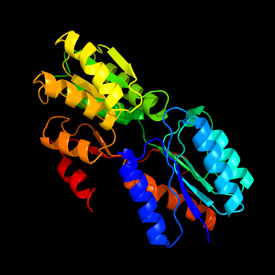
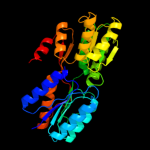
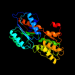
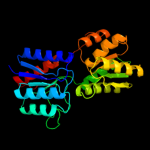
PDB header: signaling proteinChain: A: PDB Molecule: twin-arginine translocation pathway signal protein;PDBTitle: crystal structure of twin-arginine translocation pathway signal2 protein from burkholderia phytofirmans
2 d1jx6a_
100.0
13
Fold: Periplasmic binding protein-like ISuperfamily: Periplasmic binding protein-like IFamily: L-arabinose binding protein-like3 c3ma0A_
100.0
18
PDB header: sugar binding proteinChain: A: PDB Molecule: d-xylose-binding periplasmic protein;PDBTitle: closed liganded crystal structure of xylose binding protein from2 escherichia coli
4 c3g1wB_
100.0
20
PDB header: transport proteinChain: B: PDB Molecule: sugar abc transporter;PDBTitle: crystal structure of sugar abc transporter (sugar-binding protein)2 from bacillus halodurans
5 c2qvcC_
100.0
21
PDB header: transport proteinChain: C: PDB Molecule: sugar abc transporter, periplasmic sugar-bindingPDBTitle: crystal structure of a periplasmic sugar abc transporter2 from thermotoga maritima
6 d1tjya_
100.0
20
Fold: Periplasmic binding protein-like ISuperfamily: Periplasmic binding protein-like IFamily: L-arabinose binding protein-like7 c3ksmA_
100.0
19
PDB header: transport proteinChain: A: PDB Molecule: abc-type sugar transport system, periplasmic component;PDBTitle: crystal structure of abc-type sugar transport system, periplasmic2 component from hahella chejuensis
8 c3o1hB_
100.0
15
PDB header: signaling proteinChain: B: PDB Molecule: periplasmic protein tort;PDBTitle: crystal structure of the tors sensor domain - tort complex in the2 presence of tmao
9 c3d02A_
100.0
17
PDB header: sugar binding proteinChain: A: PDB Molecule: putative laci-type transcriptional regulator;PDBTitle: crystal structure of periplasmic sugar-binding protein2 (yp_001338366.1) from klebsiella pneumoniae subsp. pneumoniae mgh3 78578 at 1.30 a resolution
10 c2x7xA_
100.0
16
PDB header: transferaseChain: A: PDB Molecule: sensor protein;PDBTitle: fructose binding periplasmic domain of hybrid two component2 system bt1754
11 d1guda_
100.0
19
Fold: Periplasmic binding protein-like ISuperfamily: Periplasmic binding protein-like IFamily: L-arabinose binding protein-like12 c3gbvB_
100.0
13
PDB header: transcription regulatorChain: B: PDB Molecule: putative laci-family transcriptional regulator;PDBTitle: crystal structure of a putative laci transcriptional regulator from2 bacteroides fragilis
13 c3l49D_
100.0
18
PDB header: transport proteinChain: D: PDB Molecule: abc sugar (ribose) transporter, periplasmicPDBTitle: crystal structure of abc sugar transporter subunit from2 rhodobacter sphaeroides 2.4.1
14 c2fn9A_
100.0
21
PDB header: sugar binding proteinChain: A: PDB Molecule: ribose abc transporter, periplasmic ribose-binding protein;PDBTitle: thermotoga maritima ribose binding protein unliganded form
15 d8abpa_
100.0
15
Fold: Periplasmic binding protein-like ISuperfamily: Periplasmic binding protein-like IFamily: L-arabinose binding protein-like16 d1gcaa_
100.0
20
Fold: Periplasmic binding protein-like ISuperfamily: Periplasmic binding protein-like IFamily: L-arabinose binding protein-like17 c3brsA_
100.0
19
PDB header: transport proteinChain: A: PDB Molecule: periplasmic binding protein/laci transcriptional regulator;PDBTitle: crystal structure of sugar transporter from clostridium2 phytofermentans
18 c3h75A_
100.0
14
PDB header: sugar binding proteinChain: A: PDB Molecule: periplasmic sugar-binding domain protein;PDBTitle: crystal structure of a periplasmic sugar-binding protein from the2 pseudomonas fluorescens
19 c3rotA_
100.0
17
PDB header: transport proteinChain: A: PDB Molecule: abc sugar transporter, periplasmic sugar binding protein;PDBTitle: crystal structure of abc sugar transporter (periplasmic sugar binding2 protein) from legionella pneumophila
20 d2fvya1
100.0
20
Fold: Periplasmic binding protein-like ISuperfamily: Periplasmic binding protein-like IFamily: L-arabinose binding protein-like21 c3l6uA_
not modelled
100.0
15
PDB header: transport proteinChain: A: PDB Molecule: abc-type sugar transport system periplasmicPDBTitle: crystal structure of abc-type sugar transport system,2 periplasmic component from exiguobacterium sibiricum
22 c2vk2A_
not modelled
100.0
18
PDB header: transport proteinChain: A: PDB Molecule: abc transporter periplasmic-binding protein ytfq;PDBTitle: crystal structure of a galactofuranose binding protein
23 c2ioyB_
not modelled
100.0
21
PDB header: sugar binding proteinChain: B: PDB Molecule: periplasmic sugar-binding protein;PDBTitle: crystal structure of thermoanaerobacter tengcongensis2 ribose binding protein
24 d2nzug1
not modelled
100.0
17
Fold: Periplasmic binding protein-like ISuperfamily: Periplasmic binding protein-like IFamily: L-arabinose binding protein-like25 c3h5oB_
not modelled
100.0
13
PDB header: transcription regulatorChain: B: PDB Molecule: transcriptional regulator gntr;PDBTitle: the crystal structure of transcription regulator gntr from2 chromobacterium violaceum
26 c2iksA_
not modelled
100.0
16
PDB header: transcriptionChain: A: PDB Molecule: dna-binding transcriptional dual regulator;PDBTitle: crystal structure of n-terminal truncated dna-binding transcriptional2 dual regulator from escherichia coli k12
27 c3c3kA_
not modelled
100.0
14
PDB header: isomeraseChain: A: PDB Molecule: alanine racemase;PDBTitle: crystal structure of an uncharacterized protein from actinobacillus2 succinogenes
28 d2dria_
not modelled
100.0
18
Fold: Periplasmic binding protein-like ISuperfamily: Periplasmic binding protein-like IFamily: L-arabinose binding protein-like29 c3d8uA_
not modelled
100.0
12
PDB header: transcription regulatorChain: A: PDB Molecule: purr transcriptional regulator;PDBTitle: the crystal structure of a purr family transcriptional regulator from2 vibrio parahaemolyticus rimd 2210633
30 c3hcwB_
not modelled
100.0
15
PDB header: rna binding proteinChain: B: PDB Molecule: maltose operon transcriptional repressor;PDBTitle: crystal structure of probable maltose operon transcriptional repressor2 malr from staphylococcus areus
31 c3e3mA_
not modelled
100.0
15
PDB header: transcriptionChain: A: PDB Molecule: transcriptional regulator, laci family;PDBTitle: crystal structure of a laci family transcriptional2 regulator from silicibacter pomeroyi
32 c3mizB_
not modelled
100.0
12
PDB header: transcription regulatorChain: B: PDB Molecule: putative transcriptional regulator protein, laciPDBTitle: crystal structure of a putative transcriptional regulator2 protein, lacl family from rhizobium etli
33 c3k9cA_
not modelled
100.0
17
PDB header: transcription regulatorChain: A: PDB Molecule: transcriptional regulator, laci family protein;PDBTitle: crystal structure of laci transcriptional regulator from rhodococcus2 species.
34 c3o74A_
not modelled
100.0
12
PDB header: transcriptionChain: A: PDB Molecule: fructose transport system repressor frur;PDBTitle: crystal structure of cra transcriptional dual regulator from2 pseudomonas putida
35 c3k4hA_
not modelled
100.0
13
PDB header: transcription regulatorChain: A: PDB Molecule: putative transcriptional regulator;PDBTitle: crystal structure of putative transcriptional regulator laci from2 bacillus cereus subsp. cytotoxis nvh 391-98
36 c2rgyA_
not modelled
100.0
12
PDB header: transcription regulatorChain: A: PDB Molecule: transcriptional regulator, laci family;PDBTitle: crystal structure of transcriptional regulator of laci family from2 burkhoderia phymatum
37 c3dbiA_
not modelled
100.0
12
PDB header: transcription regulatorChain: A: PDB Molecule: sugar-binding transcriptional regulator, laci family;PDBTitle: crystal structure of sugar-binding transcriptional regulator (laci2 family) from escherichia coli complexed with phosphate
38 d1tlfa_
not modelled
100.0
13
Fold: Periplasmic binding protein-like ISuperfamily: Periplasmic binding protein-like IFamily: L-arabinose binding protein-like39 c3bblA_
not modelled
100.0
14
PDB header: regulatory proteinChain: A: PDB Molecule: regulatory protein of laci family;PDBTitle: crystal structure of a regulatory protein of laci family from2 chloroflexus aggregans
40 c3egcF_
not modelled
100.0
17
PDB header: structural genomics, unknown functionChain: F: PDB Molecule: putative ribose operon repressor;PDBTitle: crystal structure of a putative ribose operon repressor from2 burkholderia thailandensis
41 c3qk7C_
not modelled
100.0
11
PDB header: transcription regulatorChain: C: PDB Molecule: transcriptional regulators;PDBTitle: crystal structure of putative transcriptional regulator from yersinia2 pestis biovar microtus str. 91001
42 c2o20H_
not modelled
100.0
14
PDB header: transcriptionChain: H: PDB Molecule: catabolite control protein a;PDBTitle: crystal structure of transcription regulator ccpa of lactococcus2 lactis
43 c1jyeA_
not modelled
100.0
13
PDB header: transcriptionChain: A: PDB Molecule: lactose operon repressor;PDBTitle: structure of a dimeric lac repressor with c-terminal deletion and k84l2 substitution
44 d1jyea_
not modelled
100.0
13
Fold: Periplasmic binding protein-like ISuperfamily: Periplasmic binding protein-like IFamily: L-arabinose binding protein-like45 c3hs3A_
not modelled
100.0
15
PDB header: transcription regulatorChain: A: PDB Molecule: ribose operon repressor;PDBTitle: crystal structure of periplasmic binding ribose operon2 repressor protein from lactobacillus acidophilus
46 c3brqA_
not modelled
100.0
12
PDB header: transcription regulatorChain: A: PDB Molecule: hth-type transcriptional regulator ascg;PDBTitle: crystal structure of the escherichia coli transcriptional repressor2 ascg
47 c3jy6B_
not modelled
100.0
15
PDB header: transcription regulatorChain: B: PDB Molecule: transcriptional regulator, laci family;PDBTitle: crystal structure of laci transcriptional regulator from lactobacillus2 brevis
48 c2qu7B_
not modelled
100.0
14
PDB header: transcriptionChain: B: PDB Molecule: putative transcriptional regulator;PDBTitle: crystal structure of a putative transcription regulator2 from staphylococcus saprophyticus subsp. saprophyticus
49 c3ctpB_
not modelled
100.0
15
PDB header: transcription regulatorChain: B: PDB Molecule: periplasmic binding protein/laci transcriptional regulator;PDBTitle: crystal structure of periplasmic binding protein/laci transcriptional2 regulator from alkaliphilus metalliredigens qymf complexed with d-3 xylulofuranose
50 d1dbqa_
not modelled
100.0
13
Fold: Periplasmic binding protein-like ISuperfamily: Periplasmic binding protein-like IFamily: L-arabinose binding protein-like51 c3kkeA_
not modelled
100.0
17
PDB header: transcription regulatorChain: A: PDB Molecule: laci family transcriptional regulator;PDBTitle: crystal structure of a laci family transcriptional regulator2 from mycobacterium smegmatis
52 c3gv0A_
not modelled
100.0
14
PDB header: transcription regulatorChain: A: PDB Molecule: transcriptional regulator, laci family;PDBTitle: crystal structure of laci family transcription regulator from2 agrobacterium tumefaciens
53 c3g85A_
not modelled
100.0
15
PDB header: transcription regulatorChain: A: PDB Molecule: transcriptional regulator (laci family);PDBTitle: crystal structure of laci family transcription regulator from2 clostridium acetobutylicum
54 c1zvvA_
not modelled
100.0
16
PDB header: transcription/dnaChain: A: PDB Molecule: glucose-resistance amylase regulator;PDBTitle: crystal structure of a ccpa-crh-dna complex
55 c3cs3A_
not modelled
100.0
12
PDB header: transcription regulatorChain: A: PDB Molecule: sugar-binding transcriptional regulator, laci family;PDBTitle: crystal structure of sugar-binding transcriptional regulator (laci2 family) from enterococcus faecalis
56 c3huuC_
not modelled
100.0
12
PDB header: transcription regulatorChain: C: PDB Molecule: transcription regulator like protein;PDBTitle: crystal structure of transcription regulator like protein from2 staphylococcus haemolyticus
57 c3clkB_
not modelled
100.0
11
PDB header: transcription regulatorChain: B: PDB Molecule: transcription regulator;PDBTitle: crystal structure of a transcription regulator from lactobacillus2 plantarum
58 c3bilA_
not modelled
100.0
16
PDB header: structural genomics, unknown functionChain: A: PDB Molecule: probable laci-family transcriptional regulator;PDBTitle: crystal structure of a probable laci family transcriptional2 regulator from corynebacterium glutamicum
59 d1byka_
not modelled
100.0
12
Fold: Periplasmic binding protein-like ISuperfamily: Periplasmic binding protein-like IFamily: L-arabinose binding protein-like60 c2h0aA_
not modelled
100.0
14
PDB header: transcriptionChain: A: PDB Molecule: transcriptional regulator;PDBTitle: crystal structure of probable transcription regulator from2 thermus thermophilus
61 c3jvdA_
not modelled
100.0
15
PDB header: transcription regulatorChain: A: PDB Molecule: transcriptional regulators;PDBTitle: crystal structure of putative transcription regulation repressor (laci2 family) from corynebacterium glutamicum
62 c1bdhA_
not modelled
100.0
13
PDB header: transcription/dnaChain: A: PDB Molecule: protein (purine repressor);PDBTitle: purine repressor mutant-hypoxanthine-palindromic operator2 complex
63 c3gybB_
not modelled
100.0
14
PDB header: transcription regulatorChain: B: PDB Molecule: transcriptional regulators (laci-familyPDBTitle: crystal structure of a laci-family transcriptional2 regulatory protein from corynebacterium glutamicum
64 c2qh8A_
not modelled
100.0
15
PDB header: structural genomics, unknown functionChain: A: PDB Molecule: uncharacterized protein;PDBTitle: crystal structure of conserved domain protein from vibrio2 cholerae o1 biovar eltor str. n16961
65 c3kjxD_
not modelled
100.0
15
PDB header: transcription regulatorChain: D: PDB Molecule: transcriptional regulator, laci family;PDBTitle: crystal structure of a transcriptional regulator, lacl2 family protein from silicibacter pomeroyi
66 c3lftA_
not modelled
100.0
15
PDB header: structure genomics, unknown functionChain: A: PDB Molecule: uncharacterized protein;PDBTitle: the crystal structure of the abc domain in complex with l-trp from2 streptococcus pneumonia to 1.35a
67 c3e61A_
not modelled
100.0
18
PDB header: structural genomics, unknown functionChain: A: PDB Molecule: putative transcriptional repressor of ribose operon;PDBTitle: crystal structure of a putative transcriptional repressor of ribose2 operon from staphylococcus saprophyticus subsp. saprophyticus
68 c3h5tA_
not modelled
99.9
16
PDB header: transcription regulatorChain: A: PDB Molecule: transcriptional regulator, laci family;PDBTitle: crystal structure of a transcriptional regulator, lacl2 family protein from corynebacterium glutamicum
69 c2hqbA_
not modelled
99.9
14
PDB header: transcriptionChain: A: PDB Molecule: transcriptional activator of comk gene;PDBTitle: crystal structure of a transcriptional activator of comk2 gene from bacillus halodurans
70 c2fqxA_
not modelled
99.9
14
PDB header: transport proteinChain: A: PDB Molecule: membrane lipoprotein tmpc;PDBTitle: pnra from treponema pallidum complexed with guanosine
71 c3s99A_
not modelled
99.8
11
PDB header: lipid binding proteinChain: A: PDB Molecule: basic membrane lipoprotein;PDBTitle: crystal structure of a basic membrane lipoprotein from brucella2 melitensis, iodide soak
72 c3i45A_
not modelled
99.0
15
PDB header: signaling proteinChain: A: PDB Molecule: twin-arginine translocation pathway signal protein;PDBTitle: crystal structure of putative twin-arginine translocation pathway2 signal protein from rhodospirillum rubrum atcc 11170
73 c3eafA_
not modelled
98.9
9
PDB header: transport proteinChain: A: PDB Molecule: abc transporter, substrate binding protein;PDBTitle: crystal structure of abc transporter, substrate binding protein2 aeropyrum pernix
74 c3t0nA_
not modelled
98.8
13
PDB header: signaling proteinChain: A: PDB Molecule: twin-arginine translocation pathway signal;PDBTitle: crystal structure of twin-arginine translocation pathway signal from2 rhodopseudomonas palustris bisb5
75 c3ip5A_
not modelled
98.8
12
PDB header: transport proteinChain: A: PDB Molecule: abc transporter, substrate binding protein (amino acid);PDBTitle: structure of atu2422-gaba receptor in complex with alanine
76 c3i09A_
not modelled
98.8
11
PDB header: transport proteinChain: A: PDB Molecule: periplasmic branched-chain amino acid-binding protein;PDBTitle: crystal structure of a periplasmic binding protein (bma2936) from2 burkholderia mallei at 1.80 a resolution
77 d1qo0a_
not modelled
98.8
11
Fold: Periplasmic binding protein-like ISuperfamily: Periplasmic binding protein-like IFamily: L-arabinose binding protein-like78 c3h5lB_
not modelled
98.8
12
PDB header: transport proteinChain: B: PDB Molecule: putative branched-chain amino acid abcPDBTitle: crystal structure of a putative branched-chain amino acid2 abc transporter from silicibacter pomeroyi
79 c3sg0A_
not modelled
98.7
15
PDB header: signaling proteinChain: A: PDB Molecule: extracellular ligand-binding receptor;PDBTitle: the crystal structure of an extracellular ligand-binding receptor from2 rhodopseudomonas palustris haa2
80 c3n0wA_
not modelled
98.7
13
PDB header: transport proteinChain: A: PDB Molecule: abc branched chain amino acid family transporter,PDBTitle: crystal structure of a branched chain amino acid abc transporter2 periplasmic ligand-binding protein (bxe_c0949) from burkholderia3 xenovorans lb400 at 1.88 a resolution
81 c3lkbB_
not modelled
98.7
12
PDB header: transport proteinChain: B: PDB Molecule: probable branched-chain amino acid abcPDBTitle: crystal structure of a branched chain amino acid abc2 transporter from thermus thermophilus with bound valine
82 c3snrA_
not modelled
98.7
14
PDB header: transport proteinChain: A: PDB Molecule: extracellular ligand-binding receptor;PDBTitle: rpd_1889 protein, an extracellular ligand-binding receptor from2 rhodopseudomonas palustris.
83 d2liva_
not modelled
98.7
10
Fold: Periplasmic binding protein-like ISuperfamily: Periplasmic binding protein-like IFamily: L-arabinose binding protein-like84 c3hutA_
not modelled
98.6
10
PDB header: transport proteinChain: A: PDB Molecule: putative branched-chain amino acid abcPDBTitle: crystal structure of a putative branched-chain amino acid2 abc transporter from rhodospirillum rubrum
85 c3td9A_
not modelled
98.6
14
PDB header: transport proteinChain: A: PDB Molecule: branched chain amino acid abc transporter, periplasmicPDBTitle: crystal structure of a leucine binding protein livk (tm1135) from2 thermotoga maritima msb8 at 1.90 a resolution
86 d1usga_
not modelled
98.5
10
Fold: Periplasmic binding protein-like ISuperfamily: Periplasmic binding protein-like IFamily: L-arabinose binding protein-like87 c3n0xA_
not modelled
98.4
13
PDB header: transport proteinChain: A: PDB Molecule: possible substrate binding protein of abc transporterPDBTitle: crystal structure of an abc-type branched-chain amino acid transporter2 (rpa4397) from rhodopseudomonas palustris cga009 at 1.50 a resolution
88 c1jdpA_
not modelled
98.4
16
PDB header: signaling proteinChain: A: PDB Molecule: atrial natriuretic peptide clearance receptor;PDBTitle: crystal structure of hormone/receptor complex
89 d1jdpa_
not modelled
98.4
16
Fold: Periplasmic binding protein-like ISuperfamily: Periplasmic binding protein-like IFamily: L-arabinose binding protein-like90 c3lopA_
not modelled
98.0
10
PDB header: substrate binding proteinChain: A: PDB Molecule: substrate binding periplasmic protein;PDBTitle: crystal structure of substrate-binding periplasmic protein2 (pbp) from ralstonia solanacearum
91 c1yk1B_
not modelled
97.9
16
PDB header: hormone/growth factor receptorChain: B: PDB Molecule: atrial natriuretic peptide clearance receptor;PDBTitle: structure of natriuretic peptide receptor-c complexed with brain2 natriuretic peptide
92 d1ewka_
not modelled
97.4
11
Fold: Periplasmic binding protein-like ISuperfamily: Periplasmic binding protein-like IFamily: L-arabinose binding protein-like93 c3qi7A_
not modelled
97.2
10
PDB header: transcriptionChain: A: PDB Molecule: putative transcriptional regulator;PDBTitle: crystal structure of a putative transcriptional regulator2 (yp_001089212.1) from clostridium difficile 630 at 1.86 a resolution
94 c3h6hB_
not modelled
97.2
11
PDB header: membrane proteinChain: B: PDB Molecule: glutamate receptor, ionotropic kainate 2;PDBTitle: crystal structure of the glur6 amino terminal domain dimer assembly2 mpd form
95 c3q41B_
not modelled
96.6
17
PDB header: transport proteinChain: B: PDB Molecule: glutamate [nmda] receptor subunit zeta-1;PDBTitle: crystal structure of the glun1 n-terminal domain (ntd)
96 c3sm9A_
not modelled
96.5
12
PDB header: signaling proteinChain: A: PDB Molecule: metabotropic glutamate receptor 3;PDBTitle: crystal structure of metabotropic glutamate receptor 3 precursor in2 presence of ly341495 antagonist
97 d3ckma1
not modelled
96.5
12
Fold: Periplasmic binding protein-like ISuperfamily: Periplasmic binding protein-like IFamily: L-arabinose binding protein-like98 c3sajB_
not modelled
96.0
8
PDB header: transport proteinChain: B: PDB Molecule: glutamate receptor 1;PDBTitle: crystal structure of glutamate receptor glua1 amino terminal domain
99 c3om1A_
not modelled
96.0
13
PDB header: membrane proteinChain: A: PDB Molecule: glutamate receptor gluk5 (ka2);PDBTitle: crystal structure of the gluk5 (ka2) atd dimer at 1.7 angstrom2 resolution
100 d1dp4a_
not modelled
95.6
13
Fold: Periplasmic binding protein-like ISuperfamily: Periplasmic binding protein-like IFamily: L-arabinose binding protein-like101 c2uygF_
not modelled
94.7
15
PDB header: lyaseChain: F: PDB Molecule: 3-dehydroquinate dehydratase;PDBTitle: crystallogaphic structure of the typeii 3-dehydroquinase2 from thermus thermophilus
102 c3u80A_
not modelled
94.1
11
PDB header: unknown functionChain: A: PDB Molecule: 3-dehydroquinate dehydratase, type ii;PDBTitle: 1.60 angstrom resolution crystal structure of a 3-dehydroquinate2 dehydratase-like protein from bifidobacterium longum
103 c3kg2A_
not modelled
93.2
9
PDB header: membrane protein, transport proteinChain: A: PDB Molecule: glutamate receptor 2;PDBTitle: ampa subtype ionotropic glutamate receptor in complex with competitive2 antagonist zk 200775
104 c3opyG_
not modelled
93.2
14
PDB header: transferaseChain: G: PDB Molecule: 6-phosphofructo-1-kinase alpha-subunit;PDBTitle: crystal structure of pichia pastoris phosphofructokinase in the t-2 state
105 c2wjxA_
not modelled
93.0
8
PDB header: transport proteinChain: A: PDB Molecule: glutamate receptor 2;PDBTitle: crystal structure of the human ionotropic glutamate2 receptor glur2 atd region at 4.1 a resolution
106 d1gqoa_
not modelled
92.7
15
Fold: Flavodoxin-likeSuperfamily: Type II 3-dehydroquinate dehydrataseFamily: Type II 3-dehydroquinate dehydratase107 c3p3wC_
not modelled
92.7
7
PDB header: transport proteinChain: C: PDB Molecule: glutamate receptor 3;PDBTitle: structure of a dimeric glua3 n-terminal domain (ntd) at 4.2 a2 resolution
108 d1gtza_
not modelled
92.4
20
Fold: Flavodoxin-likeSuperfamily: Type II 3-dehydroquinate dehydrataseFamily: Type II 3-dehydroquinate dehydratase109 c3opyE_
not modelled
92.3
12
PDB header: transferaseChain: E: PDB Molecule: 6-phosphofructo-1-kinase alpha-subunit;PDBTitle: crystal structure of pichia pastoris phosphofructokinase in the t-2 state
110 d2c4va1
not modelled
92.1
22
Fold: Flavodoxin-likeSuperfamily: Type II 3-dehydroquinate dehydrataseFamily: Type II 3-dehydroquinate dehydratase111 c3lwzC_
not modelled
92.0
17
PDB header: lyaseChain: C: PDB Molecule: 3-dehydroquinate dehydratase;PDBTitle: 1.65 angstrom resolution crystal structure of type ii 3-2 dehydroquinate dehydratase (aroq) from yersinia pestis
112 c3n8kG_
not modelled
91.0
11
PDB header: lyaseChain: G: PDB Molecule: 3-dehydroquinate dehydratase;PDBTitle: type ii dehydroquinase from mycobacterium tuberculosis complexed with2 citrazinic acid
113 c2e4zA_
not modelled
90.7
15
PDB header: signaling proteinChain: A: PDB Molecule: metabotropic glutamate receptor 7;PDBTitle: crystal structure of the ligand-binding region of the group iii2 metabotropic glutamate receptor
114 d1h05a_
not modelled
90.7
11
Fold: Flavodoxin-likeSuperfamily: Type II 3-dehydroquinate dehydrataseFamily: Type II 3-dehydroquinate dehydratase115 d1uqra_
not modelled
90.6
17
Fold: Flavodoxin-likeSuperfamily: Type II 3-dehydroquinate dehydrataseFamily: Type II 3-dehydroquinate dehydratase116 c3kipU_
not modelled
90.5
18
PDB header: lyaseChain: U: PDB Molecule: 3-dehydroquinase, type ii;PDBTitle: crystal structure of type-ii 3-dehydroquinase from c. albicans
117 d2ajta2
not modelled
90.5
8
Fold: FucI/AraA N-terminal and middle domainsSuperfamily: FucI/AraA N-terminal and middle domainsFamily: AraA N-terminal and middle domain-like118 d1fuia2
not modelled
89.7
14
Fold: FucI/AraA N-terminal and middle domainsSuperfamily: FucI/AraA N-terminal and middle domainsFamily: L-fucose isomerase, N-terminal and second domains119 c3o8nA_
not modelled
88.8
15
PDB header: transferaseChain: A: PDB Molecule: 6-phosphofructokinase, muscle type;PDBTitle: structure of phosphofructokinase from rabbit skeletal muscle
120 d1b0aa2
not modelled
88.3
10
Fold: Aminoacid dehydrogenase-like, N-terminal domainSuperfamily: Aminoacid dehydrogenase-like, N-terminal domainFamily: Tetrahydrofolate dehydrogenase/cyclohydrolase