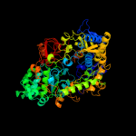
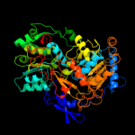
1 c2nyaF_
100.0
100
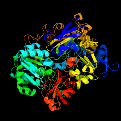
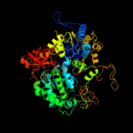
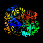
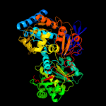
PDB header: oxidoreductaseChain: F: PDB Molecule: periplasmic nitrate reductase;PDBTitle: crystal structure of the periplasmic nitrate reductase2 (nap) from escherichia coli
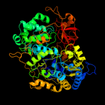
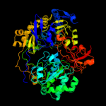
2 c1ogyA_
100.0
68
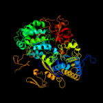
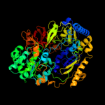
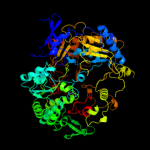
PDB header: oxidoreductaseChain: A: PDB Molecule: periplasmic nitrate reductase;PDBTitle: crystal structure of the heterodimeric nitrate reductase2 from rhodobacter sphaeroides
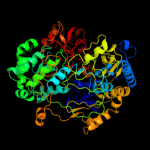
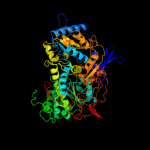
3 c1kqgA_
100.0
22
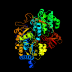
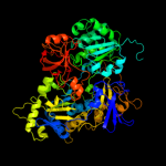
PDB header: oxidoreductaseChain: A: PDB Molecule: formate dehydrogenase, nitrate-inducible, major subunit;PDBTitle: formate dehydrogenase n from e. coli
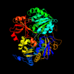
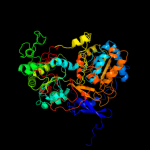
4 c1h0hA_
100.0
22
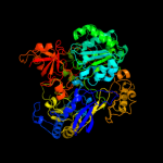
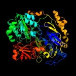
PDB header: dehydrogenaseChain: A: PDB Molecule: formate dehydrogenase (large subunit);PDBTitle: tungsten containing formate dehydrogenase from2 desulfovibrio gigas
5 c2v45A_
100.0
41
PDB header: oxidoreductaseChain: A: PDB Molecule: periplasmic nitrate reductase;PDBTitle: a new catalytic mechanism of periplasmic nitrate reductase2 from desulfovibrio desulfuricans atcc 27774 from3 crystallographic and epr data and based on detailed4 analysis of the sixth ligand
6 c1y5iA_
100.0
20
PDB header: oxidoreductaseChain: A: PDB Molecule: respiratory nitrate reductase 1 alpha chain;PDBTitle: the crystal structure of the narghi mutant nari-k86a
7 c2ivfA_
100.0
16
PDB header: oxidoreductaseChain: A: PDB Molecule: ethylbenzene dehydrogenase alpha-subunit;PDBTitle: ethylbenzene dehydrogenase from aromatoleum aromaticum
8 c2e7zA_
100.0
19
PDB header: lyaseChain: A: PDB Molecule: acetylene hydratase ahy;PDBTitle: acetylene hydratase from pelobacter acetylenicus
9 c2vpyE_
100.0
22
PDB header: oxidoreductaseChain: E: PDB Molecule: thiosulfate reductase;PDBTitle: polysulfide reductase with bound quinone inhibitor,2 pentachlorophenol (pcp)
10 c2iv2X_
100.0
25
PDB header: oxidoreductaseChain: X: PDB Molecule: formate dehydrogenase h;PDBTitle: reinterpretation of reduced form of formate dehydrogenase h2 from e. coli
11 c1tmoA_
100.0
17
PDB header: oxidoreductaseChain: A: PDB Molecule: trimethylamine n-oxide reductase;PDBTitle: trimethylamine n-oxide reductase from shewanella massilia
12 c1h5nC_
100.0
17
PDB header: oxidoreductaseChain: C: PDB Molecule: dmso reductase;PDBTitle: dmso reductase modified by the presence of dms and air
13 c1g8jC_
100.0
21
PDB header: oxidoreductaseChain: C: PDB Molecule: arsenite oxidase;PDBTitle: crystal structure analysis of arsenite oxidase from2 alcaligenes faecalis
14 c1eu1A_
100.0
17
PDB header: oxidoreductaseChain: A: PDB Molecule: dimethyl sulfoxide reductase;PDBTitle: the crystal structure of rhodobacter sphaeroides dimethylsulfoxide2 reductase reveals two distinct molybdenum coordination environments.
15 d1ogya2
100.0
69
Fold: Formate dehydrogenase/DMSO reductase, domains 1-3Superfamily: Formate dehydrogenase/DMSO reductase, domains 1-3Family: Formate dehydrogenase/DMSO reductase, domains 1-316 c1vlfQ_
100.0
19
PDB header: oxidoreductaseChain: Q: PDB Molecule: pyrogallol hydroxytransferase large subunit;PDBTitle: crystal structure of pyrogallol-phloroglucinol2 transhydroxylase from pelobacter acidigallici complexed3 with inhibitor 1,2,4,5-tetrahydroxy-benzene
17 d1kqfa2
100.0
20
Fold: Formate dehydrogenase/DMSO reductase, domains 1-3Superfamily: Formate dehydrogenase/DMSO reductase, domains 1-3Family: Formate dehydrogenase/DMSO reductase, domains 1-318 d1h0ha2
100.0
20
Fold: Formate dehydrogenase/DMSO reductase, domains 1-3Superfamily: Formate dehydrogenase/DMSO reductase, domains 1-3Family: Formate dehydrogenase/DMSO reductase, domains 1-319 d2jioa2
100.0
40
Fold: Formate dehydrogenase/DMSO reductase, domains 1-3Superfamily: Formate dehydrogenase/DMSO reductase, domains 1-3Family: Formate dehydrogenase/DMSO reductase, domains 1-320 d1y5ia2
100.0
17
Fold: Formate dehydrogenase/DMSO reductase, domains 1-3Superfamily: Formate dehydrogenase/DMSO reductase, domains 1-3Family: Formate dehydrogenase/DMSO reductase, domains 1-321 d2iv2x2
not modelled
100.0
25
Fold: Formate dehydrogenase/DMSO reductase, domains 1-3Superfamily: Formate dehydrogenase/DMSO reductase, domains 1-3Family: Formate dehydrogenase/DMSO reductase, domains 1-322 d1g8ka2
not modelled
100.0
20
Fold: Formate dehydrogenase/DMSO reductase, domains 1-3Superfamily: Formate dehydrogenase/DMSO reductase, domains 1-3Family: Formate dehydrogenase/DMSO reductase, domains 1-323 d1tmoa2
not modelled
100.0
15
Fold: Formate dehydrogenase/DMSO reductase, domains 1-3Superfamily: Formate dehydrogenase/DMSO reductase, domains 1-3Family: Formate dehydrogenase/DMSO reductase, domains 1-324 d1dmra2
not modelled
100.0
16
Fold: Formate dehydrogenase/DMSO reductase, domains 1-3Superfamily: Formate dehydrogenase/DMSO reductase, domains 1-3Family: Formate dehydrogenase/DMSO reductase, domains 1-325 d1vlfm2
not modelled
100.0
19
Fold: Formate dehydrogenase/DMSO reductase, domains 1-3Superfamily: Formate dehydrogenase/DMSO reductase, domains 1-3Family: Formate dehydrogenase/DMSO reductase, domains 1-326 d1eu1a2
not modelled
100.0
16
Fold: Formate dehydrogenase/DMSO reductase, domains 1-3Superfamily: Formate dehydrogenase/DMSO reductase, domains 1-3Family: Formate dehydrogenase/DMSO reductase, domains 1-327 c2fugC_
not modelled
100.0
23
PDB header: oxidoreductaseChain: C: PDB Molecule: nadh-quinone oxidoreductase chain 3;PDBTitle: crystal structure of the hydrophilic domain of respiratory complex i2 from thermus thermophilus
28 d2fug32
not modelled
100.0
22
Fold: Formate dehydrogenase/DMSO reductase, domains 1-3Superfamily: Formate dehydrogenase/DMSO reductase, domains 1-3Family: Formate dehydrogenase/DMSO reductase, domains 1-329 d2jioa1
not modelled
100.0
48
Fold: Double psi beta-barrelSuperfamily: ADC-likeFamily: Formate dehydrogenase/DMSO reductase, C-terminal domain30 d1h0ha1
not modelled
100.0
33
Fold: Double psi beta-barrelSuperfamily: ADC-likeFamily: Formate dehydrogenase/DMSO reductase, C-terminal domain31 d1ogya1
not modelled
100.0
64
Fold: Double psi beta-barrelSuperfamily: ADC-likeFamily: Formate dehydrogenase/DMSO reductase, C-terminal domain32 d1kqfa1
not modelled
100.0
27
Fold: Double psi beta-barrelSuperfamily: ADC-likeFamily: Formate dehydrogenase/DMSO reductase, C-terminal domain33 d1g8ka1
not modelled
99.9
24
Fold: Double psi beta-barrelSuperfamily: ADC-likeFamily: Formate dehydrogenase/DMSO reductase, C-terminal domain34 d1dmra1
not modelled
99.9
24
Fold: Double psi beta-barrelSuperfamily: ADC-likeFamily: Formate dehydrogenase/DMSO reductase, C-terminal domain35 d1eu1a1
not modelled
99.9
23
Fold: Double psi beta-barrelSuperfamily: ADC-likeFamily: Formate dehydrogenase/DMSO reductase, C-terminal domain36 d1tmoa1
not modelled
99.9
27
Fold: Double psi beta-barrelSuperfamily: ADC-likeFamily: Formate dehydrogenase/DMSO reductase, C-terminal domain37 d1vlfm1
not modelled
99.9
26
Fold: Double psi beta-barrelSuperfamily: ADC-likeFamily: Formate dehydrogenase/DMSO reductase, C-terminal domain38 d2iv2x1
not modelled
99.9
30
Fold: Double psi beta-barrelSuperfamily: ADC-likeFamily: Formate dehydrogenase/DMSO reductase, C-terminal domain39 d1y5ia1
not modelled
99.9
17
Fold: Double psi beta-barrelSuperfamily: ADC-likeFamily: Formate dehydrogenase/DMSO reductase, C-terminal domain40 c2ki8A_
not modelled
99.8
24
PDB header: oxidoreductaseChain: A: PDB Molecule: tungsten formylmethanofuran dehydrogenase,PDBTitle: solution nmr structure of tungsten formylmethanofuran2 dehydrogenase subunit d from archaeoglobus fulgidus,3 northeast structural genomics consortium target att7
41 c2pq4B_
97.2
100
PDB header: chaperone/oxidoreductaseChain: B: PDB Molecule: periplasmic nitrate reductase precursor;PDBTitle: nmr solution structure of napd in complex with napa1-352 signal peptide
42 c3etnD_
not modelled
96.7
12
PDB header: isomeraseChain: D: PDB Molecule: putative phosphosugar isomerase involved in capsulePDBTitle: crystal structure of putative phosphosugar isomerase involved in2 capsule formation (yp_209877.1) from bacteroides fragilis nctc 93433 at 1.70 a resolution
43 d1tk9a_
not modelled
96.2
13
Fold: SIS domainSuperfamily: SIS domainFamily: mono-SIS domain44 d2ji7a1
not modelled
95.9
9
Fold: DHS-like NAD/FAD-binding domainSuperfamily: DHS-like NAD/FAD-binding domainFamily: Pyruvate oxidase and decarboxylase, middle domain45 c2yvaB_
not modelled
95.9
13
PDB header: dna binding proteinChain: B: PDB Molecule: dnaa initiator-associating protein diaa;PDBTitle: crystal structure of escherichia coli diaa
46 d1ozha1
not modelled
95.8
11
Fold: DHS-like NAD/FAD-binding domainSuperfamily: DHS-like NAD/FAD-binding domainFamily: Pyruvate oxidase and decarboxylase, middle domain47 d2fug31
not modelled
95.8
32
Fold: Double psi beta-barrelSuperfamily: ADC-likeFamily: Formate dehydrogenase/DMSO reductase, C-terminal domain48 d1x92a_
not modelled
95.6
13
Fold: SIS domainSuperfamily: SIS domainFamily: mono-SIS domain49 d2ihta1
not modelled
95.6
10
Fold: DHS-like NAD/FAD-binding domainSuperfamily: DHS-like NAD/FAD-binding domainFamily: Pyruvate oxidase and decarboxylase, middle domain50 d1ybha1
not modelled
95.5
8
Fold: DHS-like NAD/FAD-binding domainSuperfamily: DHS-like NAD/FAD-binding domainFamily: Pyruvate oxidase and decarboxylase, middle domain51 c3euaD_
not modelled
95.3
10
PDB header: isomeraseChain: D: PDB Molecule: putative fructose-aminoacid-6-phosphate deglycase;PDBTitle: crystal structure of a putative phosphosugar isomerase (bsu32610) from2 bacillus subtilis at 1.90 a resolution
52 c2pjhB_
not modelled
95.3
21
PDB header: transport proteinChain: B: PDB Molecule: transitional endoplasmic reticulum atpase;PDBTitle: strctural model of the p97 n domain- npl4 ubd complex
53 c1cz5A_
not modelled
94.6
15
PDB header: hydrolaseChain: A: PDB Molecule: vcp-like atpase;PDBTitle: nmr structure of vat-n: the n-terminal domain of vat (vcp-2 like atpase of thermoplasma)
54 c2x3yA_
not modelled
94.5
9
PDB header: isomeraseChain: A: PDB Molecule: phosphoheptose isomerase;PDBTitle: crystal structure of gmha from burkholderia pseudomallei
55 d1m3sa_
not modelled
94.1
14
Fold: SIS domainSuperfamily: SIS domainFamily: mono-SIS domain56 d1q6za1
not modelled
94.1
11
Fold: DHS-like NAD/FAD-binding domainSuperfamily: DHS-like NAD/FAD-binding domainFamily: Pyruvate oxidase and decarboxylase, middle domain57 d1jeoa_
not modelled
94.1
18
Fold: SIS domainSuperfamily: SIS domainFamily: mono-SIS domain58 c3k35D_
not modelled
93.9
13
PDB header: hydrolaseChain: D: PDB Molecule: nad-dependent deacetylase sirtuin-6;PDBTitle: crystal structure of human sirt6
59 d1x94a_
not modelled
93.9
13
Fold: SIS domainSuperfamily: SIS domainFamily: mono-SIS domain60 c3pkiF_
not modelled
93.8
10
PDB header: hydrolaseChain: F: PDB Molecule: nad-dependent deacetylase sirtuin-6;PDBTitle: human sirt6 crystal structure in complex with adp ribose
61 d2ez9a1
not modelled
93.8
11
Fold: DHS-like NAD/FAD-binding domainSuperfamily: DHS-like NAD/FAD-binding domainFamily: Pyruvate oxidase and decarboxylase, middle domain62 d1vima_
not modelled
92.9
15
Fold: SIS domainSuperfamily: SIS domainFamily: mono-SIS domain63 d1e32a1
not modelled
92.8
20
Fold: Double psi beta-barrelSuperfamily: ADC-likeFamily: Cdc48 N-terminal domain-like64 d1zpda1
not modelled
92.5
12
Fold: DHS-like NAD/FAD-binding domainSuperfamily: DHS-like NAD/FAD-binding domainFamily: Pyruvate oxidase and decarboxylase, middle domain65 d1cz5a1
not modelled
92.4
18
Fold: Double psi beta-barrelSuperfamily: ADC-likeFamily: Cdc48 N-terminal domain-like66 d1ovma1
not modelled
92.1
10
Fold: DHS-like NAD/FAD-binding domainSuperfamily: DHS-like NAD/FAD-binding domainFamily: Pyruvate oxidase and decarboxylase, middle domain67 d1m2ka_
not modelled
92.0
10
Fold: DHS-like NAD/FAD-binding domainSuperfamily: DHS-like NAD/FAD-binding domainFamily: Sir2 family of transcriptional regulators68 c2a3nA_
not modelled
91.8
10
PDB header: sugar binding proteinChain: A: PDB Molecule: putative glucosamine-fructose-6-phosphate aminotransferase;PDBTitle: crystal structure of a putative glucosamine-fructose-6-phosphate2 aminotransferase (stm4540.s) from salmonella typhimurium lt2 at 1.353 a resolution
69 d1ma3a_
not modelled
91.8
13
Fold: DHS-like NAD/FAD-binding domainSuperfamily: DHS-like NAD/FAD-binding domainFamily: Sir2 family of transcriptional regulators70 c3shoA_
not modelled
91.5
11
PDB header: transcription regulatorChain: A: PDB Molecule: transcriptional regulator, rpir family;PDBTitle: crystal structure of rpir transcription factor from sphaerobacter2 thermophilus (sugar isomerase domain)
71 d2djia1
not modelled
91.0
10
Fold: DHS-like NAD/FAD-binding domainSuperfamily: DHS-like NAD/FAD-binding domainFamily: Pyruvate oxidase and decarboxylase, middle domain72 c3trjC_
not modelled
90.6
11
PDB header: isomeraseChain: C: PDB Molecule: phosphoheptose isomerase;PDBTitle: structure of a phosphoheptose isomerase from francisella tularensis
73 d2b4ya1
not modelled
90.3
13
Fold: DHS-like NAD/FAD-binding domainSuperfamily: DHS-like NAD/FAD-binding domainFamily: Sir2 family of transcriptional regulators74 d1yc5a1
not modelled
90.3
13
Fold: DHS-like NAD/FAD-binding domainSuperfamily: DHS-like NAD/FAD-binding domainFamily: Sir2 family of transcriptional regulators75 c3fkjA_
not modelled
90.2
11
PDB header: isomeraseChain: A: PDB Molecule: putative phosphosugar isomerases;PDBTitle: crystal structure of a putative phosphosugar isomerase (stm_0572) from2 salmonella typhimurium lt2 at 2.12 a resolution
76 c2e76D_
not modelled
89.8
29
PDB header: photosynthesisChain: D: PDB Molecule: cytochrome b6-f complex iron-sulfur subunit;PDBTitle: crystal structure of the cytochrome b6f complex with tridecyl-2 stigmatellin (tds) from m.laminosus
77 c1s3sA_
not modelled
88.1
20
PDB header: protein bindingChain: A: PDB Molecule: transitional endoplasmic reticulum atpase (terPDBTitle: crystal structure of aaa atpase p97/vcp nd1 in complex with2 p47 c
78 c3lq1A_
not modelled
87.5
7
PDB header: transferaseChain: A: PDB Molecule: 2-succinyl-5-enolpyruvyl-6-hydroxy-3-cyclohexene-PDBTitle: crystal structure of 2-succinyl-6-hydroxy-2,4-cyclohexadiene2 1-carboxylic acid synthase/2-oxoglutarate decarboxylase3 from listeria monocytogenes str. 4b f2365
79 c1ozhD_
not modelled
86.9
12
PDB header: lyaseChain: D: PDB Molecule: acetolactate synthase, catabolic;PDBTitle: the crystal structure of klebsiella pneumoniae acetolactate2 synthase with enzyme-bound cofactor and with an unusual3 intermediate.
80 c3jwpA_
not modelled
86.6
7
PDB header: transcriptionChain: A: PDB Molecule: transcriptional regulatory protein sir2 homologue;PDBTitle: crystal structure of plasmodium falciparum sir2a (pf13_0152) in2 complex with amp
81 d1t9ba1
not modelled
85.7
10
Fold: DHS-like NAD/FAD-binding domainSuperfamily: DHS-like NAD/FAD-binding domainFamily: Pyruvate oxidase and decarboxylase, middle domain82 c2xhzC_
not modelled
85.5
12
PDB header: isomeraseChain: C: PDB Molecule: arabinose 5-phosphate isomerase;PDBTitle: probing the active site of the sugar isomerase domain from e. coli2 arabinose-5-phosphate isomerase via x-ray crystallography
83 c2x7jA_
not modelled
83.9
8
PDB header: transferaseChain: A: PDB Molecule: 2-succinyl-5-enolpyruvyl-6-hydroxy-3-cyclohexenePDBTitle: structure of the menaquinone biosynthesis protein mend from2 bacillus subtilis
84 c3cf1C_
not modelled
83.4
20
PDB header: transport proteinChain: C: PDB Molecule: transitional endoplasmic reticulum atpase;PDBTitle: structure of p97/vcp in complex with adp/adp.alfx
85 c1wlfA_
not modelled
83.3
30
PDB header: protein transportChain: A: PDB Molecule: peroxisome biogenesis factor 1;PDBTitle: structure of the n-terminal domain of pex1 aaa-atpase:2 characterization of a putative adaptor-binding domain
86 c1p84E_
not modelled
83.0
13
PDB header: oxidoreductaseChain: E: PDB Molecule: ubiquinol-cytochrome c reductase iron-sulfurPDBTitle: hdbt inhibited yeast cytochrome bc1 complex
87 c3hu2C_
not modelled
82.8
20
PDB header: transport proteinChain: C: PDB Molecule: transitional endoplasmic reticulum atpase;PDBTitle: structure of p97 n-d1 r86a mutant in complex with atpgs
88 c2fynO_
not modelled
82.8
35
PDB header: oxidoreductaseChain: O: PDB Molecule: ubiquinol-cytochrome c reductase iron-sulfurPDBTitle: crystal structure analysis of the double mutant rhodobacter2 sphaeroides bc1 complex
89 d1ylea1
not modelled
80.1
22
Fold: Acyl-CoA N-acyltransferases (Nat)Superfamily: Acyl-CoA N-acyltransferases (Nat)Family: AstA-like90 c1jscA_
not modelled
80.0
11
PDB header: lyaseChain: A: PDB Molecule: acetohydroxy-acid synthase;PDBTitle: crystal structure of the catalytic subunit of yeast2 acetohydroxyacid synthase: a target for herbicidal3 inhibitors
91 d1s5pa_
not modelled
79.5
12
Fold: DHS-like NAD/FAD-binding domainSuperfamily: DHS-like NAD/FAD-binding domainFamily: Sir2 family of transcriptional regulators92 c3fxaA_
not modelled
78.9
13
PDB header: sugar binding proteinChain: A: PDB Molecule: sis domain protein;PDBTitle: crystal structure of a putative sugar-phosphate isomerase2 (lmof2365_0531) from listeria monocytogenes str. 4b f2365 at 1.60 a3 resolution
93 c2ji6B_
not modelled
78.7
5
PDB header: lyaseChain: B: PDB Molecule: oxalyl-coa decarboxylase;PDBTitle: x-ray structure of oxalyl-coa decarboxylase in complex with2 3-deaza-thdp and oxalyl-coa
94 d1wlfa2
not modelled
77.2
30
Fold: Double psi beta-barrelSuperfamily: ADC-likeFamily: Cdc48 N-terminal domain-like95 c2puwA_
not modelled
76.8
9
PDB header: transferaseChain: A: PDB Molecule: isomerase domain of glutamine-fructose-6-phosphatePDBTitle: the crystal structure of isomerase domain of glucosamine-6-phosphate2 synthase from candida albicans
96 c3knzA_
not modelled
75.4
11
PDB header: sugar binding proteinChain: A: PDB Molecule: putative sugar binding protein;PDBTitle: crystal structure of putative sugar binding protein (np_459565.1) from2 salmonella typhimurium lt2 at 2.50 a resolution
97 c1yi1A_
not modelled
75.4
8
PDB header: transferaseChain: A: PDB Molecule: acetolactate synthase;PDBTitle: crystal structure of arabidopsis thaliana acetohydroxyacid synthase in2 complex with a sulfonylurea herbicide, tribenuron methyl
98 c2zj3A_
not modelled
75.2
12
PDB header: transferaseChain: A: PDB Molecule: glucosamine--fructose-6-phosphatePDBTitle: isomerase domain of human glucose:fructose-6-phosphate2 amidotransferase
99 d1pvda1
not modelled
75.1
13
Fold: DHS-like NAD/FAD-binding domainSuperfamily: DHS-like NAD/FAD-binding domainFamily: Pyruvate oxidase and decarboxylase, middle domain100 c3fj1A_
not modelled
74.8
9
PDB header: isomeraseChain: A: PDB Molecule: putative phosphosugar isomerase;PDBTitle: crystal structure of putative phosphosugar isomerase (yp_167080.1)2 from silicibacter pomeroyi dss-3 at 1.75 a resolution
101 c1t9dB_
not modelled
74.2
13
PDB header: transferaseChain: B: PDB Molecule: acetolactate synthase, mitochondrial;PDBTitle: crystal structure of yeast acetohydroxyacid synthase in2 complex with a sulfonylurea herbicide, metsulfuron methyl
102 c3g68A_
not modelled
73.2
10
PDB header: isomeraseChain: A: PDB Molecule: putative phosphosugar isomerase;PDBTitle: crystal structure of a putative phosphosugar isomerase (cd3275) from2 clostridium difficile 630 at 1.80 a resolution
103 c2fyuE_
not modelled
73.0
14
PDB header: oxidoreductaseChain: E: PDB Molecule: ubiquinol-cytochrome c reductase iron-sulfur subunit,PDBTitle: crystal structure of bovine heart mitochondrial bc1 with jg1442 inhibitor
104 c3cvjB_
not modelled
72.6
12
PDB header: isomeraseChain: B: PDB Molecule: putative phosphoheptose isomerase;PDBTitle: crystal structure of a putative phosphoheptose isomerase (bh3325) from2 bacillus halodurans c-125 at 2.00 a resolution
105 c1zpdA_
not modelled
71.5
11
PDB header: alcohol fermentationChain: A: PDB Molecule: pyruvate decarboxylase;PDBTitle: pyruvate decarboxylase from zymomonas mobilis
106 c2vbiF_
not modelled
71.3
16
PDB header: lyaseChain: F: PDB Molecule: pyruvate decarboxylase;PDBTitle: holostructure of pyruvate decarboxylase from acetobacter2 pasteurianus
107 c2ag1A_
not modelled
69.9
11
PDB header: lyaseChain: A: PDB Molecule: benzaldehyde lyase;PDBTitle: crystal structure of benzaldehyde lyase (bal)- semet
108 c3glsC_
not modelled
69.3
9
PDB header: hydrolaseChain: C: PDB Molecule: nad-dependent deacetylase sirtuin-3,PDBTitle: crystal structure of human sirt3
109 c2dwcB_
not modelled
62.6
16
PDB header: transferaseChain: B: PDB Molecule: 433aa long hypothetical phosphoribosylglycinamide formylPDBTitle: crystal structure of probable phosphoribosylglycinamide formyl2 transferase from pyrococcus horikoshii ot3 complexed with adp
110 c3cf4G_
not modelled
62.3
8
PDB header: oxidoreductaseChain: G: PDB Molecule: acetyl-coa decarboxylase/synthase epsilon subunit;PDBTitle: structure of the codh component of the m. barkeri acds complex
111 c2panF_
not modelled
60.9
11
PDB header: lyaseChain: F: PDB Molecule: glyoxylate carboligase;PDBTitle: crystal structure of e. coli glyoxylate carboligase
112 c2l66B_
not modelled
59.5
20
PDB header: transcription regulatorChain: B: PDB Molecule: transcriptional regulator, abrb family;PDBTitle: the dna-recognition fold of sso7c4 suggests a new member of spovt-abrb2 superfamily from archaea.
113 c2pgnA_
not modelled
57.3
9
PDB header: hydrolaseChain: A: PDB Molecule: cyclohexane-1,2-dione hydrolase (cdh);PDBTitle: the crystal structure of fad and thdp-dependent cyclohexane-1,2-dione2 hydrolase in complex with cyclohexane-1,2-dione
114 c2vbgB_
not modelled
57.2
10
PDB header: lyaseChain: B: PDB Molecule: branched-chain alpha-ketoacid decarboxylase;PDBTitle: the complex structure of the branched-chain keto acid2 decarboxylase (kdca) from lactococcus lactis with 2r-1-3 hydroxyethyl-deazathdp
115 c3f2hA_
not modelled
56.9
15
PDB header: lyaseChain: A: PDB Molecule: alkylmercury lyase;PDBTitle: crystal structure of the mercury-bound form of merb mutant2 c160s, the organomercurial lyase involved in a bacterial3 mercury resistance system
116 d1mvfd_
not modelled
56.6
26
Fold: Double-split beta-barrelSuperfamily: AbrB/MazE/MraZ-likeFamily: Kis/PemI addiction antidote117 c2amlB_
not modelled
56.2
14
PDB header: transferaseChain: B: PDB Molecule: sis domain protein;PDBTitle: crystal structure of lmo0035 protein (46906266) from listeria2 monocytogenes 4b f2365 at 1.50 a resolution
118 d1s6la2
not modelled
55.8
15
Fold: NosL/MerB-likeSuperfamily: NosL/MerB-likeFamily: MerB-like119 d2nx2a1
not modelled
55.7
15
Fold: MCP/YpsA-likeSuperfamily: MCP/YpsA-likeFamily: YpsA-like120 c3m7aA_
not modelled
54.5
38
PDB header: structural genomics, unknown functionChain: A: PDB Molecule: uncharacterized protein;PDBTitle: crystal structure of saro_0823 (yp_496102.1) a protein of2 unknown function from novosphingobium aromaticivorans dsm3 12444 at 1.22 a resolution