1 c3p1lA_
100.0
100
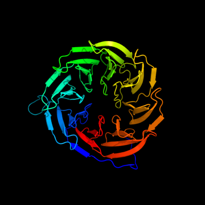
PDB header: protein bindingChain: A: PDB Molecule: lipoprotein yfgl;PDBTitle: crystal structure of escherichia coli bamb, a lipoprotein component of2 the beta-barrel assembly machinery complex, native crystals.
2 c1kv9A_
100.0
21
PDB header: oxidoreductaseChain: A: PDB Molecule: type ii quinohemoprotein alcohol dehydrogenase;PDBTitle: structure at 1.9 a resolution of a quinohemoprotein alcohol2 dehydrogenase from pseudomonas putida hk5
3 c1yiqA_
100.0
20
PDB header: oxidoreductaseChain: A: PDB Molecule: quinohemoprotein alcohol dehydrogenase;PDBTitle: molecular cloning and structural analysis of2 quinohemoprotein alcohol dehydrogenase adhiig from3 pseudomonas putida hk5. compariison to the other4 quinohemoprotein alcohol dehydrogenase adhiib found in the5 same microorganism.
4 c1kb0A_
100.0
20
PDB header: oxidoreductaseChain: A: PDB Molecule: quinohemoprotein alcohol dehydrogenase;PDBTitle: crystal structure of quinohemoprotein alcohol dehydrogenase from2 comamonas testosteroni
5 d1flga_
100.0
23
Fold: 8-bladed beta-propellerSuperfamily: Quinoprotein alcohol dehydrogenase-likeFamily: Quinoprotein alcohol dehydrogenase-like6 d1kv9a2
100.0
17
Fold: 8-bladed beta-propellerSuperfamily: Quinoprotein alcohol dehydrogenase-likeFamily: Quinoprotein alcohol dehydrogenase-like7 d2ad6a1
100.0
22
Fold: 8-bladed beta-propellerSuperfamily: Quinoprotein alcohol dehydrogenase-likeFamily: Quinoprotein alcohol dehydrogenase-like8 d1kb0a2
100.0
21
Fold: 8-bladed beta-propellerSuperfamily: Quinoprotein alcohol dehydrogenase-likeFamily: Quinoprotein alcohol dehydrogenase-like9 d1lrwa_
100.0
15
Fold: 8-bladed beta-propellerSuperfamily: Quinoprotein alcohol dehydrogenase-likeFamily: Quinoprotein alcohol dehydrogenase-like10 d1w6sa_
100.0
17
Fold: 8-bladed beta-propellerSuperfamily: Quinoprotein alcohol dehydrogenase-likeFamily: Quinoprotein alcohol dehydrogenase-like11 c3hxjA_
99.9
15
PDB header: oxidoreductaseChain: A: PDB Molecule: pyrrolo-quinoline quinone;PDBTitle: crystal structure of pyrrolo-quinoline quinone (pqq_dh) from2 methanococcus maripaludis, northeast structural genomics consortium3 target mrr86
12 c2be1A_
99.8
16
PDB header: transcriptionChain: A: PDB Molecule: serine/threonine-protein kinase/endoribonuclease ire1;PDBTitle: structure of the compact lumenal domain of yeast ire1
13 c2hz6A_
99.8
13
PDB header: signaling proteinChain: A: PDB Molecule: endoplasmic reticulum to nucleus signalling 1PDBTitle: the crystal structure of human ire1-alpha luminal domain
14 d1jmxb_
99.7
8
Fold: 7-bladed beta-propellerSuperfamily: YVTN repeat-like/Quinoprotein amine dehydrogenaseFamily: Quinohemoprotein amine dehydrogenase B chain15 c3no2A_
99.7
11
PDB header: unknown functionChain: A: PDB Molecule: uncharacterized protein;PDBTitle: crystal structure of a protein of unknown function (baccac_01654) from2 bacteroides caccae at 1.35 a resolution
16 c3dm0A_
99.6
13
PDB header: sugar binding protein,signaling proteinChain: A: PDB Molecule: maltose-binding periplasmic protein fused withPDBTitle: maltose binding protein fusion with rack1 from a. thaliana
17 d1l0qa2
99.6
9
Fold: 7-bladed beta-propellerSuperfamily: YVTN repeat-like/Quinoprotein amine dehydrogenaseFamily: YVTN repeat18 d2madh_
99.6
13
Fold: 7-bladed beta-propellerSuperfamily: YVTN repeat-like/Quinoprotein amine dehydrogenaseFamily: Methylamine dehydrogenase, H-chain19 d1pbyb_
99.6
11
Fold: 7-bladed beta-propellerSuperfamily: YVTN repeat-like/Quinoprotein amine dehydrogenaseFamily: Quinohemoprotein amine dehydrogenase B chain20 c1nexD_
99.6
12
PDB header: ligase, cell cycleChain: D: PDB Molecule: cdc4 protein;PDBTitle: crystal structure of scskp1-sccdc4-cpd peptide complex
21 d2bbkh_
not modelled
99.5
11
Fold: 7-bladed beta-propellerSuperfamily: YVTN repeat-like/Quinoprotein amine dehydrogenaseFamily: Methylamine dehydrogenase, H-chain22 c2ovqB_
not modelled
99.5
10
PDB header: transcription/cell cycleChain: B: PDB Molecule: f-box/wd repeat protein 7;PDBTitle: structure of the skp1-fbw7-cyclinedegc complex
23 c3bwsA_
not modelled
99.5
9
PDB header: unknown functionChain: A: PDB Molecule: protein lp49;PDBTitle: crystal structure of the leptospiral antigen lp49
24 c2i0tB_
not modelled
99.5
11
PDB header: oxidoreductaseChain: B: PDB Molecule: aromatic amine dehydrogenase;PDBTitle: crystal structure of phenylacetaldehyde derived r-2 carbinolamine adduct of aromatic amine dehydrogenase
25 c1gq1B_
not modelled
99.5
9
PDB header: oxidoreductaseChain: B: PDB Molecule: cytochrome cd1 nitrite reductase;PDBTitle: cytochrome cd1 nitrite reductase, y25s mutant, oxidised2 form
26 c2w18A_
not modelled
99.5
10
PDB header: nuclear proteinChain: A: PDB Molecule: partner and localizer of brca2;PDBTitle: crystal structure of the c-terminal wd40 domain of human2 palb2
27 d1nexb2
not modelled
99.4
12
Fold: 7-bladed beta-propellerSuperfamily: WD40 repeat-likeFamily: WD40-repeat28 c3vh0C_
not modelled
99.4
11
PDB header: protein binding/dnaChain: C: PDB Molecule: uncharacterized protein ynce;PDBTitle: crystal structure of e. coli ynce complexed with dna
29 c2j57J_
not modelled
99.4
11
PDB header: oxidoreductaseChain: J: PDB Molecule: methylamine dehydrogenase heavy chain;PDBTitle: x-ray reduced paraccocus denitrificans methylamine2 dehydrogenase n-quinol in complex with amicyanin.
30 c2h47F_
not modelled
99.4
10
PDB header: oxidoreductase/electron transportChain: F: PDB Molecule: aromatic amine dehydrogenase;PDBTitle: crystal structure of an electron transfer complex between2 aromatic amine dephydrogenase and azurin from alcaligenes3 faecalis (form 1)
31 c3c75J_
not modelled
99.4
10
PDB header: oxidoreductaseChain: J: PDB Molecule: methylamine dehydrogenase heavy chain;PDBTitle: paracoccus versutus methylamine dehydrogenase in complex2 with amicyanin
32 c1nnoA_
not modelled
99.4
8
PDB header: oxidoreductaseChain: A: PDB Molecule: nitrite reductase;PDBTitle: conformational changes occurring upon no binding in nitrite2 reductase from pseudomonas aeruginosa
33 c3mbrX_
not modelled
99.3
14
PDB header: transferaseChain: X: PDB Molecule: glutamine cyclotransferase;PDBTitle: crystal structure of the glutaminyl cyclase from xanthomonas2 campestris
34 c3jzhA_
not modelled
99.3
10
PDB header: gene regulationChain: A: PDB Molecule: polycomb protein eed;PDBTitle: eed-h3k79me3
35 c3mkqA_
not modelled
99.2
10
PDB header: transport proteinChain: A: PDB Molecule: coatomer beta'-subunit;PDBTitle: crystal structure of yeast alpha/betaprime-cop subcomplex of the copi2 vesicular coat
36 c1l0qC_
not modelled
99.2
9
PDB header: protein bindingChain: C: PDB Molecule: surface layer protein;PDBTitle: tandem yvtn beta-propeller and pkd domains from an archaeal surface2 layer protein
37 c3nolA_
not modelled
99.2
12
PDB header: transferaseChain: A: PDB Molecule: glutamine cyclotransferase;PDBTitle: crystal structure of zymomonas mobilis glutaminyl cyclase (trigonal2 form)
38 c3iytG_
not modelled
99.2
13
PDB header: apoptosisChain: G: PDB Molecule: apoptotic protease-activating factor 1;PDBTitle: structure of an apoptosome-procaspase-9 card complex
39 d1nira2
not modelled
99.2
10
Fold: 8-bladed beta-propellerSuperfamily: C-terminal (heme d1) domain of cytochrome cd1-nitrite reductaseFamily: C-terminal (heme d1) domain of cytochrome cd1-nitrite reductase40 d1nr0a1
not modelled
99.1
10
Fold: 7-bladed beta-propellerSuperfamily: WD40 repeat-likeFamily: WD40-repeat41 c3dw8B_
not modelled
99.1
8
PDB header: hydrolase/hydrolase inhibitorChain: B: PDB Molecule: serine/threonine-protein phosphatase 2a 55 kda regulatoryPDBTitle: structure of a protein phosphatase 2a holoenzyme with b55 subunit
42 c2qxvA_
not modelled
99.1
9
PDB header: gene regulationChain: A: PDB Molecule: embryonic ectoderm development;PDBTitle: structural basis of ezh2 recognition by eed
43 c3dsmA_
not modelled
99.1
9
PDB header: structural genomics, unknown functionChain: A: PDB Molecule: uncharacterized protein bacuni_02894;PDBTitle: crystal structure of the surface layer protein bacuni_02894 from2 bacteroides uniformis, northeast structural genomics consortium3 target btr193d.
44 c4a11B_
not modelled
99.1
10
PDB header: dna binding proteinChain: B: PDB Molecule: dna excision repair protein ercc-8;PDBTitle: structure of the hsddb1-hscsa complex
45 c3u4yA_
not modelled
99.1
12
PDB header: structural genomics, unknown functionChain: A: PDB Molecule: uncharacterized protein;PDBTitle: the crystal structure of a functionally unknown protein (dtox_1751)2 from desulfotomaculum acetoxidans dsm 771.
46 c3lrvA_
not modelled
99.1
13
PDB header: splicingChain: A: PDB Molecule: pre-mrna-splicing factor 19;PDBTitle: the prp19 wd40 domain contains a conserved protein interaction region2 essential for its function.
47 d1yfqa_
not modelled
99.1
9
Fold: 7-bladed beta-propellerSuperfamily: WD40 repeat-likeFamily: Cell cycle arrest protein BUB348 c2oajA_
not modelled
99.1
12
PDB header: endocytosis/exocytosisChain: A: PDB Molecule: protein sni1;PDBTitle: crystal structure of sro7 from s. cerevisiae
49 c3odtB_
not modelled
99.1
11
PDB header: nuclear proteinChain: B: PDB Molecule: protein doa1;PDBTitle: crystal structure of wd40 beta propeller domain of doa1
50 c2pbiB_
not modelled
99.0
11
PDB header: signaling proteinChain: B: PDB Molecule: guanine nucleotide-binding protein subunit beta 5;PDBTitle: the multifunctional nature of gbeta5/rgs9 revealed from its crystal2 structure
51 c3nokB_
not modelled
99.0
12
PDB header: transferaseChain: B: PDB Molecule: glutaminyl cyclase;PDBTitle: crystal structure of myxococcus xanthus glutaminyl cyclase
52 c1r5mA_
not modelled
99.0
10
PDB header: transcriptionChain: A: PDB Molecule: sir4-interacting protein sif2;PDBTitle: crystal structure of the c-terminal wd40 domain of sif2
53 c3i2nA_
not modelled
99.0
13
PDB header: transcriptionChain: A: PDB Molecule: wd repeat-containing protein 92;PDBTitle: crystal structure of wd40 repeats protein wdr92
54 c2gnqA_
not modelled
99.0
12
PDB header: transcriptionChain: A: PDB Molecule: wd-repeat protein 5;PDBTitle: structure of wdr5
55 d1qksa2
not modelled
99.0
12
Fold: 8-bladed beta-propellerSuperfamily: C-terminal (heme d1) domain of cytochrome cd1-nitrite reductaseFamily: C-terminal (heme d1) domain of cytochrome cd1-nitrite reductase56 c3acpA_
not modelled
99.0
12
PDB header: chaperoneChain: A: PDB Molecule: wd repeat-containing protein ygl004c;PDBTitle: crystal structure of yeast rpn14, a chaperone of the 19s regulatory2 particle of the proteasome
57 d2ovrb2
not modelled
98.9
11
Fold: 7-bladed beta-propellerSuperfamily: WD40 repeat-likeFamily: WD40-repeat58 d1fwxa2
not modelled
98.9
11
Fold: 7-bladed beta-propellerSuperfamily: Nitrous oxide reductase, N-terminal domainFamily: Nitrous oxide reductase, N-terminal domain59 c1vyhT_
not modelled
98.9
10
PDB header: hydrolaseChain: T: PDB Molecule: platelet-activating factor acetylhydrolase ibPDBTitle: paf-ah holoenzyme: lis1/alfa2
60 d1vyhc1
not modelled
98.9
9
Fold: 7-bladed beta-propellerSuperfamily: WD40 repeat-likeFamily: WD40-repeat61 c3mmyE_
not modelled
98.9
10
PDB header: nuclear proteinChain: E: PDB Molecule: mrna export factor;PDBTitle: structural and functional analysis of the interaction between the2 nucleoporin nup98 and the mrna export factor rae1
62 d1tbga_
not modelled
98.9
8
Fold: 7-bladed beta-propellerSuperfamily: WD40 repeat-likeFamily: WD40-repeat63 c2iwaA_
not modelled
98.9
10
PDB header: transferaseChain: A: PDB Molecule: glutamine cyclotransferase;PDBTitle: unbound glutaminyl cyclotransferase from carica papaya.
64 c3eg6A_
not modelled
98.9
11
PDB header: protein bindingChain: A: PDB Molecule: wd repeat-containing protein 5;PDBTitle: structure of wdr5 bound to mll1 peptide
65 c3ei4D_
not modelled
98.9
13
PDB header: dna binding proteinChain: D: PDB Molecule: dna damage-binding protein 2;PDBTitle: structure of the hsddb1-hsddb2 complex
66 c1p22A_
not modelled
98.8
9
PDB header: signaling proteinChain: A: PDB Molecule: f-box/wd-repeat protein 1a;PDBTitle: structure of a beta-trcp1-skp1-beta-catenin complex:2 destruction motif binding and lysine specificity on the3 scfbeta-trcp1 ubiquitin ligase
67 d1k32a3
not modelled
98.8
10
Fold: 7-bladed beta-propellerSuperfamily: Tricorn protease domain 2Family: Tricorn protease domain 268 d1erja_
not modelled
98.8
9
Fold: 7-bladed beta-propellerSuperfamily: WD40 repeat-likeFamily: WD40-repeat69 c3ei3B_
not modelled
98.8
6
PDB header: dna binding proteinChain: B: PDB Molecule: dna damage-binding protein 2;PDBTitle: structure of the hsddb1-drddb2 complex
70 d1pgua1
not modelled
98.7
14
Fold: 7-bladed beta-propellerSuperfamily: WD40 repeat-likeFamily: WD40-repeat71 c3frxB_
not modelled
98.7
9
PDB header: signaling proteinChain: B: PDB Molecule: guanine nucleotide-binding protein subunit beta-PDBTitle: crystal structure of the yeast orthologue of rack1, asc1.
72 c2pm9A_
not modelled
98.7
8
PDB header: protein transportChain: A: PDB Molecule: protein transport protein sec31;PDBTitle: crystal structure of yeast sec13/31 vertex element of the2 copii vesicular coat
73 c3ow8A_
not modelled
98.7
11
PDB header: transcriptionChain: A: PDB Molecule: wd repeat-containing protein 61;PDBTitle: crystal structure of the wd repeat-containing protein 61
74 c3e5zA_
not modelled
98.7
11
PDB header: structural genomics, unknown functionChain: A: PDB Molecule: putative gluconolactonase;PDBTitle: x-ray structure of the putative gluconolactonase in protein family2 pf08450. northeast structural genomics consortium target drr130.
75 c3g4hB_
not modelled
98.6
10
PDB header: hydrolaseChain: B: PDB Molecule: regucalcin;PDBTitle: crystal structure of human senescence marker protein-30 (zinc bound)
76 c1pi6A_
not modelled
98.6
8
PDB header: protein bindingChain: A: PDB Molecule: actin interacting protein 1;PDBTitle: yeast actin interacting protein 1 (aip1), orthorhombic crystal form
77 c2xznR_
not modelled
98.6
8
PDB header: ribosomeChain: R: PDB Molecule: rack1;PDBTitle: crystal structure of the eukaryotic 40s ribosomal2 subunit in complex with initiation factor 1. this file3 contains the 40s subunit and initiation factor for4 molecule 2
78 c3iz6a_
not modelled
98.5
11
PDB header: ribosomeChain: A: PDB Molecule: 40s ribosomal protein sa (s2p);PDBTitle: localization of the small subunit ribosomal proteins into a 5.5 a2 cryo-em map of triticum aestivum translating 80s ribosome
79 c1fwxB_
not modelled
98.5
11
PDB header: oxidoreductaseChain: B: PDB Molecule: nitrous oxide reductase;PDBTitle: crystal structure of nitrous oxide reductase from p. denitrificans
80 c1n6dE_
not modelled
98.5
14
PDB header: hydrolaseChain: E: PDB Molecule: tricorn protease;PDBTitle: tricorn protease in complex with tetrapeptide chloromethyl2 ketone derivative
81 c1k32E_
not modelled
98.5
14
PDB header: hydrolaseChain: E: PDB Molecule: tricorn protease;PDBTitle: crystal structure of the tricorn protease
82 c3hx6A_
not modelled
98.5
19
PDB header: cell adhesionChain: A: PDB Molecule: type 4 fimbrial biogenesis protein pily1;PDBTitle: crystal structure of pseudomonas aeruginosa pily1 c-terminal2 domain
83 c2j04B_
not modelled
98.4
11
PDB header: transcriptionChain: B: PDB Molecule: ydr362cp;PDBTitle: the tau60-tau91 subcomplex of yeast transcription factor2 iiic
84 d1sq9a_
not modelled
98.4
12
Fold: 7-bladed beta-propellerSuperfamily: WD40 repeat-likeFamily: WD40-repeat85 c3jroA_
not modelled
98.4
8
PDB header: transport protein, structural proteinChain: A: PDB Molecule: fusion protein of protein transport protein sec13PDBTitle: nup84-nup145c-sec13 edge element of the npc lattice
86 d1qnia2
not modelled
98.3
17
Fold: 7-bladed beta-propellerSuperfamily: Nitrous oxide reductase, N-terminal domainFamily: Nitrous oxide reductase, N-terminal domain87 d1ri6a_
not modelled
98.3
12
Fold: 7-bladed beta-propellerSuperfamily: Putative isomerase YbhEFamily: Putative isomerase YbhE88 c1nr0A_
not modelled
98.2
9
PDB header: structural proteinChain: A: PDB Molecule: actin interacting protein 1;PDBTitle: two seven-bladed beta-propeller domains revealed by the2 structure of a c. elegans homologue of yeast actin3 interacting protein 1 (aip1).
89 d1pjxa_
not modelled
98.1
10
Fold: 6-bladed beta-propellerSuperfamily: Calcium-dependent phosphotriesteraseFamily: SGL-like90 d1gxra_
not modelled
98.1
9
Fold: 7-bladed beta-propellerSuperfamily: WD40 repeat-likeFamily: WD40-repeat91 c3pe7A_
not modelled
98.1
8
PDB header: lyaseChain: A: PDB Molecule: oligogalacturonate lyase;PDBTitle: oligogalacturonate lyase in complex with manganese
92 c3jrpA_
not modelled
98.1
8
PDB header: transport protein, structural proteinChain: A: PDB Molecule: fusion protein of protein transport protein sec13PDBTitle: sec13 with nup145c (aa109-179) insertion blade
93 c2hesX_
not modelled
98.0
7
PDB header: biosynthetic proteinChain: X: PDB Molecule: ydr267cp;PDBTitle: cytosolic iron-sulphur assembly protein- 1
94 c3fhcA_
not modelled
98.0
12
PDB header: transport protein/hydrolaseChain: A: PDB Molecule: nuclear pore complex protein nup214;PDBTitle: crystal structure of human dbp5 in complex with nup214
95 c3greA_
not modelled
98.0
10
PDB header: signaling protein,protein bindingChain: A: PDB Molecule: serine/threonine-protein kinase vps15;PDBTitle: crystal structure of saccharomyces cerevisiae vps15 wd2 repeat domain
96 c1qniE_
not modelled
98.0
12
PDB header: oxidoreductaseChain: E: PDB Molecule: nitrous-oxide reductase;PDBTitle: crystal structure of nitrous oxide reductase from2 pseudomonas nautica, at 2.4a resolution
97 c3dr2A_
not modelled
98.0
10
PDB header: hydrolaseChain: A: PDB Molecule: exported gluconolactonase;PDBTitle: structural and functional analyses of xc5397 from2 xanthomonas campestris: a gluconolactonase important in3 glucose secondary metabolic pathways
98 d1ospo_
not modelled
98.0
9
Fold: open-sided beta-meanderSuperfamily: Outer surface proteinFamily: Outer surface protein99 c2aq5A_
not modelled
98.0
14
PDB header: structural proteinChain: A: PDB Molecule: coronin-1a;PDBTitle: crystal structure of murine coronin-1
100 d1nr0a2
not modelled
97.9
12
Fold: 7-bladed beta-propellerSuperfamily: WD40 repeat-likeFamily: WD40-repeat101 d2ghsa1
not modelled
97.9
14
Fold: 6-bladed beta-propellerSuperfamily: Calcium-dependent phosphotriesteraseFamily: SGL-like102 c2ghsA_
not modelled
97.9
14
PDB header: calcium-binding proteinChain: A: PDB Molecule: agr_c_1268p;PDBTitle: crystal structure of a calcium-binding protein, regucalcin2 (agr_c_1268) from agrobacterium tumefaciens str. c58 at 1.55 a3 resolution
103 c2g8sB_
not modelled
97.9
8
PDB header: sugar binding proteinChain: B: PDB Molecule: glucose/sorbosone dehydrogenases;PDBTitle: crystal structure of the soluble aldose sugar dehydrogenase2 (asd) from escherichia coli in the apo-form
104 c2fp8A_
not modelled
97.8
10
PDB header: lyaseChain: A: PDB Molecule: strictosidine synthase;PDBTitle: structure of strictosidine synthase, the biosynthetic entry to the2 monoterpenoid indole alkaloid family
105 c3fgbB_
not modelled
97.8
12
PDB header: structural genomics, unknown functionChain: B: PDB Molecule: uncharacterized protein q89zh8_bactn;PDBTitle: crystal structure of the q89zh8_bactn protein from2 bacteroides thetaiotaomicron. northeast structural3 genomics consortium target btr289b.
106 d1k8kc_
not modelled
97.8
10
Fold: 7-bladed beta-propellerSuperfamily: WD40 repeat-likeFamily: WD40-repeat107 c3sbrF_
not modelled
97.8
11
PDB header: oxidoreductaseChain: F: PDB Molecule: nitrous-oxide reductase;PDBTitle: pseudomonas stutzeri nitrous oxide reductase, p1 crystal form with2 substrate
108 c3dwlH_
not modelled
97.7
7
PDB header: structural proteinChain: H: PDB Molecule: actin-related protein 2/3 complex subunit 1;PDBTitle: crystal structure of fission yeast arp2/3 complex lacking the arp22 subunit
109 c3ottB_
not modelled
97.6
12
PDB header: transcriptionChain: B: PDB Molecule: two-component system sensor histidine kinase;PDBTitle: crystal structure of the extracellular domain of the putative one2 component system bt4673 from b. thetaiotaomicron
110 c2pm7B_
not modelled
97.6
10
PDB header: protein transportChain: B: PDB Molecule: protein transport protein sec13;PDBTitle: crystal structure of yeast sec13/31 edge element of the2 copii vesicular coat, selenomethionine version
111 d1xfda1
not modelled
97.5
10
Fold: 8-bladed beta-propellerSuperfamily: DPP6 N-terminal domain-likeFamily: DPP6 N-terminal domain-like112 d1p22a2
not modelled
97.5
9
Fold: 7-bladed beta-propellerSuperfamily: WD40 repeat-likeFamily: WD40-repeat113 c3fm0A_
not modelled
97.4
7
PDB header: biosynthetic proteinChain: A: PDB Molecule: protein ciao1;PDBTitle: crystal structure of wd40 protein ciao1
114 d2p4oa1
not modelled
97.2
12
Fold: 6-bladed beta-propellerSuperfamily: Calcium-dependent phosphotriesteraseFamily: All0351-like115 c2vduB_
not modelled
97.2
9
PDB header: transferaseChain: B: PDB Molecule: trna (guanine-n(7)-)-methyltransferase-PDBTitle: structure of trm8-trm82, the yeast trna m7g methylation2 complex
116 c3dasA_
not modelled
97.1
10
PDB header: oxidoreductaseChain: A: PDB Molecule: putative oxidoreductase;PDBTitle: structure of the pqq-bound form of aldose sugar2 dehydrogenase (adh) from streptomyces coelicolor
117 c2ojhA_
not modelled
97.1
9
PDB header: structural genomics, unknown functionChain: A: PDB Molecule: uncharacterized protein atu1656/agr_c_3050;PDBTitle: the structure of putative tolb from agrobacterium tumefaciens
118 d1pgua2
not modelled
97.0
6
Fold: 7-bladed beta-propellerSuperfamily: WD40 repeat-likeFamily: WD40-repeat119 d1k32a2
not modelled
97.0
10
Fold: 6-bladed beta-propellerSuperfamily: Tricorn protease N-terminal domainFamily: Tricorn protease N-terminal domain120 c2zkqa_
not modelled
96.9
12
PDB header: ribosomal protein/rnaChain: A: PDB Molecule: 18s ribosomal rna;PDBTitle: structure of a mammalian ribosomal 40s subunit within an2 80s complex obtained by docking homology models of the rna3 and proteins into an 8.7 a cryo-em map