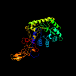
1 c3e6eC_
100.0
17
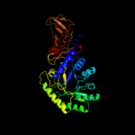
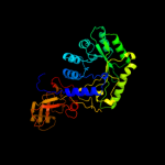
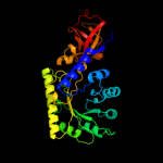
PDB header: isomeraseChain: C: PDB Molecule: alanine racemase;PDBTitle: crystal structure of alanine racemase from e.faecalis2 complex with cycloserine
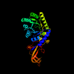
2 c1niuA_
100.0
18
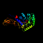
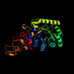
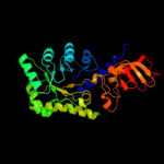
PDB header: isomeraseChain: A: PDB Molecule: alanine racemase;PDBTitle: alanine racemase with bound inhibitor derived from l-2 cycloserine
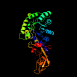
3 c1vftA_
100.0
18
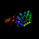
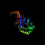
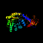
PDB header: isomeraseChain: A: PDB Molecule: alanine racemase;PDBTitle: crystal structure of l-cycloserine-bound form of alanine2 racemase from d-cycloserine-producing streptomyces3 lavendulae
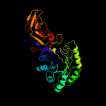
4 c3hurA_
100.0
15
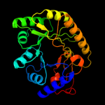
PDB header: isomeraseChain: A: PDB Molecule: alanine racemase;PDBTitle: crystal structure of alanine racemase from oenococcus oeni
5 c3mubB_
100.0
18
PDB header: isomeraseChain: B: PDB Molecule: alanine racemase;PDBTitle: the crystal structure of alanine racemase from streptococcus2 pneumoniae
6 c3kw3B_
100.0
14
PDB header: isomeraseChain: B: PDB Molecule: alanine racemase;PDBTitle: crystal structure of alanine racemase from bartonella henselae with2 covalently bound pyridoxal phosphate
7 c3oo2A_
100.0
14
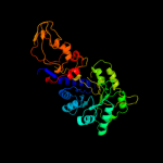
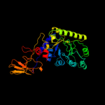
PDB header: isomeraseChain: A: PDB Molecule: alanine racemase 1;PDBTitle: 2.37 angstrom resolution crystal structure of an alanine racemase2 (alr) from staphylococcus aureus subsp. aureus col
8 c1xfcB_
100.0
16
PDB header: isomeraseChain: B: PDB Molecule: alanine racemase;PDBTitle: the 1.9 a crystal structure of alanine racemase from mycobacterium2 tuberculosis contains a conserved entryway into the active site
9 c2rjgC_
100.0
16
PDB header: isomeraseChain: C: PDB Molecule: alanine racemase;PDBTitle: crystal structure of biosynthetic alaine racemase from escherichia2 coli
10 c2odoC_
100.0
15
PDB header: isomeraseChain: C: PDB Molecule: alanine racemase;PDBTitle: crystal structure of pseudomonas fluorescens alanine racemase
11 c3oo2B_
100.0
15
PDB header: isomeraseChain: B: PDB Molecule: alanine racemase 1;PDBTitle: 2.37 angstrom resolution crystal structure of an alanine racemase2 (alr) from staphylococcus aureus subsp. aureus col
12 c3co8B_
100.0
14
PDB header: isomeraseChain: B: PDB Molecule: alanine racemase;PDBTitle: crystal structure of alanine racemase from oenococcus oeni
13 c2dy3B_
100.0
17
PDB header: isomeraseChain: B: PDB Molecule: alanine racemase;PDBTitle: crystal structure of alanine racemase from corynebacterium glutamicum
14 c2vd9A_
100.0
19
PDB header: isomeraseChain: A: PDB Molecule: alanine racemase;PDBTitle: the crystal structure of alanine racemase from bacillus2 anthracis (ba0252) with bound l-ala-p
15 c3anuA_
100.0
17
PDB header: lyaseChain: A: PDB Molecule: d-serine dehydratase;PDBTitle: crystal structure of d-serine dehydratase from chicken kidney
16 c3llxA_
100.0
18
PDB header: isomeraseChain: A: PDB Molecule: predicted amino acid aldolase or racemase;PDBTitle: crystal structure of an ala racemase-like protein (il1761) from2 idiomarina loihiensis at 1.50 a resolution
17 c3gwqB_
100.0
14
PDB header: lyaseChain: B: PDB Molecule: d-serine deaminase;PDBTitle: crystal structure of a putative d-serine deaminase (bxe_a4060) from2 burkholderia xenovorans lb400 at 2.00 a resolution
18 d1vfsa2
100.0
20
Fold: TIM beta/alpha-barrelSuperfamily: PLP-binding barrelFamily: Alanine racemase-like, N-terminal domain19 d1bd0a2
100.0
19
Fold: TIM beta/alpha-barrelSuperfamily: PLP-binding barrelFamily: Alanine racemase-like, N-terminal domain20 d1rcqa2
100.0
15
Fold: TIM beta/alpha-barrelSuperfamily: PLP-binding barrelFamily: Alanine racemase-like, N-terminal domain21 c1w8gA_
not modelled
100.0
16
PDB header: plp-binding proteinChain: A: PDB Molecule: hypothetical upf0001 protein yggs;PDBTitle: crystal structure of e. coli k-12 yggs
22 c2p3eA_
not modelled
100.0
16
PDB header: lyaseChain: A: PDB Molecule: diaminopimelate decarboxylase;PDBTitle: crystal structure of aq1208 from aquifex aeolicus
23 c3n2bD_
not modelled
100.0
16
PDB header: lyaseChain: D: PDB Molecule: diaminopimelate decarboxylase;PDBTitle: 1.8 angstrom resolution crystal structure of diaminopimelate2 decarboxylase (lysa) from vibrio cholerae.
24 c2qghA_
not modelled
100.0
10
PDB header: lyaseChain: A: PDB Molecule: diaminopimelate decarboxylase;PDBTitle: crystal structure of diaminopimelate decarboxylase from helicobacter2 pylori complexed with l-lysine
25 c2j66A_
not modelled
100.0
14
PDB header: lyaseChain: A: PDB Molecule: btrk;PDBTitle: structural characterisation of btrk decarboxylase from2 butirosin biosynthesis
26 c3r79B_
not modelled
100.0
19
PDB header: structure genomics, unknown functionChain: B: PDB Molecule: uncharacterized protein;PDBTitle: crystal structure of an uncharactertized protein from agrobacterium2 tumefaciens
27 c3cpgA_
not modelled
100.0
18
PDB header: structural genomics, unknown functionChain: A: PDB Molecule: uncharacterized protein;PDBTitle: crystal structure of an unknown protein from bifidobacterium2 adolescentis
28 d1ct5a_
not modelled
100.0
13
Fold: TIM beta/alpha-barrelSuperfamily: PLP-binding barrelFamily: "Hypothetical" protein ybl036c29 c1njjC_
not modelled
100.0
13
PDB header: lyaseChain: C: PDB Molecule: ornithine decarboxylase;PDBTitle: crystal structure determination of t. brucei ornithine2 decarboxylase bound to d-ornithine and to g418
30 c1tufA_
not modelled
100.0
13
PDB header: lyaseChain: A: PDB Molecule: diaminopimelate decarboxylase;PDBTitle: crystal structure of diaminopimelate decarboxylase from m.2 jannaschi
31 c2nvaH_
not modelled
100.0
10
PDB header: lyaseChain: H: PDB Molecule: arginine decarboxylase, a207r protein;PDBTitle: the x-ray crystal structure of the paramecium bursaria2 chlorella virus arginine decarboxylase bound to agmatine
32 c2pljA_
not modelled
100.0
18
PDB header: lyaseChain: A: PDB Molecule: lysine/ornithine decarboxylase;PDBTitle: crystal structure of lysine/ornithine decarboxylase complexed with2 putrescine from vibrio vulnificus
33 c2o0tB_
not modelled
100.0
17
PDB header: lyaseChain: B: PDB Molecule: diaminopimelate decarboxylase;PDBTitle: the three dimensional structure of diaminopimelate decarboxylase from2 mycobacterium tuberculosis reveals a tetrameric enzyme organisation
34 c2yxxA_
not modelled
100.0
14
PDB header: lyaseChain: A: PDB Molecule: diaminopimelate decarboxylase;PDBTitle: crystal structure analysis of diaminopimelate decarboxylate (lysa)
35 c1knwA_
not modelled
100.0
15
PDB header: lyaseChain: A: PDB Molecule: diaminopimelate decarboxylase;PDBTitle: crystal structure of diaminopimelate decarboxylase
36 c2on3A_
not modelled
100.0
17
PDB header: lyaseChain: A: PDB Molecule: ornithine decarboxylase;PDBTitle: a structural insight into the inhibition of human and2 leishmania donovani ornithine decarboxylases by 3-aminooxy-3 1-aminopropane
37 c3btnA_
not modelled
99.9
14
PDB header: biosynthetic proteinChain: A: PDB Molecule: antizyme inhibitor 1;PDBTitle: crystal structure of antizyme inhibitor, an ornithine2 decarboxylase homologous protein
38 c1d7kB_
not modelled
99.9
17
PDB header: lyaseChain: B: PDB Molecule: human ornithine decarboxylase;PDBTitle: crystal structure of human ornithine decarboxylase at 2.12 angstroms resolution
39 d1hkva2
not modelled
99.9
17
Fold: TIM beta/alpha-barrelSuperfamily: PLP-binding barrelFamily: Alanine racemase-like, N-terminal domain40 d1f3ta2
not modelled
99.9
16
Fold: TIM beta/alpha-barrelSuperfamily: PLP-binding barrelFamily: Alanine racemase-like, N-terminal domain41 d1twia2
not modelled
99.9
13
Fold: TIM beta/alpha-barrelSuperfamily: PLP-binding barrelFamily: Alanine racemase-like, N-terminal domain42 d7odca2
not modelled
99.9
15
Fold: TIM beta/alpha-barrelSuperfamily: PLP-binding barrelFamily: Alanine racemase-like, N-terminal domain43 c3n29A_
not modelled
99.9
15
PDB header: lyaseChain: A: PDB Molecule: carboxynorspermidine decarboxylase;PDBTitle: crystal structure of carboxynorspermidine decarboxylase complexed with2 norspermidine from campylobacter jejuni
44 c3nzqB_
not modelled
99.9
13
PDB header: lyaseChain: B: PDB Molecule: biosynthetic arginine decarboxylase;PDBTitle: crystal structure of biosynthetic arginine decarboxylase adc (spea)2 from escherichia coli, northeast structural genomics consortium3 target er600
45 d1d7ka2
not modelled
99.9
15
Fold: TIM beta/alpha-barrelSuperfamily: PLP-binding barrelFamily: Alanine racemase-like, N-terminal domain46 c3mt1B_
not modelled
99.9
12
PDB header: lyaseChain: B: PDB Molecule: putative carboxynorspermidine decarboxylase protein;PDBTitle: crystal structure of putative carboxynorspermidine decarboxylase2 protein from sinorhizobium meliloti
47 c3nzpA_
not modelled
99.8
16
PDB header: lyaseChain: A: PDB Molecule: arginine decarboxylase;PDBTitle: crystal structure of the biosynthetic arginine decarboxylase spea from2 campylobacter jejuni, northeast structural genomics consortium target3 br53
48 c3n2oA_
not modelled
99.8
12
PDB header: lyaseChain: A: PDB Molecule: biosynthetic arginine decarboxylase;PDBTitle: x-ray crystal structure of arginine decarboxylase complexed with2 arginine from vibrio vulnificus
49 d1knwa2
not modelled
99.8
16
Fold: TIM beta/alpha-barrelSuperfamily: PLP-binding barrelFamily: Alanine racemase-like, N-terminal domain50 d1bd0a1
not modelled
99.7
13
Fold: Domain of alpha and beta subunits of F1 ATP synthase-likeSuperfamily: Alanine racemase C-terminal domain-likeFamily: Alanine racemase51 d1vfsa1
not modelled
99.6
15
Fold: Domain of alpha and beta subunits of F1 ATP synthase-likeSuperfamily: Alanine racemase C-terminal domain-likeFamily: Alanine racemase52 d1rcqa1
not modelled
99.6
22
Fold: Domain of alpha and beta subunits of F1 ATP synthase-likeSuperfamily: Alanine racemase C-terminal domain-likeFamily: Alanine racemase53 c2y85D_
not modelled
97.1
17
PDB header: isomeraseChain: D: PDB Molecule: phosphoribosyl isomerase a;PDBTitle: crystal structure of mycobacterium tuberculosis phosphoribosyl2 isomerase with bound rcdrp
54 c3qjaA_
not modelled
96.6
14
PDB header: lyaseChain: A: PDB Molecule: indole-3-glycerol phosphate synthase;PDBTitle: crystal structure of the mycobacterium tuberculosis indole-3-glycerol2 phosphate synthase (trpc) in apo form
55 d1vc4a_
not modelled
96.1
16
Fold: TIM beta/alpha-barrelSuperfamily: Ribulose-phoshate binding barrelFamily: Tryptophan biosynthesis enzymes56 d1rpxa_
not modelled
96.1
14
Fold: TIM beta/alpha-barrelSuperfamily: Ribulose-phoshate binding barrelFamily: D-ribulose-5-phosphate 3-epimerase57 c2c3zA_
not modelled
95.6
17
PDB header: lyaseChain: A: PDB Molecule: indole-3-glycerol phosphate synthase;PDBTitle: crystal structure of a truncated variant of indole-3-2 glycerol phosphate synthase from sulfolobus solfataricus
58 d1y0ea_
not modelled
94.6
13
Fold: TIM beta/alpha-barrelSuperfamily: Ribulose-phoshate binding barrelFamily: NanE-like59 d1tqja_
not modelled
94.5
16
Fold: TIM beta/alpha-barrelSuperfamily: Ribulose-phoshate binding barrelFamily: D-ribulose-5-phosphate 3-epimerase60 c3inpA_
not modelled
94.4
14
PDB header: isomeraseChain: A: PDB Molecule: d-ribulose-phosphate 3-epimerase;PDBTitle: 2.05 angstrom resolution crystal structure of d-ribulose-phosphate 3-2 epimerase from francisella tularensis.
61 d1h5ya_
not modelled
94.1
10
Fold: TIM beta/alpha-barrelSuperfamily: Ribulose-phoshate binding barrelFamily: Histidine biosynthesis enzymes62 c3ct7E_
not modelled
92.9
13
PDB header: isomeraseChain: E: PDB Molecule: d-allulose-6-phosphate 3-epimerase;PDBTitle: crystal structure of d-allulose 6-phosphate 3-epimerase2 from escherichia coli k-12
63 c3ivuB_
not modelled
91.9
17
PDB header: transferaseChain: B: PDB Molecule: homocitrate synthase, mitochondrial;PDBTitle: homocitrate synthase lys4 bound to 2-og
64 d1izca_
not modelled
91.9
17
Fold: TIM beta/alpha-barrelSuperfamily: Phosphoenolpyruvate/pyruvate domainFamily: HpcH/HpaI aldolase65 c1izcA_
not modelled
91.9
17
PDB header: lyaseChain: A: PDB Molecule: macrophomate synthase intermolecular diels-alderase;PDBTitle: crystal structure analysis of macrophomate synthase
66 d1j5ta_
not modelled
91.5
16
Fold: TIM beta/alpha-barrelSuperfamily: Ribulose-phoshate binding barrelFamily: Tryptophan biosynthesis enzymes67 d2flia1
not modelled
90.9
15
Fold: TIM beta/alpha-barrelSuperfamily: Ribulose-phoshate binding barrelFamily: D-ribulose-5-phosphate 3-epimerase68 d1znna1
not modelled
90.5
10
Fold: TIM beta/alpha-barrelSuperfamily: Ribulose-phoshate binding barrelFamily: PdxS-like69 d1vzwa1
not modelled
90.4
16
Fold: TIM beta/alpha-barrelSuperfamily: Ribulose-phoshate binding barrelFamily: Histidine biosynthesis enzymes70 c1znnF_
not modelled
90.0
10
PDB header: biosynthetic proteinChain: F: PDB Molecule: plp synthase;PDBTitle: structure of the synthase subunit of plp synthase
71 c2w6rA_
not modelled
88.1
11
PDB header: lyaseChain: A: PDB Molecule: imidazole glycerol phosphate synthase subunitPDBTitle: crystal structure of an artificial (ba)8-barrel protein2 designed from identical half barrels
72 d1thfd_
not modelled
88.1
14
Fold: TIM beta/alpha-barrelSuperfamily: Ribulose-phoshate binding barrelFamily: Histidine biosynthesis enzymes73 c3igsB_
not modelled
87.6
19
PDB header: isomeraseChain: B: PDB Molecule: n-acetylmannosamine-6-phosphate 2-epimerase 2;PDBTitle: structure of the salmonella enterica n-acetylmannosamine-6-phosphate2 2-epimerase
74 d1i4na_
not modelled
86.1
16
Fold: TIM beta/alpha-barrelSuperfamily: Ribulose-phoshate binding barrelFamily: Tryptophan biosynthesis enzymes75 c3qz6A_
not modelled
85.9
14
PDB header: lyaseChain: A: PDB Molecule: hpch/hpai aldolase;PDBTitle: the crystal structure of hpch/hpai aldolase from desulfitobacterium2 hafniense dcb-2
76 d1ka9f_
not modelled
83.3
10
Fold: TIM beta/alpha-barrelSuperfamily: Ribulose-phoshate binding barrelFamily: Histidine biosynthesis enzymes77 c2ftpA_
not modelled
81.1
15
PDB header: lyaseChain: A: PDB Molecule: hydroxymethylglutaryl-coa lyase;PDBTitle: crystal structure of hydroxymethylglutaryl-coa lyase from pseudomonas2 aeruginosa
78 d1a53a_
not modelled
80.5
17
Fold: TIM beta/alpha-barrelSuperfamily: Ribulose-phoshate binding barrelFamily: Tryptophan biosynthesis enzymes79 d1mxsa_
not modelled
77.7
13
Fold: TIM beta/alpha-barrelSuperfamily: AldolaseFamily: Class I aldolase80 c2wyhA_
77.0
15
PDB header: hydrolaseChain: A: PDB Molecule: alpha-mannosidase;PDBTitle: structure of the streptococcus pyogenes family gh38 alpha-2 mannosidase
81 c1ydoC_
not modelled
77.0
18
PDB header: lyaseChain: C: PDB Molecule: hmg-coa lyase;PDBTitle: crystal structure of the bacillis subtilis hmg-coa lyase, northeast2 structural genomics target sr181.
82 d1qo2a_
not modelled
76.7
14
Fold: TIM beta/alpha-barrelSuperfamily: Ribulose-phoshate binding barrelFamily: Histidine biosynthesis enzymes83 d1dxea_
not modelled
76.7
16
Fold: TIM beta/alpha-barrelSuperfamily: Phosphoenolpyruvate/pyruvate domainFamily: HpcH/HpaI aldolase84 d1h1ya_
not modelled
76.2
14
Fold: TIM beta/alpha-barrelSuperfamily: Ribulose-phoshate binding barrelFamily: D-ribulose-5-phosphate 3-epimerase85 d1tqxa_
not modelled
76.1
11
Fold: TIM beta/alpha-barrelSuperfamily: Ribulose-phoshate binding barrelFamily: D-ribulose-5-phosphate 3-epimerase86 c2h9aA_
not modelled
73.6
20
PDB header: oxidoreductaseChain: A: PDB Molecule: carbon monoxide dehydrogenase corrinoid/iron-PDBTitle: corrinoid iron-sulfur protein
87 c2vefB_
not modelled
73.3
19
PDB header: transferaseChain: B: PDB Molecule: dihydropteroate synthase;PDBTitle: dihydropteroate synthase from streptococcus pneumoniae
88 d7reqa2
not modelled
72.2
18
Fold: Flavodoxin-likeSuperfamily: Cobalamin (vitamin B12)-binding domainFamily: Cobalamin (vitamin B12)-binding domain89 d1eyea_
not modelled
72.2
15
Fold: TIM beta/alpha-barrelSuperfamily: Dihydropteroate synthetase-likeFamily: Dihydropteroate synthetase90 c2bdqA_
not modelled
70.7
15
PDB header: metal transportChain: A: PDB Molecule: copper homeostasis protein cutc;PDBTitle: crystal structure of the putative copper homeostasis2 protein cutc from streptococcus agalactiae, northeast3 strucural genomics target sar15.
91 c2h90A_
not modelled
70.0
13
PDB header: oxidoreductaseChain: A: PDB Molecule: xenobiotic reductase a;PDBTitle: xenobiotic reductase a in complex with coumarin
92 c2v82A_
not modelled
67.7
14
PDB header: lyaseChain: A: PDB Molecule: 2-dehydro-3-deoxy-6-phosphogalactonate aldolase;PDBTitle: kdpgal complexed to kdpgal
93 c1x1oC_
not modelled
67.7
15
PDB header: transferaseChain: C: PDB Molecule: nicotinate-nucleotide pyrophosphorylase;PDBTitle: crystal structure of project id tt0268 from thermus thermophilus hb8
94 c3q58A_
not modelled
66.5
16
PDB header: isomeraseChain: A: PDB Molecule: n-acetylmannosamine-6-phosphate 2-epimerase;PDBTitle: structure of n-acetylmannosamine-6-phosphate epimerase from salmonella2 enterica
95 c3bolB_
not modelled
66.1
20
PDB header: transferaseChain: B: PDB Molecule: 5-methyltetrahydrofolate s-homocysteinePDBTitle: cobalamin-dependent methionine synthase (1-566) from2 thermotoga maritima complexed with zn2+
96 c3ghfA_
not modelled
65.7
20
PDB header: cell cycleChain: A: PDB Molecule: septum site-determining protein minc;PDBTitle: crystal structure of the septum site-determining protein2 minc from salmonella typhimurium
97 d1ps9a1
not modelled
65.3
12
Fold: TIM beta/alpha-barrelSuperfamily: FMN-linked oxidoreductasesFamily: FMN-linked oxidoreductases98 c3qc3B_
not modelled
64.9
14
PDB header: isomeraseChain: B: PDB Molecule: d-ribulose-5-phosphate-3-epimerase;PDBTitle: crystal structure of a d-ribulose-5-phosphate-3-epimerase (np_954699)2 from homo sapiens at 2.20 a resolution
99 c1k1yA_
not modelled
64.5
21
PDB header: transferaseChain: A: PDB Molecule: 4-alpha-glucanotransferase;PDBTitle: crystal structure of thermococcus litoralis 4-alpha-glucanotransferase2 complexed with acarbose
100 d1yxya1
not modelled
64.5
13
Fold: TIM beta/alpha-barrelSuperfamily: Ribulose-phoshate binding barrelFamily: NanE-like101 c3cu2A_
not modelled
64.4
17
PDB header: isomeraseChain: A: PDB Molecule: ribulose-5-phosphate 3-epimerase;PDBTitle: crystal structure of ribulose-5-phosphate 3-epimerase (yp_718263.1)2 from haemophilus somnus 129pt at 1.91 a resolution
102 c3rfaA_
not modelled
61.5
7
PDB header: oxidoreductaseChain: A: PDB Molecule: ribosomal rna large subunit methyltransferase n;PDBTitle: x-ray structure of rlmn from escherichia coli in complex with s-2 adenosylmethionine
103 c2b7pA_
not modelled
61.2
14
PDB header: transferaseChain: A: PDB Molecule: probable nicotinate-nucleotide pyrophosphorylase;PDBTitle: crystal structure of quinolinic acid phosphoribosyltransferase from2 helicobacter pylori
104 c2qv5A_
not modelled
60.9
16
PDB header: structural genomics, unknown functionChain: A: PDB Molecule: uncharacterized protein atu2773;PDBTitle: crystal structure of uncharacterized protein atu2773 from2 agrobacterium tumefaciens c58
105 d1olta_
not modelled
60.5
17
Fold: TIM beta/alpha-barrelSuperfamily: Radical SAM enzymesFamily: Oxygen-independent coproporphyrinogen III oxidase HemN106 d1lt7a_
not modelled
60.0
17
Fold: TIM beta/alpha-barrelSuperfamily: Homocysteine S-methyltransferaseFamily: Homocysteine S-methyltransferase107 d1umya_
not modelled
59.9
17
Fold: TIM beta/alpha-barrelSuperfamily: Homocysteine S-methyltransferaseFamily: Homocysteine S-methyltransferase108 d1piia2
not modelled
58.8
15
Fold: TIM beta/alpha-barrelSuperfamily: Ribulose-phoshate binding barrelFamily: Tryptophan biosynthesis enzymes109 c3hf3A_
not modelled
57.8
17
PDB header: oxidoreductaseChain: A: PDB Molecule: chromate reductase;PDBTitle: old yellow enzyme from thermus scotoductus sa-01
110 c1qapA_
not modelled
57.6
14
PDB header: glycosyltransferaseChain: A: PDB Molecule: quinolinic acid phosphoribosyltransferase;PDBTitle: quinolinic acid phosphoribosyltransferase with bound2 quinolinic acid
111 d1gvfa_
not modelled
57.0
15
Fold: TIM beta/alpha-barrelSuperfamily: AldolaseFamily: Class II FBP aldolase112 c3bdkB_
not modelled
55.7
17
PDB header: lyaseChain: B: PDB Molecule: d-mannonate dehydratase;PDBTitle: crystal structure of streptococcus suis mannonate2 dehydratase complexed with substrate analogue
113 c3kruC_
not modelled
54.1
11
PDB header: oxidoreductaseChain: C: PDB Molecule: nadh:flavin oxidoreductase/nadh oxidase;PDBTitle: crystal structure of the thermostable old yellow enzyme from2 thermoanaerobacter pseudethanolicus e39
114 c1ydnA_
not modelled
53.4
15
PDB header: lyaseChain: A: PDB Molecule: hydroxymethylglutaryl-coa lyase;PDBTitle: crystal structure of the hmg-coa lyase from brucella melitensis,2 northeast structural genomics target lr35.
115 d1xcfa_
not modelled
52.5
10
Fold: TIM beta/alpha-barrelSuperfamily: Ribulose-phoshate binding barrelFamily: Tryptophan biosynthesis enzymes116 d1njjb1
not modelled
52.4
12
Fold: Domain of alpha and beta subunits of F1 ATP synthase-likeSuperfamily: Alanine racemase C-terminal domain-likeFamily: Eukaryotic ODC-like117 d1wa3a1
not modelled
50.2
16
Fold: TIM beta/alpha-barrelSuperfamily: AldolaseFamily: Class I aldolase118 d1vcfa1
not modelled
50.0
14
Fold: TIM beta/alpha-barrelSuperfamily: FMN-linked oxidoreductasesFamily: FMN-linked oxidoreductases119 c3gnnA_
not modelled
49.8
10
PDB header: transferaseChain: A: PDB Molecule: nicotinate-nucleotide pyrophosphorylase;PDBTitle: crystal structure of nicotinate-nucleotide2 pyrophosphorylase from burkholderi pseudomallei
120 c2dzaA_
not modelled
47.7
20
PDB header: transferaseChain: A: PDB Molecule: dihydropteroate synthase;PDBTitle: crystal structure of dihydropteroate synthase from thermus2 thermophilus hb8 in complex with 4-aminobenzoate