1 c3ojaB_
96.4
13
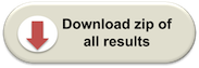
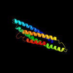
PDB header: protein bindingChain: B: PDB Molecule: anopheles plasmodium-responsive leucine-rich repeat proteinPDBTitle: crystal structure of lrim1/apl1c complex
2 c3ghgK_
96.3
7
PDB header: blood clottingChain: K: PDB Molecule: fibrinogen beta chain;PDBTitle: crystal structure of human fibrinogen
3 c1deqO_
93.6
9
PDB header: PDB COMPND: 4 c2efrB_
93.2
13
PDB header: contractile proteinChain: B: PDB Molecule: general control protein gcn4 and tropomyosin 1 alpha chain;PDBTitle: crystal structure of the c-terminal tropomyosin fragment with n- and2 c-terminal extensions of the leucine zipper at 1.8 angstroms3 resolution
5 c1ei3E_
92.9
10
PDB header: PDB COMPND: 6 c2d3eD_
92.8
13
PDB header: contractile proteinChain: D: PDB Molecule: general control protein gcn4 and tropomyosin 1PDBTitle: crystal structure of the c-terminal fragment of rabbit2 skeletal alpha-tropomyosin
7 c2fxmB_
92.4
14
PDB header: contractile proteinChain: B: PDB Molecule: myosin heavy chain, cardiac muscle beta isoform;PDBTitle: structure of the human beta-myosin s2 fragment
8 c2b9cA_
91.1
14
PDB header: contractile proteinChain: A: PDB Molecule: striated-muscle alpha tropomyosin;PDBTitle: structure of tropomyosin's mid-region: bending and binding2 sites for actin
9 c1deqF_
90.6
13
PDB header: PDB COMPND: 10 c2rd0B_
89.9
12
PDB header: transferase/oncoproteinChain: B: PDB Molecule: phosphatidylinositol 3-kinase regulatory subunit alpha;PDBTitle: structure of a human p110alpha/p85alpha complex
11 c3o0zD_
88.5
12
PDB header: transferaseChain: D: PDB Molecule: rho-associated protein kinase 1;PDBTitle: crystal structure of a coiled-coil domain from human rock i
12 c3na7A_
87.9
12
PDB header: gene regulation, chaperoneChain: A: PDB Molecule: hp0958;PDBTitle: 2.2 angstrom structure of the hp0958 protein from helicobacter pylori2 ccug 17874
13 c1bf5A_
85.4
8
PDB header: gene regulation/dnaChain: A: PDB Molecule: signal transducer and activator of transcriptionPDBTitle: tyrosine phosphorylated stat-1/dna complex
14 c3ojaA_
85.4
10
PDB header: protein bindingChain: A: PDB Molecule: leucine-rich immune molecule 1;PDBTitle: crystal structure of lrim1/apl1c complex
15 c3cwgA_
83.4
10
PDB header: transcriptionChain: A: PDB Molecule: signal transducer and activator of transcriptionPDBTitle: unphosphorylated mouse stat3 core fragment
16 c1ciiA_
80.5
10
PDB header: transmembrane proteinChain: A: PDB Molecule: colicin ia;PDBTitle: colicin ia
17 c2v71A_
80.0
10
PDB header: nuclear proteinChain: A: PDB Molecule: nuclear distribution protein nude-like 1;PDBTitle: coiled-coil region of nudel
18 c1l8dB_
79.9
10
PDB header: replicationChain: B: PDB Molecule: dna double-strand break repair rad50 atpase;PDBTitle: rad50 coiled-coil zn hook
19 c4a55B_
79.2
13
PDB header: transferaseChain: B: PDB Molecule: phosphatidylinositol 3-kinase regulatory subunit alpha;PDBTitle: crystal structure of p110alpha in complex with ish2 of p85alpha and2 the inhibitor pik-108
20 c3dtpA_
70.7
13
PDB header: contractile proteinChain: A: PDB Molecule: myosin 2 heavy chain chimera of smooth andPDBTitle: tarantula heavy meromyosin obtained by flexible docking to2 tarantula muscle thick filament cryo-em 3d-map
21 c3ipkA_
not modelled
69.5
16
PDB header: cell adhesionChain: A: PDB Molecule: agi/ii;PDBTitle: crystal structure of a3vp1 of agi/ii of streptococcus mutans
22 c1y4cA_
not modelled
67.9
7
PDB header: de novo proteinChain: A: PDB Molecule: maltose binding protein fused with designedPDBTitle: designed helical protein fusion mbp
23 c3hizB_
not modelled
66.7
15
PDB header: transferase/oncoproteinChain: B: PDB Molecule: phosphatidylinositol 3-kinase regulatory subunitPDBTitle: crystal structure of p110alpha h1047r mutant in complex with2 nish2 of p85alpha
24 d1vmaa1
not modelled
66.1
13
Fold: Four-helical up-and-down bundleSuperfamily: Domain of the SRP/SRP receptor G-proteinsFamily: Domain of the SRP/SRP receptor G-proteins25 c3ol1A_
not modelled
64.0
11
PDB header: structural proteinChain: A: PDB Molecule: vimentin;PDBTitle: crystal structure of vimentin (fragment 144-251) from homo sapiens,2 northeast structural genomics consortium target hr4796b
26 c3u59C_
not modelled
63.5
12
PDB header: contractile proteinChain: C: PDB Molecule: tropomyosin beta chain;PDBTitle: n-terminal 98-aa fragment of smooth muscle tropomyosin beta
27 c1c1gA_
not modelled
59.8
14
PDB header: contractile proteinChain: A: PDB Molecule: tropomyosin;PDBTitle: crystal structure of tropomyosin at 7 angstroms resolution2 in the spermine-induced crystal form
28 c3iv1F_
not modelled
57.8
11
PDB header: hydrolaseChain: F: PDB Molecule: tumor susceptibility gene 101 protein;PDBTitle: coiled-coil domain of tumor susceptibility gene 101
29 c2jeeA_
not modelled
53.2
19
PDB header: cell cycleChain: A: PDB Molecule: yiiu;PDBTitle: xray structure of e. coli yiiu
30 c1ei3C_
not modelled
52.4
15
PDB header: PDB COMPND: 31 c2v66C_
not modelled
52.2
7
PDB header: structural proteinChain: C: PDB Molecule: nuclear distribution protein nude-like 1;PDBTitle: crystal structure of the coiled-coil domain of ndel1 (a.a.2 58 to 169)c
32 c1bg1A_
not modelled
51.7
12
PDB header: transcription/dnaChain: A: PDB Molecule: protein (transcription factor stat3b);PDBTitle: transcription factor stat3b/dna complex
33 c3hnwB_
not modelled
43.5
12
PDB header: structural genomics, unknown functionChain: B: PDB Molecule: uncharacterized protein;PDBTitle: crystal structure of a basic coiled-coil protein of unknown function2 from eubacterium eligens atcc 27750
34 c1deqD_
not modelled
42.5
5
PDB header: PDB COMPND: 35 c2gl2B_
not modelled
42.2
13
PDB header: cell adhesionChain: B: PDB Molecule: adhesion a;PDBTitle: crystal structure of the tetra muntant (t66g,r67g,f68g,2 y69g) of bacterial adhesin fada
36 c2y3aB_
not modelled
42.2
12
PDB header: transferaseChain: B: PDB Molecule: phosphatidylinositol 3-kinase regulatory subunit beta;PDBTitle: crystal structure of p110beta in complex with icsh2 of p85beta and2 the drug gdc-0941
37 c2v1yB_
not modelled
37.9
13
PDB header: transferaseChain: B: PDB Molecule: phosphatidylinositol 3-kinase regulatory subunit alpha;PDBTitle: structure of a phosphoinositide 3-kinase alpha adaptor-2 binding domain (abd) in a complex with the ish2 domain3 from p85 alpha
38 c3movB_
not modelled
35.7
11
PDB header: structural proteinChain: B: PDB Molecule: lamin-b1;PDBTitle: crystal structure of human lamin-b1 coil 2 segment
39 c3kltB_
not modelled
33.8
7
PDB header: structural proteinChain: B: PDB Molecule: vimentin;PDBTitle: crystal structure of a vimentin fragment
40 c3u1aC_
not modelled
31.6
9
PDB header: contractile proteinChain: C: PDB Molecule: smooth muscle tropomyosin alpha;PDBTitle: n-terminal 81-aa fragment of smooth muscle tropomyosin alpha
41 c1yvlB_
not modelled
29.1
12
PDB header: signaling proteinChain: B: PDB Molecule: signal transducer and activator of transcriptionPDBTitle: structure of unphosphorylated stat1
42 c1gk4A_
not modelled
28.3
11
PDB header: vimentinChain: A: PDB Molecule: vimentin;PDBTitle: human vimentin coil 2b fragment (cys2)
43 d1fxka_
not modelled
27.9
13
Fold: Long alpha-hairpinSuperfamily: PrefoldinFamily: Prefoldin44 d2gqba1
not modelled
25.3
27
Fold: RPA2825-likeSuperfamily: RPA2825-likeFamily: RPA2825-like45 c3a7pB_
not modelled
21.9
9
PDB header: protein transportChain: B: PDB Molecule: autophagy protein 16;PDBTitle: the crystal structure of saccharomyces cerevisiae atg16
46 c1vmaA_
not modelled
21.7
13
PDB header: protein transportChain: A: PDB Molecule: cell division protein ftsy;PDBTitle: crystal structure of cell division protein ftsy (tm0570) from2 thermotoga maritima at 1.60 a resolution
47 c1x8yA_
not modelled
18.7
10
PDB header: structural proteinChain: A: PDB Molecule: lamin a/c;PDBTitle: human lamin coil 2b
48 d1r05a_
not modelled
17.4
16
Fold: HLH-likeSuperfamily: HLH, helix-loop-helix DNA-binding domainFamily: HLH, helix-loop-helix DNA-binding domain49 d2daha1
not modelled
16.9
36
Fold: RuvA C-terminal domain-likeSuperfamily: UBA-likeFamily: UBA domain50 d2bwba1
not modelled
15.6
18
Fold: RuvA C-terminal domain-likeSuperfamily: UBA-likeFamily: UBA domain51 c1jchC_
not modelled
15.4
7
PDB header: ribosome inhibitor, hydrolaseChain: C: PDB Molecule: colicin e3;PDBTitle: crystal structure of colicin e3 in complex with its immunity protein
52 c2xdjF_
not modelled
14.3
9
PDB header: unknown functionChain: F: PDB Molecule: uncharacterized protein ybgf;PDBTitle: crystal structure of the n-terminal domain of e.coli ybgf
53 c1junB_
not modelled
13.6
17
PDB header: transcription regulationChain: B: PDB Molecule: c-jun homodimer;PDBTitle: nmr study of c-jun homodimer
54 c2zvfG_
not modelled
13.4
13
PDB header: ligaseChain: G: PDB Molecule: alanyl-trna synthetase;PDBTitle: crystal structure of archaeoglobus fulgidus alanyl-trna2 synthetase c-terminal dimerization domain
55 c3n4xB_
not modelled
13.4
10
PDB header: replicationChain: B: PDB Molecule: monopolin complex subunit csm1;PDBTitle: structure of csm1 full-length
56 c1m1jA_
not modelled
13.4
13
PDB header: blood clottingChain: A: PDB Molecule: fibrinogen alpha subunit;PDBTitle: crystal structure of native chicken fibrinogen with two different2 bound ligands
57 c2dnaA_
not modelled
12.7
32
PDB header: structural genomics, unknown functionChain: A: PDB Molecule: unnamed protein product;PDBTitle: solution structure of rsgi ruh-056, a uba domain from mouse2 cdna
58 c2qa7B_
not modelled
11.7
13
PDB header: actin bindingChain: B: PDB Molecule: huntingtin-interacting protein 1;PDBTitle: crystal structure of huntingtin-interacting protein 12 (hip1) coiled-coil domain with a basic surface suitable3 for hip-protein interactor (hippi)
59 c1gk7A_
not modelled
11.6
20
PDB header: vimentinChain: A: PDB Molecule: vimentin;PDBTitle: human vimentin coil 1a fragment (1a)
60 d2cp8a1
not modelled
11.2
50
Fold: RuvA C-terminal domain-likeSuperfamily: UBA-likeFamily: UBA domain61 c3qh9A_
not modelled
11.2
14
PDB header: structural proteinChain: A: PDB Molecule: liprin-beta-2;PDBTitle: human liprin-beta2 coiled-coil
62 c3he4A_
not modelled
10.9
14
PDB header: de novo proteinChain: A: PDB Molecule: synzip6;PDBTitle: heterospecific coiled-coil pair synzip5:synzip6
63 c3m9bK_
not modelled
10.4
10
PDB header: chaperoneChain: K: PDB Molecule: proteasome-associated atpase;PDBTitle: crystal structure of the amino terminal coiled coil domain and the2 inter domain of the mycobacterium tuberculosis proteasomal atpase mpa
64 c1ic2B_
not modelled
9.8
19
PDB header: contractile proteinChain: B: PDB Molecule: tropomyosin alpha chain, skeletal muscle;PDBTitle: deciphering the design of the tropomyosin molecule
65 d1z0kb1
not modelled
9.7
9
Fold: Long alpha-hairpinSuperfamily: Rabenosyn-5 Rab-binding domain-likeFamily: Rabenosyn-5 Rab-binding domain-like66 c1ci6A_
not modelled
9.7
12
PDB header: transcriptionChain: A: PDB Molecule: transcription factor atf-4;PDBTitle: transcription factor atf4-c/ebp beta bzip heterodimer
67 c2jy5A_
not modelled
9.7
40
PDB header: signaling proteinChain: A: PDB Molecule: ubiquilin-1;PDBTitle: nmr structure of ubiquilin 1 uba domain
68 c1fosF_
not modelled
9.6
10
PDB header: transcription/dnaChain: F: PDB Molecule: c-jun proto-oncogene protein;PDBTitle: two human c-fos:c-jun:dna complexes
69 c2hpcF_
not modelled
9.3
12
PDB header: blood clottingChain: F: PDB Molecule: fibrinogen, gamma polypeptide;PDBTitle: crystal structure of fragment d from human fibrinogen complexed with2 gly-pro-arg-pro-amide.
70 c1sjjB_
not modelled
9.2
6
PDB header: contractile proteinChain: B: PDB Molecule: actinin;PDBTitle: cryo-em structure of chicken gizzard smooth muscle alpha-2 actinin
71 c2wukD_
not modelled
9.2
18
PDB header: cell cycleChain: D: PDB Molecule: septum site-determining protein diviva;PDBTitle: diviva n-terminal domain, f17a mutant
72 c3layF_
not modelled
9.0
11
PDB header: metal binding proteinChain: F: PDB Molecule: zinc resistance-associated protein;PDBTitle: alpha-helical barrel formed by the decamer of the zinc resistance-2 associated protein (stm4172) from salmonella enterica subsp. enterica3 serovar typhimurium str. lt2
73 d1lvfa_
not modelled
8.9
18
Fold: STAT-likeSuperfamily: t-snare proteinsFamily: t-snare proteins74 c3p8cD_
not modelled
8.8
10
PDB header: protein bindingChain: D: PDB Molecule: wiskott-aldrich syndrome protein family member 1;PDBTitle: structure and control of the actin regulatory wave complex
75 c2kxoA_
not modelled
8.8
14
PDB header: cell cycleChain: A: PDB Molecule: cell division topological specificity factor;PDBTitle: solution nmr structure of the cell division regulator mine protein2 from neisseria gonorrhoeae
76 c1jccC_
not modelled
8.7
13
PDB header: membrane proteinChain: C: PDB Molecule: major outer membrane lipoprotein;PDBTitle: crystal structure of a novel alanine-zipper trimer at 1.7 a2 resolution, v13a,l16a,v20a,l23a,v27a,m30a,v34a mutations
77 c1gd2G_
not modelled
8.6
15
PDB header: transcription/dnaChain: G: PDB Molecule: transcription factor pap1;PDBTitle: crystal structure of bzip transcription factor pap1 bound2 to dna
78 d2dnaa1
not modelled
8.6
30
Fold: RuvA C-terminal domain-likeSuperfamily: UBA-likeFamily: UBA domain79 d1fxkc_
not modelled
8.5
19
Fold: Long alpha-hairpinSuperfamily: PrefoldinFamily: Prefoldin80 c3og02_
not modelled
8.4
33
PDB header: ribosomeChain: 2: PDB Molecule: 50s ribosomal protein l34;PDBTitle: crystal structure of the e. coli ribosome bound to clindamycin. this2 file contains the 50s subunit of the second 70s ribosome.
81 c1t2kD_
not modelled
8.4
15
PDB header: transcription/dnaChain: D: PDB Molecule: cyclic-amp-dependent transcription factor atf-2;PDBTitle: structure of the dna binding domains of irf3, atf-2 and jun2 bound to dna
82 c1novE_
not modelled
8.3
21
PDB header: virusChain: E: PDB Molecule: nodamura virus coat proteins;PDBTitle: nodamura virus
83 d1k8ib2
not modelled
8.1
38
Fold: MHC antigen-recognition domainSuperfamily: MHC antigen-recognition domainFamily: MHC antigen-recognition domain84 c1htmB_
not modelled
8.1
13
PDB header: viral proteinChain: B: PDB Molecule: hemagglutinin ha2 chain;PDBTitle: structure of influenza haemagglutinin at the ph of membrane2 fusion
85 c3qw3B_
not modelled
8.1
17
PDB header: transferase, lyaseChain: B: PDB Molecule: orotidine-5-phosphate decarboxylase/orotatePDBTitle: structure of leishmania donovani omp decarboxylase
86 c2dahA_
not modelled
8.1
40
PDB header: structural genomics, unknown functionChain: A: PDB Molecule: ubiquilin-3;PDBTitle: solution structure of the c-terminal uba domain in the2 human ubiquilin 3
87 d1yzma1
not modelled
8.1
8
Fold: Long alpha-hairpinSuperfamily: Rabenosyn-5 Rab-binding domain-likeFamily: Rabenosyn-5 Rab-binding domain-like88 c2zdiC_
not modelled
8.0
13
PDB header: chaperoneChain: C: PDB Molecule: prefoldin subunit alpha;PDBTitle: crystal structure of prefoldin from pyrococcus horikoshii2 ot3
89 c2no2A_
not modelled
7.9
14
PDB header: cell adhesionChain: A: PDB Molecule: huntingtin-interacting protein 1;PDBTitle: crystal structure of the dllrkn-containing coiled-coil2 domain of huntingtin-interacting protein 1
90 c3cvfA_
not modelled
7.7
18
PDB header: signaling proteinChain: A: PDB Molecule: homer protein homolog 3;PDBTitle: crystal structure of the carboxy terminus of homer3
91 c3orb2_
not modelled
7.7
33
PDB header: ribosomeChain: 2: PDB Molecule: 50s ribosomal protein l34;PDBTitle: crystal structure of the e. coli ribosome bound to cem-101. this file2 contains the 50s subunit of the first 70s ribosome bound to cem-101.
92 c3onjA_
not modelled
7.6
13
PDB header: protein transportChain: A: PDB Molecule: t-snare vti1;PDBTitle: crystal structure of yeast vti1p_habc domain
93 c3g67A_
not modelled
7.6
11
PDB header: signaling proteinChain: A: PDB Molecule: methyl-accepting chemotaxis protein;PDBTitle: crystal structure of a soluble chemoreceptor from thermotoga2 maritima
94 c2cwbA_
not modelled
7.5
30
PDB header: protein bindingChain: A: PDB Molecule: chimera of immunoglobulin g binding protein gPDBTitle: solution structure of the ubiquitin-associated domain of2 human bmsc-ubp and its complex with ubiquitin
95 d1khia2
not modelled
7.5
10
Fold: OB-foldSuperfamily: Nucleic acid-binding proteinsFamily: Cold shock DNA-binding domain-like96 c1kddC_
not modelled
7.4
19
PDB header: de novo proteinChain: C: PDB Molecule: gcn4 acid base heterodimer acid-d12la16i;PDBTitle: x-ray structure of the coiled coil gcn4 acid base2 heterodimer acid-d12la16i base-d12la16l
97 d1vcsa1
not modelled
7.4
7
Fold: STAT-likeSuperfamily: t-snare proteinsFamily: t-snare proteins98 c1wr1B_
not modelled
7.3
30
PDB header: signaling proteinChain: B: PDB Molecule: ubiquitin-like protein dsk2;PDBTitle: the complex sturcture of dsk2p uba with ubiquitin
99 d1veja1
not modelled
7.3
30
Fold: RuvA C-terminal domain-likeSuperfamily: UBA-likeFamily: UBA domain