| 1 | c1b0aA_
|
|
 |
100.0 |
100 |
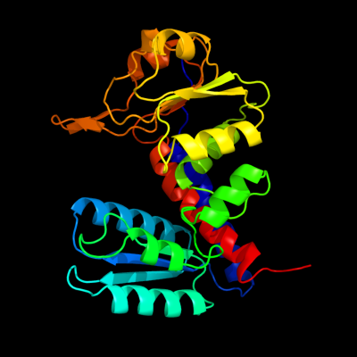
PDB header:oxidoreductase,hydrolase
Chain: A: PDB Molecule:protein (fold bifunctional protein);
PDBTitle: 5,10, methylene-tetrahydropholate2 dehydrogenase/cyclohydrolase from e coli.
|
| 2 | c4a5oB_
|
|
 |
100.0 |
67 |
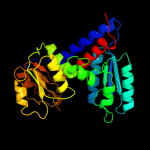
PDB header:oxidoreductase
Chain: B: PDB Molecule:bifunctional protein fold;
PDBTitle: crystal structure of pseudomonas aeruginosa n5, n10-2 methylenetetrahydrofolate dehydrogenase-cyclohydrolase (fold)
|
| 3 | c2c2xB_
|
|
 |
100.0 |
43 |
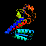
PDB header:oxidoreductase
Chain: B: PDB Molecule:methylenetetrahydrofolate dehydrogenase-
PDBTitle: three dimensional structure of bifunctional2 methylenetetrahydrofolate dehydrogenase-cyclohydrolase3 from mycobacterium tuberculosis
|
| 4 | c3l07B_
|
|
 |
100.0 |
53 |
PDB header:oxidoreductase,hydrolase
Chain: B: PDB Molecule:bifunctional protein fold;
PDBTitle: methylenetetrahydrofolate dehydrogenase/methenyltetrahydrofolate2 cyclohydrolase, putative bifunctional protein fold from francisella3 tularensis.
|
| 5 | c1a4iB_
|
|
 |
100.0 |
44 |
PDB header:oxidoreductase
Chain: B: PDB Molecule:methylenetetrahydrofolate dehydrogenase /
PDBTitle: human tetrahydrofolate dehydrogenase / cyclohydrolase
|
| 6 | c3p2oB_
|
|
 |
100.0 |
53 |
PDB header:oxidoreductase, hydrolase
Chain: B: PDB Molecule:bifunctional protein fold;
PDBTitle: crystal structure of fold bifunctional protein from campylobacter2 jejuni
|
| 7 | c3p2oA_
|
|
 |
100.0 |
53 |
PDB header:oxidoreductase, hydrolase
Chain: A: PDB Molecule:bifunctional protein fold;
PDBTitle: crystal structure of fold bifunctional protein from campylobacter2 jejuni
|
| 8 | c3nglA_
|
|
 |
100.0 |
35 |
PDB header:oxidoreductase, hydrolase
Chain: A: PDB Molecule:bifunctional protein fold;
PDBTitle: crystal structure of bifunctional 5,10-methylenetetrahydrofolate2 dehydrogenase / cyclohydrolase from thermoplasma acidophilum
|
| 9 | c4a26B_
|
|
 |
100.0 |
47 |
PDB header:oxidoreductase
Chain: B: PDB Molecule:putative c-1-tetrahydrofolate synthase, cytoplasmic;
PDBTitle: the crystal structure of leishmania major n5,n10-2 methylenetetrahydrofolate dehydrogenase/cyclohydrolase
|
| 10 | c1edzA_
|
|
 |
100.0 |
28 |
PDB header:oxidoreductase
Chain: A: PDB Molecule:5,10-methylenetetrahydrofolate dehydrogenase;
PDBTitle: structure of the nad-dependent 5,10-2 methylenetetrahydrofolate dehydrogenase from saccharomyces3 cerevisiae
|
| 11 | d1b0aa1
|
|
 |
100.0 |
100 |
Fold:NAD(P)-binding Rossmann-fold domains
Superfamily:NAD(P)-binding Rossmann-fold domains
Family:Aminoacid dehydrogenase-like, C-terminal domain |
| 12 | d1a4ia1
|
|
 |
100.0 |
47 |
Fold:NAD(P)-binding Rossmann-fold domains
Superfamily:NAD(P)-binding Rossmann-fold domains
Family:Aminoacid dehydrogenase-like, C-terminal domain |
| 13 | d1edza1
|
|
 |
100.0 |
31 |
Fold:NAD(P)-binding Rossmann-fold domains
Superfamily:NAD(P)-binding Rossmann-fold domains
Family:Aminoacid dehydrogenase-like, C-terminal domain |
| 14 | d1edza2
|
|
 |
100.0 |
22 |
Fold:Aminoacid dehydrogenase-like, N-terminal domain
Superfamily:Aminoacid dehydrogenase-like, N-terminal domain
Family:Tetrahydrofolate dehydrogenase/cyclohydrolase |
| 15 | d1b0aa2
|
|
 |
100.0 |
100 |
Fold:Aminoacid dehydrogenase-like, N-terminal domain
Superfamily:Aminoacid dehydrogenase-like, N-terminal domain
Family:Tetrahydrofolate dehydrogenase/cyclohydrolase |
| 16 | d1a4ia2
|
|
 |
100.0 |
40 |
Fold:Aminoacid dehydrogenase-like, N-terminal domain
Superfamily:Aminoacid dehydrogenase-like, N-terminal domain
Family:Tetrahydrofolate dehydrogenase/cyclohydrolase |
| 17 | c3d4oA_
|
|
 |
100.0 |
19 |
PDB header:oxidoreductase
Chain: A: PDB Molecule:dipicolinate synthase subunit a;
PDBTitle: crystal structure of dipicolinate synthase subunit a (np_243269.1)2 from bacillus halodurans at 2.10 a resolution
|
| 18 | c2rirA_
|
|
 |
100.0 |
17 |
PDB header:oxidoreductase
Chain: A: PDB Molecule:dipicolinate synthase, a chain;
PDBTitle: crystal structure of dipicolinate synthase, a chain, from bacillus2 subtilis
|
| 19 | c1v8bA_
|
|
 |
100.0 |
16 |
PDB header:hydrolase
Chain: A: PDB Molecule:adenosylhomocysteinase;
PDBTitle: crystal structure of a hydrolase
|
| 20 | c3oneA_
|
|
 |
99.6 |
20 |
PDB header:hydrolase/hydrolase substrate
Chain: A: PDB Molecule:adenosylhomocysteinase;
PDBTitle: crystal structure of lupinus luteus s-adenosyl-l-homocysteine2 hydrolase in complex with adenine
|
| 21 | d1li4a1 |
|
not modelled |
99.3 |
20 |
Fold:NAD(P)-binding Rossmann-fold domains
Superfamily:NAD(P)-binding Rossmann-fold domains
Family:Formate/glycerate dehydrogenases, NAD-domain |
| 22 | c3gvpB_ |
|
not modelled |
99.3 |
23 |
PDB header:hydrolase
Chain: B: PDB Molecule:adenosylhomocysteinase 3;
PDBTitle: human sahh-like domain of human adenosylhomocysteinase 3
|
| 23 | d1v8ba1 |
|
not modelled |
99.3 |
21 |
Fold:NAD(P)-binding Rossmann-fold domains
Superfamily:NAD(P)-binding Rossmann-fold domains
Family:Formate/glycerate dehydrogenases, NAD-domain |
| 24 | c1d4fD_ |
|
not modelled |
99.3 |
22 |
PDB header:hydrolase
Chain: D: PDB Molecule:s-adenosylhomocysteine hydrolase;
PDBTitle: crystal structure of recombinant rat-liver d244e mutant s-2 adenosylhomocysteine hydrolase
|
| 25 | c3d64A_ |
|
not modelled |
99.2 |
18 |
PDB header:hydrolase
Chain: A: PDB Molecule:adenosylhomocysteinase;
PDBTitle: crystal structure of s-adenosyl-l-homocysteine hydrolase from2 burkholderia pseudomallei
|
| 26 | c3n58D_ |
|
not modelled |
99.2 |
26 |
PDB header:hydrolase
Chain: D: PDB Molecule:adenosylhomocysteinase;
PDBTitle: crystal structure of s-adenosyl-l-homocysteine hydrolase from brucella2 melitensis in ternary complex with nad and adenosine, orthorhombic3 form
|
| 27 | c3dhyC_ |
|
not modelled |
99.1 |
20 |
PDB header:hydrolase
Chain: C: PDB Molecule:adenosylhomocysteinase;
PDBTitle: crystal structures of mycobacterium tuberculosis s-adenosyl-l-2 homocysteine hydrolase in ternary complex with substrate and3 inhibitors
|
| 28 | c2eklA_ |
|
not modelled |
98.9 |
20 |
PDB header:oxidoreductase
Chain: A: PDB Molecule:d-3-phosphoglycerate dehydrogenase;
PDBTitle: structure of st1218 protein from sulfolobus tokodaii
|
| 29 | c3pgjB_ |
|
not modelled |
98.9 |
20 |
PDB header:oxidoreductase
Chain: B: PDB Molecule:shikimate dehydrogenase;
PDBTitle: 2.49 angstrom resolution crystal structure of shikimate 5-2 dehydrogenase (aroe) from vibrio cholerae o1 biovar eltor str. n169613 in complex with shikimate
|
| 30 | c2j6iC_ |
|
not modelled |
98.9 |
20 |
PDB header:oxidoreductase
Chain: C: PDB Molecule:formate dehydrogenase;
PDBTitle: candida boidinii formate dehydrogenase (fdh) c-terminal2 mutant
|
| 31 | c3o8qB_ |
|
not modelled |
98.8 |
20 |
PDB header:oxidoreductase
Chain: B: PDB Molecule:shikimate 5-dehydrogenase i alpha;
PDBTitle: 1.45 angstrom resolution crystal structure of shikimate 5-2 dehydrogenase (aroe) from vibrio cholerae
|
| 32 | c1gpjA_ |
|
not modelled |
98.8 |
27 |
PDB header:reductase
Chain: A: PDB Molecule:glutamyl-trna reductase;
PDBTitle: glutamyl-trna reductase from methanopyrus kandleri
|
| 33 | c2nloA_ |
|
not modelled |
98.8 |
18 |
PDB header:oxidoreductase
Chain: A: PDB Molecule:shikimate dehydrogenase;
PDBTitle: crystal structure of the quinate dehydrogenase from corynebacterium2 glutamicum
|
| 34 | c2hk8B_ |
|
not modelled |
98.8 |
21 |
PDB header:oxidoreductase
Chain: B: PDB Molecule:shikimate dehydrogenase;
PDBTitle: crystal structure of shikimate dehydrogenase from aquifex2 aeolicus at 2.35 angstrom resolution
|
| 35 | c3n7uD_ |
|
not modelled |
98.8 |
18 |
PDB header:oxidoreductase
Chain: D: PDB Molecule:formate dehydrogenase;
PDBTitle: nad-dependent formate dehydrogenase from higher-plant arabidopsis2 thaliana in complex with nad and azide
|
| 36 | c2cukC_ |
|
not modelled |
98.8 |
22 |
PDB header:oxidoreductase
Chain: C: PDB Molecule:glycerate dehydrogenase/glyoxylate reductase;
PDBTitle: crystal structure of tt0316 protein from thermus thermophilus hb8
|
| 37 | c3fbtB_ |
|
not modelled |
98.8 |
19 |
PDB header:oxidoreductase, lyase
Chain: B: PDB Molecule:chorismate mutase and shikimate 5-dehydrogenase
PDBTitle: crystal structure of a chorismate mutase/shikimate 5-2 dehydrogenase fusion protein from clostridium3 acetobutylicum
|
| 38 | c2dbqA_ |
|
not modelled |
98.7 |
17 |
PDB header:oxidoreductase
Chain: A: PDB Molecule:glyoxylate reductase;
PDBTitle: crystal structure of glyoxylate reductase (ph0597) from pyrococcus2 horikoshii ot3, complexed with nadp (i41)
|
| 39 | c2eggA_ |
|
not modelled |
98.7 |
25 |
PDB header:oxidoreductase
Chain: A: PDB Molecule:shikimate 5-dehydrogenase;
PDBTitle: crystal structure of shikimate 5-dehydrogenase (aroe) from2 geobacillus kaustophilus
|
| 40 | c1gdhA_ |
|
not modelled |
98.7 |
13 |
PDB header:oxidoreductase(choh (d)-nad(p)+ (a))
Chain: A: PDB Molecule:d-glycerate dehydrogenase;
PDBTitle: crystal structure of a nad-dependent d-glycerate2 dehydrogenase at 2.4 angstroms resolution
|
| 41 | c3tozA_ |
|
not modelled |
98.7 |
18 |
PDB header:oxidoreductase
Chain: A: PDB Molecule:shikimate dehydrogenase;
PDBTitle: 2.2 angstrom crystal structure of shikimate 5-dehydrogenase from2 listeria monocytogenes in complex with nad.
|
| 42 | c3donA_ |
|
not modelled |
98.7 |
18 |
PDB header:oxidoreductase
Chain: A: PDB Molecule:shikimate dehydrogenase;
PDBTitle: crystal structure of shikimate dehydrogenase from staphylococcus2 epidermidis
|
| 43 | d1l7da1 |
|
not modelled |
98.7 |
21 |
Fold:NAD(P)-binding Rossmann-fold domains
Superfamily:NAD(P)-binding Rossmann-fold domains
Family:Formate/glycerate dehydrogenases, NAD-domain |
| 44 | c1p74B_ |
|
not modelled |
98.7 |
18 |
PDB header:oxidoreductase
Chain: B: PDB Molecule:shikimate 5-dehydrogenase;
PDBTitle: crystal structure of shikimate dehydrogenase (aroe) from2 haemophilus influenzae
|
| 45 | d1gpja2 |
|
not modelled |
98.7 |
23 |
Fold:NAD(P)-binding Rossmann-fold domains
Superfamily:NAD(P)-binding Rossmann-fold domains
Family:Aminoacid dehydrogenase-like, C-terminal domain |
| 46 | c3bazA_ |
|
not modelled |
98.7 |
18 |
PDB header:oxidoreductase
Chain: A: PDB Molecule:hydroxyphenylpyruvate reductase;
PDBTitle: structure of hydroxyphenylpyruvate reductase from coleus blumei in2 complex with nadp+
|
| 47 | c2pi1C_ |
|
not modelled |
98.6 |
22 |
PDB header:oxidoreductase
Chain: C: PDB Molecule:d-lactate dehydrogenase;
PDBTitle: crystal structure of d-lactate dehydrogenase from aquifex2 aeolicus complexed with nad and lactic acid
|
| 48 | c1pjcA_ |
|
not modelled |
98.6 |
31 |
PDB header:oxidoreductase
Chain: A: PDB Molecule:protein (l-alanine dehydrogenase);
PDBTitle: l-alanine dehydrogenase complexed with nad
|
| 49 | c3evtA_ |
|
not modelled |
98.6 |
13 |
PDB header:oxidoreductase
Chain: A: PDB Molecule:phosphoglycerate dehydrogenase;
PDBTitle: crystal structure of phosphoglycerate dehydrogenase from2 lactobacillus plantarum
|
| 50 | c3k5pA_ |
|
not modelled |
98.6 |
20 |
PDB header:oxidoreductase
Chain: A: PDB Molecule:d-3-phosphoglycerate dehydrogenase;
PDBTitle: crystal structure of amino acid-binding act: d-isomer specific 2-2 hydroxyacid dehydrogenase catalytic domain from brucella melitensis
|
| 51 | d1pjca1 |
|
not modelled |
98.6 |
26 |
Fold:NAD(P)-binding Rossmann-fold domains
Superfamily:NAD(P)-binding Rossmann-fold domains
Family:Formate/glycerate dehydrogenases, NAD-domain |
| 52 | c1wwkA_ |
|
not modelled |
98.6 |
22 |
PDB header:oxidoreductase
Chain: A: PDB Molecule:phosphoglycerate dehydrogenase;
PDBTitle: crystal structure of phosphoglycerate dehydrogenase from pyrococcus2 horikoshii ot3
|
| 53 | c1nytC_ |
|
not modelled |
98.6 |
20 |
PDB header:oxidoreductase
Chain: C: PDB Molecule:shikimate 5-dehydrogenase;
PDBTitle: shikimate dehydrogenase aroe complexed with nadp+
|
| 54 | c2o4cB_ |
|
not modelled |
98.6 |
20 |
PDB header:oxidoreductase
Chain: B: PDB Molecule:erythronate-4-phosphate dehydrogenase;
PDBTitle: crystal structure of d-erythronate-4-phosphate dehydrogenase complexed2 with nad
|
| 55 | d1ygya1 |
|
not modelled |
98.6 |
19 |
Fold:NAD(P)-binding Rossmann-fold domains
Superfamily:NAD(P)-binding Rossmann-fold domains
Family:Formate/glycerate dehydrogenases, NAD-domain |
| 56 | c2eezG_ |
|
not modelled |
98.6 |
34 |
PDB header:oxidoreductase
Chain: G: PDB Molecule:alanine dehydrogenase;
PDBTitle: crystal structure of alanine dehydrogenase from themus thermophilus
|
| 57 | c3oj0A_ |
|
not modelled |
98.6 |
16 |
PDB header:oxidoreductase
Chain: A: PDB Molecule:glutamyl-trna reductase;
PDBTitle: crystal structure of glutamyl-trna reductase from thermoplasma2 volcanium (nucleotide binding domain)
|
| 58 | c1xdwA_ |
|
not modelled |
98.6 |
24 |
PDB header:oxidoreductase
Chain: A: PDB Molecule:nad+-dependent (r)-2-hydroxyglutarate
PDBTitle: nad+-dependent (r)-2-hydroxyglutarate dehydrogenase from2 acidaminococcus fermentans
|
| 59 | c1qp8A_ |
|
not modelled |
98.6 |
19 |
PDB header:oxidoreductase
Chain: A: PDB Molecule:formate dehydrogenase;
PDBTitle: crystal structure of a putative formate dehydrogenase from2 pyrobaculum aerophilum
|
| 60 | c3oetF_ |
|
not modelled |
98.5 |
26 |
PDB header:oxidoreductase
Chain: F: PDB Molecule:erythronate-4-phosphate dehydrogenase;
PDBTitle: d-erythronate-4-phosphate dehydrogenase complexed with nad
|
| 61 | c2ev9B_ |
|
not modelled |
98.5 |
23 |
PDB header:oxidoreductase
Chain: B: PDB Molecule:shikimate 5-dehydrogenase;
PDBTitle: crystal structure of shikimate 5-dehydrogenase (aroe) from thermus2 thermophilus hb8 in complex with nadp(h) and shikimate
|
| 62 | d1mx3a1 |
|
not modelled |
98.5 |
16 |
Fold:NAD(P)-binding Rossmann-fold domains
Superfamily:NAD(P)-binding Rossmann-fold domains
Family:Formate/glycerate dehydrogenases, NAD-domain |
| 63 | c1dxyA_ |
|
not modelled |
98.5 |
19 |
PDB header:oxidoreductase
Chain: A: PDB Molecule:d-2-hydroxyisocaproate dehydrogenase;
PDBTitle: structure of d-2-hydroxyisocaproate dehydrogenase
|
| 64 | c3pwzA_ |
|
not modelled |
98.5 |
16 |
PDB header:oxidoreductase
Chain: A: PDB Molecule:shikimate dehydrogenase 3;
PDBTitle: crystal structure of an ael1 enzyme from pseudomonas putida
|
| 65 | d1j4aa1 |
|
not modelled |
98.5 |
18 |
Fold:NAD(P)-binding Rossmann-fold domains
Superfamily:NAD(P)-binding Rossmann-fold domains
Family:Formate/glycerate dehydrogenases, NAD-domain |
| 66 | c2g76A_ |
|
not modelled |
98.5 |
16 |
PDB header:oxidoreductase
Chain: A: PDB Molecule:d-3-phosphoglycerate dehydrogenase;
PDBTitle: crystal structure of human 3-phosphoglycerate dehydrogenase
|
| 67 | c1nvtA_ |
|
not modelled |
98.5 |
25 |
PDB header:oxidoreductase
Chain: A: PDB Molecule:shikimate 5'-dehydrogenase;
PDBTitle: crystal structure of shikimate dehydrogenase (aroe or2 mj1084) in complex with nadp+
|
| 68 | c3p2yA_ |
|
not modelled |
98.5 |
19 |
PDB header:oxidoreductase
Chain: A: PDB Molecule:alanine dehydrogenase/pyridine nucleotide transhydrogenase;
PDBTitle: crystal structure of alanine dehydrogenase/pyridine nucleotide2 transhydrogenase from mycobacterium smegmatis
|
| 69 | d1dxya1 |
|
not modelled |
98.5 |
19 |
Fold:NAD(P)-binding Rossmann-fold domains
Superfamily:NAD(P)-binding Rossmann-fold domains
Family:Formate/glycerate dehydrogenases, NAD-domain |
| 70 | d1vi2a1 |
|
not modelled |
98.5 |
19 |
Fold:NAD(P)-binding Rossmann-fold domains
Superfamily:NAD(P)-binding Rossmann-fold domains
Family:Aminoacid dehydrogenase-like, C-terminal domain |
| 71 | d1qp8a1 |
|
not modelled |
98.5 |
24 |
Fold:NAD(P)-binding Rossmann-fold domains
Superfamily:NAD(P)-binding Rossmann-fold domains
Family:Formate/glycerate dehydrogenases, NAD-domain |
| 72 | d1gdha1 |
|
not modelled |
98.5 |
10 |
Fold:NAD(P)-binding Rossmann-fold domains
Superfamily:NAD(P)-binding Rossmann-fold domains
Family:Formate/glycerate dehydrogenases, NAD-domain |
| 73 | c2gcgB_ |
|
not modelled |
98.5 |
16 |
PDB header:oxidoreductase
Chain: B: PDB Molecule:glyoxylate reductase/hydroxypyruvate reductase;
PDBTitle: ternary crystal structure of human glyoxylate2 reductase/hydroxypyruvate reductase
|
| 74 | c1l7eC_ |
|
not modelled |
98.5 |
15 |
PDB header:oxidoreductase
Chain: C: PDB Molecule:nicotinamide nucleotide transhydrogenase,
PDBTitle: crystal structure of r. rubrum transhydrogenase domain i2 with bound nadh
|
| 75 | c2bruB_ |
|
not modelled |
98.4 |
17 |
PDB header:oxidoreductase
Chain: B: PDB Molecule:nad(p) transhydrogenase subunit alpha;
PDBTitle: complex of the domain i and domain iii of escherichia coli2 transhydrogenase
|
| 76 | d2dlda1 |
|
not modelled |
98.4 |
18 |
Fold:NAD(P)-binding Rossmann-fold domains
Superfamily:NAD(P)-binding Rossmann-fold domains
Family:Formate/glycerate dehydrogenases, NAD-domain |
| 77 | d1nyta1 |
|
not modelled |
98.4 |
25 |
Fold:NAD(P)-binding Rossmann-fold domains
Superfamily:NAD(P)-binding Rossmann-fold domains
Family:Aminoacid dehydrogenase-like, C-terminal domain |
| 78 | d1nvta1 |
|
not modelled |
98.4 |
21 |
Fold:NAD(P)-binding Rossmann-fold domains
Superfamily:NAD(P)-binding Rossmann-fold domains
Family:Aminoacid dehydrogenase-like, C-terminal domain |
| 79 | d2naca1 |
|
not modelled |
98.4 |
16 |
Fold:NAD(P)-binding Rossmann-fold domains
Superfamily:NAD(P)-binding Rossmann-fold domains
Family:Formate/glycerate dehydrogenases, NAD-domain |
| 80 | c1j4aA_ |
|
not modelled |
98.4 |
19 |
PDB header:oxidoreductase
Chain: A: PDB Molecule:d-lactate dehydrogenase;
PDBTitle: insights into domain closure, substrate specificity and2 catalysis of d-lactate dehydrogenase from lactobacillus3 bulgaricus
|
| 81 | c3hg7A_ |
|
not modelled |
98.4 |
18 |
PDB header:oxidoreductase
Chain: A: PDB Molecule:d-isomer specific 2-hydroxyacid dehydrogenase family
PDBTitle: crystal structure of d-isomer specific 2-hydroxyacid dehydrogenase2 family protein from aeromonas salmonicida subsp. salmonicida a449
|
| 82 | c3gvxA_ |
|
not modelled |
98.4 |
16 |
PDB header:oxidoreductase
Chain: A: PDB Molecule:glycerate dehydrogenase related protein;
PDBTitle: crystal structure of glycerate dehydrogenase related2 protein from thermoplasma acidophilum
|
| 83 | c2nacA_ |
|
not modelled |
98.4 |
17 |
PDB header:oxidoreductase(aldehyde(d),nad+(a))
Chain: A: PDB Molecule:nad-dependent formate dehydrogenase;
PDBTitle: high resolution structures of holo and apo formate dehydrogenase
|
| 84 | c3gg9C_ |
|
not modelled |
98.4 |
19 |
PDB header:oxidoreductase
Chain: C: PDB Molecule:d-3-phosphoglycerate dehydrogenase oxidoreductase protein;
PDBTitle: crystal structure of putative d-3-phosphoglycerate dehydrogenase2 oxidoreductase from ralstonia solanacearum
|
| 85 | c1ybaC_ |
|
not modelled |
98.4 |
15 |
PDB header:oxidoreductase
Chain: C: PDB Molecule:d-3-phosphoglycerate dehydrogenase;
PDBTitle: the active form of phosphoglycerate dehydrogenase
|
| 86 | c1ygyA_ |
|
not modelled |
98.4 |
19 |
PDB header:oxidoreductase
Chain: A: PDB Molecule:d-3-phosphoglycerate dehydrogenase;
PDBTitle: crystal structure of d-3-phosphoglycerate dehydrogenase from2 mycobacterium tuberculosis
|
| 87 | c2omeA_ |
|
not modelled |
98.3 |
13 |
PDB header:oxidoreductase
Chain: A: PDB Molecule:c-terminal-binding protein 2;
PDBTitle: crystal structure of human ctbp2 dehydrogenase complexed with nad(h)
|
| 88 | c3kboB_ |
|
not modelled |
98.3 |
22 |
PDB header:oxidoreductase
Chain: B: PDB Molecule:glyoxylate/hydroxypyruvate reductase a;
PDBTitle: 2.14 angstrom crystal structure of putative oxidoreductase (ycdw) from2 salmonella typhimurium in complex with nadp
|
| 89 | c1vi2B_ |
|
not modelled |
98.3 |
19 |
PDB header:oxidoreductase
Chain: B: PDB Molecule:shikimate 5-dehydrogenase 2;
PDBTitle: crystal structure of shikimate-5-dehydrogenase with nad
|
| 90 | c1npyA_ |
|
not modelled |
98.3 |
20 |
PDB header:structural genomics, unknown function
Chain: A: PDB Molecule:hypothetical shikimate 5-dehydrogenase-like
PDBTitle: structure of shikimate 5-dehydrogenase-like protein hi0607
|
| 91 | c2w2kB_ |
|
not modelled |
98.3 |
18 |
PDB header:oxidoreductase
Chain: B: PDB Molecule:d-mandelate dehydrogenase;
PDBTitle: crystal structure of the apo forms of rhodotorula graminis2 d-mandelate dehydrogenase at 1.8a.
|
| 92 | d1sc6a1 |
|
not modelled |
98.2 |
16 |
Fold:NAD(P)-binding Rossmann-fold domains
Superfamily:NAD(P)-binding Rossmann-fold domains
Family:Formate/glycerate dehydrogenases, NAD-domain |
| 93 | c2vhyB_ |
|
not modelled |
98.1 |
33 |
PDB header:oxidoreductase
Chain: B: PDB Molecule:alanine dehydrogenase;
PDBTitle: crystal structure of apo l-alanine dehydrogenase from2 mycobacterium tuberculosis
|
| 94 | c2d0iC_ |
|
not modelled |
98.1 |
25 |
PDB header:oxidoreductase
Chain: C: PDB Molecule:dehydrogenase;
PDBTitle: crystal structure ph0520 protein from pyrococcus horikoshii ot3
|
| 95 | d1p77a1 |
|
not modelled |
98.1 |
21 |
Fold:NAD(P)-binding Rossmann-fold domains
Superfamily:NAD(P)-binding Rossmann-fold domains
Family:Aminoacid dehydrogenase-like, C-terminal domain |
| 96 | c3u62A_ |
|
not modelled |
98.1 |
23 |
PDB header:oxidoreductase
Chain: A: PDB Molecule:shikimate dehydrogenase;
PDBTitle: crystal structure of shikimate dehydrogenase from thermotoga maritima
|
| 97 | c2ef0A_ |
|
not modelled |
98.0 |
17 |
PDB header:transferase
Chain: A: PDB Molecule:ornithine carbamoyltransferase;
PDBTitle: crystal structure of ornithine carbamoyltransferase from thermus2 thermophilus
|
| 98 | c2p2gD_ |
|
not modelled |
98.0 |
17 |
PDB header:transferase
Chain: D: PDB Molecule:ornithine carbamoyltransferase;
PDBTitle: crystal structure of ornithine carbamoyltransferase from mycobacterium2 tuberculosis (rv1656): orthorhombic form
|
| 99 | c1luaA_ |
|
not modelled |
98.0 |
22 |
PDB header:oxidoreductase
Chain: A: PDB Molecule:methylene tetrahydromethanopterin dehydrogenase;
PDBTitle: structure of methylene-tetrahydromethanopterin dehydrogenase from2 methylobacterium extorquens am1 complexed with nadp
|
| 100 | d1np3a2 |
|
not modelled |
98.0 |
24 |
Fold:NAD(P)-binding Rossmann-fold domains
Superfamily:NAD(P)-binding Rossmann-fold domains
Family:6-phosphogluconate dehydrogenase-like, N-terminal domain |
| 101 | d1npya1 |
|
not modelled |
97.9 |
22 |
Fold:NAD(P)-binding Rossmann-fold domains
Superfamily:NAD(P)-binding Rossmann-fold domains
Family:Aminoacid dehydrogenase-like, C-terminal domain |
| 102 | c3c24A_ |
|
not modelled |
97.9 |
17 |
PDB header:oxidoreductase
Chain: A: PDB Molecule:putative oxidoreductase;
PDBTitle: crystal structure of a putative oxidoreductase (yp_511008.1) from2 jannaschia sp. ccs1 at 1.62 a resolution
|
| 103 | c1vlvA_ |
|
not modelled |
97.9 |
16 |
PDB header:transferase
Chain: A: PDB Molecule:ornithine carbamoyltransferase;
PDBTitle: crystal structure of ornithine carbamoyltransferase (tm1097) from2 thermotoga maritima at 2.25 a resolution
|
| 104 | c2f00A_ |
|
not modelled |
97.8 |
14 |
PDB header:ligase
Chain: A: PDB Molecule:udp-n-acetylmuramate--l-alanine ligase;
PDBTitle: escherichia coli murc
|
| 105 | d1luaa1 |
|
not modelled |
97.8 |
26 |
Fold:NAD(P)-binding Rossmann-fold domains
Superfamily:NAD(P)-binding Rossmann-fold domains
Family:Aminoacid dehydrogenase-like, C-terminal domain |
| 106 | c1fvoB_ |
|
not modelled |
97.7 |
16 |
PDB header:transferase
Chain: B: PDB Molecule:ornithine transcarbamylase;
PDBTitle: crystal structure of human ornithine transcarbamylase complexed with2 carbamoyl phosphate
|
| 107 | c1np3B_ |
|
not modelled |
97.7 |
21 |
PDB header:oxidoreductase
Chain: B: PDB Molecule:ketol-acid reductoisomerase;
PDBTitle: crystal structure of class i acetohydroxy acid isomeroreductase from2 pseudomonas aeruginosa
|
| 108 | c2q3eH_ |
|
not modelled |
97.7 |
17 |
PDB header:oxidoreductase
Chain: H: PDB Molecule:udp-glucose 6-dehydrogenase;
PDBTitle: structure of human udp-glucose dehydrogenase complexed with nadh and2 udp-glucose
|
| 109 | c3gd5D_ |
|
not modelled |
97.7 |
14 |
PDB header:transferase
Chain: D: PDB Molecule:ornithine carbamoyltransferase;
PDBTitle: crystal structure of ornithine carbamoyltransferase from gloeobacter2 violaceus
|
| 110 | c2y0dB_ |
|
not modelled |
97.6 |
23 |
PDB header:oxidoreductase
Chain: B: PDB Molecule:udp-glucose dehydrogenase;
PDBTitle: bcec mutation y10k
|
| 111 | c3sdsA_ |
|
not modelled |
97.6 |
13 |
PDB header:transferase
Chain: A: PDB Molecule:ornithine carbamoyltransferase, mitochondrial;
PDBTitle: crystal structure of a mitochondrial ornithine carbamoyltransferase2 from coccidioides immitis
|
| 112 | c3cumA_ |
|
not modelled |
97.5 |
26 |
PDB header:oxidoreductase
Chain: A: PDB Molecule:probable 3-hydroxyisobutyrate dehydrogenase;
PDBTitle: crystal structure of a possible 3-hydroxyisobutyrate dehydrogenase2 from pseudomonas aeruginosa pao1
|
| 113 | c1ortD_ |
|
not modelled |
97.5 |
13 |
PDB header:transferase
Chain: D: PDB Molecule:ornithine transcarbamoylase;
PDBTitle: ornithine transcarbamoylase from pseudomonas aeruginosa
|
| 114 | c2otcA_ |
|
not modelled |
97.5 |
17 |
PDB header:transferase
Chain: A: PDB Molecule:ornithine carbamoyltransferase;
PDBTitle: ornithine transcarbamoylase complexed with n-2 (phosphonacetyl)-l-ornithine
|
| 115 | c1a1sA_ |
|
not modelled |
97.5 |
14 |
PDB header:transcarbamylase
Chain: A: PDB Molecule:ornithine carbamoyltransferase;
PDBTitle: ornithine carbamoyltransferase from pyrococcus furiosus
|
| 116 | c2pv7B_ |
|
not modelled |
97.5 |
19 |
PDB header:isomerase, oxidoreductase
Chain: B: PDB Molecule:t-protein [includes: chorismate mutase (ec 5.4.99.5) (cm)
PDBTitle: crystal structure of chorismate mutase / prephenate dehydrogenase2 (tyra) (1574749) from haemophilus influenzae rd at 2.00 a resolution
|
| 117 | c3gg2B_ |
|
not modelled |
97.5 |
19 |
PDB header:oxidoreductase
Chain: B: PDB Molecule:sugar dehydrogenase, udp-glucose/gdp-mannose
PDBTitle: crystal structure of udp-glucose 6-dehydrogenase from2 porphyromonas gingivalis bound to product udp-glucuronate
|
| 118 | c3dfzB_ |
|
not modelled |
97.5 |
15 |
PDB header:oxidoreductase
Chain: B: PDB Molecule:precorrin-2 dehydrogenase;
PDBTitle: sirc, precorrin-2 dehydrogenase
|
| 119 | c3g79A_ |
|
not modelled |
97.4 |
31 |
PDB header:oxidoreductase
Chain: A: PDB Molecule:ndp-n-acetyl-d-galactosaminuronic acid dehydrogenase;
PDBTitle: crystal structure of ndp-n-acetyl-d-galactosaminuronic acid2 dehydrogenase from methanosarcina mazei go1
|
| 120 | c3ojlA_ |
|
not modelled |
97.4 |
21 |
PDB header:oxidoreductase
Chain: A: PDB Molecule:cap5o;
PDBTitle: native structure of the udp-n-acetyl-mannosamine dehydrogenase cap5o2 from staphylococcus aureus
|