Phyre Home Retrieve Phyre Job Id
Emaill.a.kelley@imperial.ac.uk
DescriptionC0Z3X9
DateThu Jan 5 10:55:44 GMT 2012
Unique Job IDbd3fcbc4bee13106

Summary 

Top model
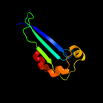
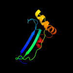
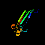
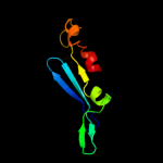
Image coloured by rainbow N → C terminus
Model (left) based on template d2qxfa1
Top template information
Fold:Ribonuclease H-like motif
Superfamily:Ribonuclease H-like
Family:DnaQ-like 3'-5' exonuclease
Confidence and coverage
Confidence:100.0% Coverage: 92%
82 residues ( 92% of your sequence) have been modelled with 100.0% confidence by the single highest scoring template.
3D viewing
Interactive 3D view in Jmol

Sequence analysis 

Secondary structure and disorder prediction 

   1........10.........20.........30.........40.........50.........60
Sequence  MTDTDKQPTFLFHDYETFGTHPALDRPAQFAAIRTDSEFNVIGEPEVFYCKPADDYLPQP
Secondary structure 


SS confidence 


Disorder  ??????


Disorder confidence 


   .........70.........80.........
Sequence  GAVLITGITPQEARAKGENEAAFAARIHS
Secondary structure 


SS confidence 


Disorder 


??


??
Disorder confidence 


Confidence Key
High(9)                    Low (0)
?Disordered
Alpha helix
Beta strand

Domain analysis 

Hover over an aligned region to see model and summary info

Please note, only up to the top 20 hits are modelled to reduce computer load

RankAligned region

PDB 2qxf chain A domain 1

3D model

Region: 7 - 88
Aligned: 82
Modelled: 82
Confidence: 100.0%
Identity: 99%
Fold: Ribonuclease H-like motif
Superfamily: Ribonuclease H-like
Family: DnaQ-like 3'-5' exonuclease

Phyre2

PDB 1y97 chain A domain 1

3D model

Region: 3 - 87
Aligned: 85
Modelled: 85
Confidence: 99.8%
Identity: 16%
Fold: Ribonuclease H-like motif
Superfamily: Ribonuclease H-like
Family: DnaQ-like 3'-5' exonuclease

Phyre2

PDB 2f96 chain A domain 1

3D model

Region: 5 - 87
Aligned: 83
Modelled: 83
Confidence: 99.8%
Identity: 24%
Fold: Ribonuclease H-like motif
Superfamily: Ribonuclease H-like
Family: DnaQ-like 3'-5' exonuclease

Phyre2

PDB 3u6f chain A

3D model

Region: 3 - 87
Aligned: 85
Modelled: 84
Confidence: 99.8%
Identity: 22%
PDB header:hydrolase/dna
Chain: A: PDB Molecule:three prime repair exonuclease 1;
PDBTitle: mouse trex1 d200n mutant

Phyre2

PDB 3tr8 chain A

3D model

Region: 5 - 87
Aligned: 82
Modelled: 83
Confidence: 99.8%
Identity: 18%
PDB header:hydrolase
Chain: A: PDB Molecule:oligoribonuclease;
PDBTitle: structure of an oligoribonuclease (orn) from coxiella burnetii

Phyre2

PDB 1j9a chain A

3D model

Region: 3 - 88
Aligned: 85
Modelled: 85
Confidence: 99.8%
Identity: 19%
Fold: Ribonuclease H-like motif
Superfamily: Ribonuclease H-like
Family: DnaQ-like 3'-5' exonuclease

Phyre2

PDB 3b6o chain A domain 1

3D model

Region: 6 - 87
Aligned: 82
Modelled: 82
Confidence: 99.8%
Identity: 22%
Fold: Ribonuclease H-like motif
Superfamily: Ribonuclease H-like
Family: DnaQ-like 3'-5' exonuclease

Phyre2

PDB 3cm6 chain A

3D model

Region: 9 - 87
Aligned: 78
Modelled: 79
Confidence: 99.8%
Identity: 24%
PDB header:hydrolase, apoptosis
Chain: A: PDB Molecule:cell death-related nuclease 4;
PDBTitle: crystal structure of cell-death related nuclease 4 (crn-4)2 bound with er

Phyre2

PDB 1w0h chain A

3D model

Region: 9 - 88
Aligned: 79
Modelled: 80
Confidence: 99.8%
Identity: 15%
Fold: Ribonuclease H-like motif
Superfamily: Ribonuclease H-like
Family: DnaQ-like 3'-5' exonuclease

Phyre2

PDB 2xri chain A

3D model

Region: 3 - 88
Aligned: 85
Modelled: 86
Confidence: 99.8%
Identity: 15%
PDB header:hydrolase
Chain: A: PDB Molecule:eri1 exoribonuclease 3;
PDBTitle: crystal structure of human eri1 exoribonuclease 3

Phyre2

PDB 2gbz chain A

3D model

Region: 7 - 88
Aligned: 81
Modelled: 82
Confidence: 99.8%
Identity: 17%
PDB header:hydrolase
Chain: A: PDB Molecule:oligoribonuclease;
PDBTitle: the crystal structure of xc847 from xanthomonas campestris: a 3-52 oligoribonuclease of dnaq fold family with a novel opposingly-shifted3 helix

Phyre2

PDB 2igi chain A domain 1

3D model

Region: 4 - 89
Aligned: 85
Modelled: 86
Confidence: 99.8%
Identity: 15%
Fold: Ribonuclease H-like motif
Superfamily: Ribonuclease H-like
Family: DnaQ-like 3'-5' exonuclease

Phyre2

PDB 1zbh chain A

3D model

Region: 9 - 88
Aligned: 79
Modelled: 80
Confidence: 99.7%
Identity: 13%
PDB header:hydrolase/rna
Chain: A: PDB Molecule:3'-5' exonuclease eri1;
PDBTitle: 3'-end specific recognition of histone mrna stem-loop by 3'-2 exonuclease

Phyre2

PDB 1zbu chain B

3D model

Region: 9 - 87
Aligned: 78
Modelled: 79
Confidence: 99.7%
Identity: 13%
PDB header:hydrolase
Chain: B: PDB Molecule:3'-5' exonuclease eri1;
PDBTitle: crystal structure of full-length 3'-exonuclease

Phyre2

PDB 2gui chain A domain 1

3D model

Region: 9 - 87
Aligned: 75
Modelled: 79
Confidence: 99.7%
Identity: 20%
Fold: Ribonuclease H-like motif
Superfamily: Ribonuclease H-like
Family: DnaQ-like 3'-5' exonuclease

Phyre2

PDB 2p1j chain B

3D model

Region: 9 - 87
Aligned: 74
Modelled: 79
Confidence: 99.7%
Identity: 23%
PDB header:transferase
Chain: B: PDB Molecule:dna polymerase iii polc-type;
PDBTitle: crystal structure of a polc-type dna polymerase iii2 exonuclease domain from thermotoga maritima

Phyre2

PDB 1wlj chain A

3D model

Region: 9 - 88
Aligned: 72
Modelled: 80
Confidence: 99.5%
Identity: 17%
Fold: Ribonuclease H-like motif
Superfamily: Ribonuclease H-like
Family: DnaQ-like 3'-5' exonuclease

Phyre2

PDB 2is3 chain B

3D model

Region: 5 - 75
Aligned: 52
Modelled: 71
Confidence: 98.3%
Identity: 13%
PDB header:hydrolase
Chain: B: PDB Molecule:ribonuclease t;
PDBTitle: crystal structure of escherichia coli rnase t

Phyre2

PDB 1uoc chain A

3D model

Region: 6 - 89
Aligned: 83
Modelled: 84
Confidence: 98.1%
Identity: 18%
Fold: Ribonuclease H-like motif
Superfamily: Ribonuclease H-like
Family: CAF1-like ribonuclease

Phyre2

PDB 1x9m chain A domain 1

3D model

Region: 10 - 38
Aligned: 29
Modelled: 29
Confidence: 97.4%
Identity: 7%
Fold: Ribonuclease H-like motif
Superfamily: Ribonuclease H-like
Family: DnaQ-like 3'-5' exonuclease

Phyre2
1

d2qxfa1
2

d1y97a1
3

d2f96a1
4

c3u6fA_
5

c3tr8A_
6

d1j9aa_
7

d3b6oa1
8

c3cm6A_
9

d1w0ha_
10

c2xriA_
11

c2gbzA_
12

d2igia1
13

c1zbhA_
14

c1zbuB_
15

d2guia1
16

c2p1jB_
17

d1wlja_
18

c2is3B_
19

d1uoca_
20

d1x9ma1
21


22


23


24


25


26


27


28


29


30


31


32


33


34


35


36


37


38


39


40


41


42


43


44


45


46


47


48


49


50


51


52


53


54


55


56


57


58


Detailed template information 

#
Template Alignment Coverage3D Model Confidence
% i.d. Template Information
1d2qxfa1


100.0 99 Fold:Ribonuclease H-like motif
Superfamily:Ribonuclease H-like
Family:DnaQ-like 3'-5' exonuclease
2d1y97a1


99.8 16 Fold:Ribonuclease H-like motif
Superfamily:Ribonuclease H-like
Family:DnaQ-like 3'-5' exonuclease
3d2f96a1


99.8 24 Fold:Ribonuclease H-like motif
Superfamily:Ribonuclease H-like
Family:DnaQ-like 3'-5' exonuclease
4c3u6fA_


99.8 22 PDB header:hydrolase/dna
Chain: A: PDB Molecule:three prime repair exonuclease 1;
PDBTitle: mouse trex1 d200n mutant
5c3tr8A_


99.8 18 PDB header:hydrolase
Chain: A: PDB Molecule:oligoribonuclease;
PDBTitle: structure of an oligoribonuclease (orn) from coxiella burnetii
6d1j9aa_


99.8 19 Fold:Ribonuclease H-like motif
Superfamily:Ribonuclease H-like
Family:DnaQ-like 3'-5' exonuclease
7d3b6oa1


99.8 22 Fold:Ribonuclease H-like motif
Superfamily:Ribonuclease H-like
Family:DnaQ-like 3'-5' exonuclease
8c3cm6A_


99.8 24 PDB header:hydrolase, apoptosis
Chain: A: PDB Molecule:cell death-related nuclease 4;
PDBTitle: crystal structure of cell-death related nuclease 4 (crn-4)2 bound with er
9d1w0ha_


99.8 15 Fold:Ribonuclease H-like motif
Superfamily:Ribonuclease H-like
Family:DnaQ-like 3'-5' exonuclease
10c2xriA_


99.8 15 PDB header:hydrolase
Chain: A: PDB Molecule:eri1 exoribonuclease 3;
PDBTitle: crystal structure of human eri1 exoribonuclease 3
11c2gbzA_


99.8 17 PDB header:hydrolase
Chain: A: PDB Molecule:oligoribonuclease;
PDBTitle: the crystal structure of xc847 from xanthomonas campestris: a 3-52 oligoribonuclease of dnaq fold family with a novel opposingly-shifted3 helix
12d2igia1


99.8 15 Fold:Ribonuclease H-like motif
Superfamily:Ribonuclease H-like
Family:DnaQ-like 3'-5' exonuclease
13c1zbhA_


99.7 13 PDB header:hydrolase/rna
Chain: A: PDB Molecule:3'-5' exonuclease eri1;
PDBTitle: 3'-end specific recognition of histone mrna stem-loop by 3'-2 exonuclease
14c1zbuB_


99.7 13 PDB header:hydrolase
Chain: B: PDB Molecule:3'-5' exonuclease eri1;
PDBTitle: crystal structure of full-length 3'-exonuclease
15d2guia1


99.7 20 Fold:Ribonuclease H-like motif
Superfamily:Ribonuclease H-like
Family:DnaQ-like 3'-5' exonuclease
16c2p1jB_


99.7 23 PDB header:transferase
Chain: B: PDB Molecule:dna polymerase iii polc-type;
PDBTitle: crystal structure of a polc-type dna polymerase iii2 exonuclease domain from thermotoga maritima
17d1wlja_


99.5 17 Fold:Ribonuclease H-like motif
Superfamily:Ribonuclease H-like
Family:DnaQ-like 3'-5' exonuclease
18c2is3B_


98.3 13 PDB header:hydrolase
Chain: B: PDB Molecule:ribonuclease t;
PDBTitle: crystal structure of escherichia coli rnase t
19d1uoca_


98.1 18 Fold:Ribonuclease H-like motif
Superfamily:Ribonuclease H-like
Family:CAF1-like ribonuclease
20d1x9ma1


97.4 7 Fold:Ribonuclease H-like motif
Superfamily:Ribonuclease H-like
Family:DnaQ-like 3'-5' exonuclease
21c2p51A_


not modelled 96.7 23 PDB header:hydrolase, gene regulation
Chain: A: PDB Molecule:spcc18.06c protein;
PDBTitle: crystal structure of the s. pombe pop2p deadenylation2 subunit
22d1kfsa1


not modelled 96.4 9 Fold:Ribonuclease H-like motif
Superfamily:Ribonuclease H-like
Family:DnaQ-like 3'-5' exonuclease
23d2d5ra1


not modelled 96.1 16 Fold:Ribonuclease H-like motif
Superfamily:Ribonuclease H-like
Family:CAF1-like ribonuclease
24d2hhva1


not modelled 96.1 9 Fold:Ribonuclease H-like motif
Superfamily:Ribonuclease H-like
Family:DnaQ-like 3'-5' exonuclease
25c2kzzA_


not modelled 95.7 19 PDB header:transferase/dna
Chain: A: PDB Molecule:protein (dna polymerase i);
PDBTitle: klenow fragment with normal substrate and zinc only
26c1njzA_


not modelled 93.8 8 PDB header:transferase/dna
Chain: A: PDB Molecule:dna polymerase i;
PDBTitle: cytosine-thymine mismatch at the polymerase active site
27c2a1sC_


not modelled 93.5 25 PDB header:hydrolase
Chain: C: PDB Molecule:poly(a)-specific ribonuclease parn;
PDBTitle: crystal structure of native parn nuclease domain
28c1tk0A_


not modelled 92.8 7 PDB header:transferase/electron transport/dna
Chain: A: PDB Molecule:dna polymerase;
PDBTitle: t7 dna polymerase ternary complex with 8 oxo guanosine and2 ddctp at the insertion site
29c3d45B_


not modelled 89.5 19 PDB header:hydrolase
Chain: B: PDB Molecule:poly(a)-specific ribonuclease parn;
PDBTitle: crystal structure of mouse parn in complex with m7gpppg
30d1wn7a1


not modelled 89.2 22 Fold:Ribonuclease H-like motif
Superfamily:Ribonuclease H-like
Family:DnaQ-like 3'-5' exonuclease
31d1d5aa1


not modelled 83.9 15 Fold:Ribonuclease H-like motif
Superfamily:Ribonuclease H-like
Family:DnaQ-like 3'-5' exonuclease
32d1tgoa1


not modelled 81.0 24 Fold:Ribonuclease H-like motif
Superfamily:Ribonuclease H-like
Family:DnaQ-like 3'-5' exonuclease
33d1qhta1


not modelled 80.0 16 Fold:Ribonuclease H-like motif
Superfamily:Ribonuclease H-like
Family:DnaQ-like 3'-5' exonuclease
34c2gv9B_


not modelled 74.5 12 PDB header:transferase
Chain: B: PDB Molecule:dna polymerase;
PDBTitle: crystal structure of the herpes simplex virus type 1 dna polymerase
35d1yt3a3


not modelled 65.2 11 Fold:Ribonuclease H-like motif
Superfamily:Ribonuclease H-like
Family:DnaQ-like 3'-5' exonuclease
36c3iayA_


not modelled 64.9 18 PDB header:transferase/dna
Chain: A: PDB Molecule:dna polymerase delta catalytic subunit;
PDBTitle: ternary complex of dna polymerase delta
37c2vwkA_


not modelled 61.4 22 PDB header:dna replication
Chain: A: PDB Molecule:dna polymerase;
PDBTitle: uracil recognition in archaeal dna polymerases captured by2 x-ray crystallography. v93q polymerase variant
38c4ktqA_


not modelled 58.8 17 PDB header:transferase/dna
Chain: A: PDB Molecule:protein (large fragment of dna polymerase i);
PDBTitle: binary complex of the large fragment of dna polymerase i2 from t. aquaticus bound to a primer/template dna
39c2e6mA_


not modelled 53.9 12 PDB header:hydrolase
Chain: A: PDB Molecule:werner syndrome atp-dependent helicase homolog;
PDBTitle: structure of mouse werner exonuclease domain
40c1yt3A_


not modelled 51.0 15 PDB header:hydrolase,translation
Chain: A: PDB Molecule:ribonuclease d;
PDBTitle: crystal structure of escherichia coli rnase d, an2 exoribonuclease involved in structured rna processing
41c1d5aA_


not modelled 45.4 20 PDB header:transferase
Chain: A: PDB Molecule:protein (dna polymerase);
PDBTitle: crystal structure of an archaebacterial dna polymerase2 d.tok. deposition of second native structure at 2.43 angstrom
42d1ih7a1


not modelled 27.7 20 Fold:Ribonuclease H-like motif
Superfamily:Ribonuclease H-like
Family:DnaQ-like 3'-5' exonuclease
43c2w0iA_


not modelled 17.6 10 PDB header:transferase
Chain: A: PDB Molecule:twinfilin-2;
PDBTitle: structure of c-terminal actin depolymerizing factor2 homology (adf-h) domain of human twinfilin-2
44c2kd5A_


not modelled 13.2 13 PDB header:hormone
Chain: A: PDB Molecule:actin severing and dynamics regulatory protein;
PDBTitle: solution structure of adf/cofilin (ldcof) from leishmania donovani
45d1ak7a_


not modelled 11.0 10 Fold:Gelsolin-like
Superfamily:Actin depolymerizing proteins
Family:Cofilin-like
46c3hhjA_


not modelled 10.7 11 PDB header:hydrolase
Chain: A: PDB Molecule:mutator mutt protein;
PDBTitle: crystal structure of mutator mutt from bartonella henselae
47c3cymA_


not modelled 10.6 22 PDB header:structural genomics, unknown function
Chain: A: PDB Molecule:uncharacterized protein bad_0989;
PDBTitle: crystal structure of protein bad_0989 from bifidobacterium2 adolescentis
48c2d3wB_


not modelled 10.2 18 PDB header:biosynthetic protein
Chain: B: PDB Molecule:probable atp-dependent transporter sufc;
PDBTitle: crystal structure of escherichia coli sufc, an atpase2 compenent of the suf iron-sulfur cluster assembly machinery
49d2dk8a1


not modelled 9.6 22 Fold:DNA/RNA-binding 3-helical bundle
Superfamily:"Winged helix" DNA-binding domain
Family:RPO3F domain-like
50c2xfaA_


not modelled 9.3 10 PDB header:protein binding
Chain: A: PDB Molecule:actin depolymerization factor 2;
PDBTitle: crystal structure of plasmodium berghei actin2 depolymerization factor 2
51d2hbka2


not modelled 8.5 15 Fold:Ribonuclease H-like motif
Superfamily:Ribonuclease H-like
Family:DnaQ-like 3'-5' exonuclease
52c2i2qA_


not modelled 8.4 10 PDB header:actin-binding protein
Chain: A: PDB Molecule:cofilin;
PDBTitle: fission yeast cofilin
53d1q8ga_


not modelled 8.1 10 Fold:Gelsolin-like
Superfamily:Actin depolymerizing proteins
Family:Cofilin-like
54d1q8ia1


not modelled 6.5 14 Fold:Ribonuclease H-like motif
Superfamily:Ribonuclease H-like
Family:DnaQ-like 3'-5' exonuclease
55c3iydB_


not modelled 6.5 24 PDB header:transcription/dna
Chain: B: PDB Molecule:dna-directed rna polymerase subunit alpha;
PDBTitle: three-dimensional em structure of an intact activator-dependent2 transcription initiation complex
56d1cnua_


not modelled 5.3 10 Fold:Gelsolin-like
Superfamily:Actin depolymerizing proteins
Family:Cofilin-like
57d1s5ja1


not modelled 5.3 12 Fold:Ribonuclease H-like motif
Superfamily:Ribonuclease H-like
Family:DnaQ-like 3'-5' exonuclease
58c1s5jA_


not modelled 5.2 20 PDB header:transferase
Chain: A: PDB Molecule:dna polymerase i;
PDBTitle: insight in dna replication: the crystal structure of dna2 polymerase b1 from the archaeon sulfolobus solfataricus

Binding site prediction 

Due to computational demand, binding site predictions are not run for batch jobs

If you want to predict binding sites, please manually submit your model of choice to 3DLigandSite


Phyre is for academic use only

Please cite: Protein structure prediction on the web: a case study using the Phyre server
Kelley LA and Sternberg MJE. Nature Protocols 4, 363 - 371 (2009) [pdf] [Import into BibTeX]
 
If you use the binding site predictions from 3DLigandSite, please also cite:
3DLigandSite: predicting ligand-binding sites using similar structures.
Wass MN, Kelley LA and Sternberg MJ Nucleic Acids Research 38, W469-73 (2010) [PubMed]
 
© Structural Bioinformatics Group
Imperial College London
Lawrence Kelley, Benjamin Jefferys
Disclaimer
Terms and Conditions
Component software
Template detection: HHpred 1.51
Secondary structure prediction: Psi-pred 2.5
Disorder prediction: Disopred 2.4
Transmembrane prediction: Memsat_SVM
Multi-template modelling and ab initio: Poing 1.0