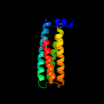
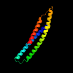
1 c2zv4O_
96.7
11
PDB header: structural proteinChain: O: PDB Molecule: major vault protein;PDBTitle: the structure of rat liver vault at 3.5 angstrom resolution
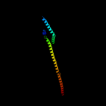
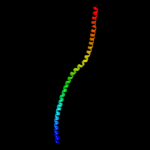
2 c2dfsA_
96.4
6
PDB header: contractile protein/transport proteinChain: A: PDB Molecule: myosin-5a;PDBTitle: 3-d structure of myosin-v inhibited state
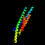
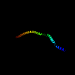
3 c1y4cA_
94.7
11
PDB header: de novo proteinChain: A: PDB Molecule: maltose binding protein fused with designedPDBTitle: designed helical protein fusion mbp
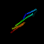
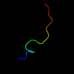
4 c3ojaB_
92.5
10
PDB header: protein bindingChain: B: PDB Molecule: anopheles plasmodium-responsive leucine-rich repeat proteinPDBTitle: crystal structure of lrim1/apl1c complex
5 c1bf5A_
89.6
6
PDB header: gene regulation/dnaChain: A: PDB Molecule: signal transducer and activator of transcriptionPDBTitle: tyrosine phosphorylated stat-1/dna complex
6 c1g8xB_
88.0
8
PDB header: structural proteinChain: B: PDB Molecule: myosin ii heavy chain fused to alpha-actinin 3;PDBTitle: structure of a genetically engineered molecular motor
7 c3ojaA_
87.7
8
PDB header: protein bindingChain: A: PDB Molecule: leucine-rich immune molecule 1;PDBTitle: crystal structure of lrim1/apl1c complex
8 c1ei3E_
78.1
7
PDB header: PDB COMPND: 9 c1f5nA_
77.4
9
PDB header: signaling proteinChain: A: PDB Molecule: interferon-induced guanylate-binding protein 1;PDBTitle: human guanylate binding protein-1 in complex with the gtp2 analogue, gmppnp.
10 d1wp1a_
65.0
15
Fold: Outer membrane efflux proteins (OEP)Superfamily: Outer membrane efflux proteins (OEP)Family: Outer membrane efflux proteins (OEP)11 c3ghgK_
63.3
6
PDB header: blood clottingChain: K: PDB Molecule: fibrinogen beta chain;PDBTitle: crystal structure of human fibrinogen
12 c1bg1A_
60.4
7
PDB header: transcription/dnaChain: A: PDB Molecule: protein (transcription factor stat3b);PDBTitle: transcription factor stat3b/dna complex
13 c1deqO_
57.6
8
PDB header: PDB COMPND: 14 c1jchC_
57.0
11
PDB header: ribosome inhibitor, hydrolaseChain: C: PDB Molecule: colicin e3;PDBTitle: crystal structure of colicin e3 in complex with its immunity protein
15 c3cwgA_
55.2
11
PDB header: transcriptionChain: A: PDB Molecule: signal transducer and activator of transcriptionPDBTitle: unphosphorylated mouse stat3 core fragment
16 c1ei3C_
54.8
8
PDB header: PDB COMPND: 17 c1deqF_
47.4
9
PDB header: PDB COMPND: 18 c3r3sD_
33.5
24
PDB header: oxidoreductaseChain: D: PDB Molecule: oxidoreductase;PDBTitle: structure of the ygha oxidoreductase from salmonella enterica
19 c3dtpA_
33.5
7
PDB header: contractile proteinChain: A: PDB Molecule: myosin 2 heavy chain chimera of smooth andPDBTitle: tarantula heavy meromyosin obtained by flexible docking to2 tarantula muscle thick filament cryo-em 3d-map
20 c3fxeA_
32.3
40
PDB header: unknown functionChain: A: PDB Molecule: protein icmq;PDBTitle: crystal structure of interacting domains of icmr and icmq (seleno-2 derivative)
21 d2au5a1
not modelled
32.0
25
Fold: EF2947-likeSuperfamily: EF2947-likeFamily: EF2947-like22 c3g33D_
not modelled
27.3
7
PDB header: cell cycleChain: D: PDB Molecule: ccnd3 protein;PDBTitle: crystal structure of cdk4/cyclin d3
23 c3a9rA_
not modelled
26.0
19
PDB header: isomeraseChain: A: PDB Molecule: d-arabinose isomerase;PDBTitle: x-ray structures of bacillus pallidus d-arabinose2 isomerasecomplex with (4r)-2-methylpentane-2,4-diol
24 c2rnmC_
not modelled
24.5
25
PDB header: protein fibrilChain: C: PDB Molecule: small s protein;PDBTitle: structure of the het-s(218-289) prion in its amyloid form2 obtained by solid-state nmr
25 c1h28B_
not modelled
23.4
17
PDB header: cell cycle/transferase substrateChain: B: PDB Molecule: cyclin a2;PDBTitle: cdk2/cyclin a in complex with an 11-residue recruitment peptide from2 p107
26 c3e0dA_
not modelled
20.5
24
PDB header: transferase/dnaChain: A: PDB Molecule: dna polymerase iii subunit alpha;PDBTitle: insights into the replisome from the crystral structure of2 the ternary complex of the eubacterial dna polymerase iii3 alpha-subunit
27 c3na7A_
not modelled
20.3
8
PDB header: gene regulation, chaperoneChain: A: PDB Molecule: hp0958;PDBTitle: 2.2 angstrom structure of the hp0958 protein from helicobacter pylori2 ccug 17874
28 d1epwa3
not modelled
18.4
24
Fold: Zincin-likeSuperfamily: Metalloproteases ("zincins"), catalytic domainFamily: Clostridium neurotoxins, catalytic domain29 c3mlqE_
not modelled
18.0
10
PDB header: transferase/transcriptionChain: E: PDB Molecule: transcription-repair coupling factor;PDBTitle: crystal structure of the thermus thermophilus transcription-repair2 coupling factor rna polymerase interacting domain with the thermus3 aquaticus rna polymerase beta1 domain
30 c3mlqH_
not modelled
16.0
8
PDB header: transferase/transcriptionChain: H: PDB Molecule: transcription-repair coupling factor;PDBTitle: crystal structure of the thermus thermophilus transcription-repair2 coupling factor rna polymerase interacting domain with the thermus3 aquaticus rna polymerase beta1 domain
31 d2cchb2
not modelled
13.9
15
Fold: Cyclin-likeSuperfamily: Cyclin-likeFamily: Cyclin32 c2l81A_
not modelled
13.8
26
PDB header: cell adhesionChain: A: PDB Molecule: enhancer of filamentation 1;PDBTitle: solution nmr structure of the serine-rich domain of hef1 (enhancer of2 filamentation 1) from homo sapiens, northeast structural genomics3 consortium target hr5554a
33 c1p8cD_
not modelled
12.6
14
PDB header: structural genomics, unknown functionChain: D: PDB Molecule: conserved hypothetical protein;PDBTitle: crystal structure of tm1620 (apc4843) from thermotoga2 maritima
34 d1k3ra1
not modelled
12.2
50
Fold: OB-foldSuperfamily: Nucleic acid-binding proteinsFamily: Hypothetical protein MTH1 (MT0001), insert domain35 c2xu8B_
not modelled
11.8
27
PDB header: structural genomicsChain: B: PDB Molecule: pa1645;PDBTitle: structure of pa1645
36 d1chka_
not modelled
11.7
24
Fold: Lysozyme-likeSuperfamily: Lysozyme-likeFamily: Chitosanase37 c2j58G_
not modelled
11.3
29
PDB header: membrane proteinChain: G: PDB Molecule: outer membrane lipoprotein wza;PDBTitle: the structure of wza
38 c3sqgE_
not modelled
11.0
17
PDB header: transferaseChain: E: PDB Molecule: methyl-coenzyme m reductase, beta subunit;PDBTitle: crystal structure of a methyl-coenzyme m reductase purified from black2 sea mats
39 c3a0fA_
not modelled
10.9
46
PDB header: hydrolaseChain: A: PDB Molecule: xyloglucanase;PDBTitle: the crystal structure of geotrichum sp. m128 xyloglucanase
40 d1v3ya_
not modelled
10.7
38
Fold: Peptide deformylaseSuperfamily: Peptide deformylaseFamily: Peptide deformylase41 d2fyuk1
not modelled
10.5
33
Fold: Single transmembrane helixSuperfamily: Subunit XI (6.4 kDa protein) of cytochrome bc1 complex (Ubiquinol-cytochrome c reductase)Family: Subunit XI (6.4 kDa protein) of cytochrome bc1 complex (Ubiquinol-cytochrome c reductase)42 d1l0nk_
not modelled
10.4
33
Fold: Single transmembrane helixSuperfamily: Subunit XI (6.4 kDa protein) of cytochrome bc1 complex (Ubiquinol-cytochrome c reductase)Family: Subunit XI (6.4 kDa protein) of cytochrome bc1 complex (Ubiquinol-cytochrome c reductase)43 c1g3nG_
not modelled
10.4
15
PDB header: cell cycle, signaling proteinChain: G: PDB Molecule: v-cyclin;PDBTitle: structure of a p18(ink4c)-cdk6-k-cyclin ternary complex
44 d1e6vb1
not modelled
10.1
22
Fold: Methyl-coenzyme M reductase alpha and beta chain C-terminal domainSuperfamily: Methyl-coenzyme M reductase alpha and beta chain C-terminal domainFamily: Methyl-coenzyme M reductase alpha and beta chain C-terminal domain45 c3hizB_
not modelled
9.9
10
PDB header: transferase/oncoproteinChain: B: PDB Molecule: phosphatidylinositol 3-kinase regulatory subunitPDBTitle: crystal structure of p110alpha h1047r mutant in complex with2 nish2 of p85alpha
46 c2v6yA_
not modelled
9.8
21
PDB header: hydrolaseChain: A: PDB Molecule: aaa family atpase, p60 katanin;PDBTitle: structure of the mit domain from a s. solfataricus vps4-2 like atpase
47 c1zy1B_
not modelled
9.6
13
PDB header: hydrolaseChain: B: PDB Molecule: peptide deformylase, mitochondrial;PDBTitle: x-ray structure of peptide deformylase from arabidopsis2 thaliana (atpdf1a) in complex with met-ala-ser
48 d1st6a6
not modelled
9.4
17
Fold: Four-helical up-and-down bundleSuperfamily: alpha-catenin/vinculin-likeFamily: alpha-catenin/vinculin49 c2ebsB_
not modelled
8.6
46
PDB header: hydrolaseChain: B: PDB Molecule: oligoxyloglucan reducing end-specificPDBTitle: crystal structure anaalysis of oligoxyloglucan reducing-end-2 specific cellobiohydrolase (oxg-rcbh) d465n mutant3 complexed with a xyloglucan heptasaccharide
50 d3ddqb2
not modelled
8.4
15
Fold: Cyclin-likeSuperfamily: Cyclin-likeFamily: Cyclin51 c1hbmE_
not modelled
8.3
20
PDB header: methanogenesisChain: E: PDB Molecule: methyl-coenzyme m reductase i beta subunit;PDBTitle: methyl-coenzyme m reductase enzyme product complex
52 d1lmea_
not modelled
8.1
17
Fold: Peptide deformylaseSuperfamily: Peptide deformylaseFamily: Peptide deformylase53 c3oqvA_
not modelled
7.9
21
PDB header: protein bindingChain: A: PDB Molecule: albc;PDBTitle: albc, a cyclodipeptide synthase from streptomyces noursei
54 c2w2uA_
not modelled
7.7
26
PDB header: hydrolase/transportChain: A: PDB Molecule: hypothetical p60 katanin;PDBTitle: structural insight into the interaction between archaeal2 escrt-iii and aaa-atpase
55 c2kxhB_
not modelled
7.7
50
PDB header: protein bindingChain: B: PDB Molecule: peptide of far upstream element-binding protein 1;PDBTitle: solution structure of the first two rrm domains of fir in the complex2 with fbp nbox peptide
56 d1hmca_
not modelled
7.5
22
Fold: 4-helical cytokinesSuperfamily: 4-helical cytokinesFamily: Short-chain cytokines57 d1yzma1
not modelled
7.4
17
Fold: Long alpha-hairpinSuperfamily: Rabenosyn-5 Rab-binding domain-likeFamily: Rabenosyn-5 Rab-binding domain-like58 c3fbvL_
not modelled
7.3
15
PDB header: transferase, hydrolaseChain: L: PDB Molecule: serine/threonine-protein kinase/endoribonuclease ire1;PDBTitle: crystal structure of the oligomer formed by the kinase-ribonuclease2 domain of ire1
59 d2eyqa1
not modelled
7.2
13
Fold: SH3-like barrelSuperfamily: CarD-likeFamily: CarD-like60 d1dmta_
not modelled
7.1
16
Fold: Zincin-likeSuperfamily: Metalloproteases ("zincins"), catalytic domainFamily: Neutral endopeptidase (neprilysin)61 c2x9qA_
not modelled
6.9
16
PDB header: ligaseChain: A: PDB Molecule: cyclodipeptide synthetase;PDBTitle: structure of the mycobacterium tuberculosis protein, rv2275,2 demonstrates that cyclodipeptide synthetases are related3 to type i trna-synthetases.
62 d1z0kb1
not modelled
6.9
22
Fold: Long alpha-hairpinSuperfamily: Rabenosyn-5 Rab-binding domain-likeFamily: Rabenosyn-5 Rab-binding domain-like63 c2w8iG_
not modelled
6.8
29
PDB header: membrane proteinChain: G: PDB Molecule: putative outer membrane lipoprotein wza;PDBTitle: crystal structure of wza24-345.
64 c3dldA_
not modelled
6.7
15
PDB header: hydrolaseChain: A: PDB Molecule: peptide deformylase;PDBTitle: crystal structure of peptide deformylase, xoo1075, from2 xanthomonas oryzae pv. oryzae kacc10331
65 d2djia3
not modelled
6.6
10
Fold: Thiamin diphosphate-binding fold (THDP-binding)Superfamily: Thiamin diphosphate-binding fold (THDP-binding)Family: Pyruvate oxidase and decarboxylase PP module66 c3g6nA_
not modelled
6.5
13
PDB header: hydrolaseChain: A: PDB Molecule: peptide deformylase;PDBTitle: crystal structure of an efpdf complex with met-ala-ser
67 c2jvwA_
not modelled
6.4
50
PDB header: structural genomics, unknown functionChain: A: PDB Molecule: uncharacterized protein;PDBTitle: solution nmr structure of uncharacterized protein q5e7h1 from vibrio2 fischeri. northeast structural genomics target vfr117
68 c2gu1A_
not modelled
6.4
9
PDB header: hydrolaseChain: A: PDB Molecule: zinc peptidase;PDBTitle: crystal structure of a zinc containing peptidase from2 vibrio cholerae
69 c1e6yE_
not modelled
6.3
17
PDB header: oxidoreductaseChain: E: PDB Molecule: methyl-coenzyme m reductase i beta subunit;PDBTitle: methyl-coenzyme m reductase from methanosarcina barkeri
70 c3dclC_
not modelled
6.2
24
PDB header: structural genomics, unknown functionChain: C: PDB Molecule: tm1086;PDBTitle: crystal structure of tm1086
71 d1hbnb1
not modelled
6.2
20
Fold: Methyl-coenzyme M reductase alpha and beta chain C-terminal domainSuperfamily: Methyl-coenzyme M reductase alpha and beta chain C-terminal domainFamily: Methyl-coenzyme M reductase alpha and beta chain C-terminal domain72 c3lkxB_
not modelled
6.2
33
PDB header: chaperoneChain: B: PDB Molecule: nascent polypeptide-associated complex subunit alpha;PDBTitle: human nac dimerization domain
73 d1pcla_
not modelled
6.1
20
Fold: Single-stranded right-handed beta-helixSuperfamily: Pectin lyase-likeFamily: Pectate lyase-like74 d3blhb1
not modelled
6.1
30
Fold: Cyclin-likeSuperfamily: Cyclin-likeFamily: Cyclin75 c2f9jP_
not modelled
6.0
18
PDB header: rna binding proteinChain: P: PDB Molecule: splicing factor 3b subunit 1;PDBTitle: 3.0 angstrom resolution structure of a y22m mutant of the spliceosomal2 protein p14 bound to a region of sf3b155
76 c3oqhB_
not modelled
6.0
24
PDB header: ligaseChain: B: PDB Molecule: putative uncharacterized protein yvmc;PDBTitle: crystal structure of b. licheniformis cdps yvmc-blic
77 d2k0bx1
not modelled
5.8
15
Fold: RuvA C-terminal domain-likeSuperfamily: UBA-likeFamily: UBA domain78 c2pjpA_
not modelled
5.8
21
PDB header: translation/rnaChain: A: PDB Molecule: selenocysteine-specific elongation factor;PDBTitle: structure of the mrna-binding domain of elongation factor2 selb from e.coli in complex with secis rna
79 d1bgfa_
not modelled
5.7
19
Fold: Transcription factor STAT-4 N-domainSuperfamily: Transcription factor STAT-4 N-domainFamily: Transcription factor STAT-4 N-domain80 c2xqoA_
not modelled
5.7
25
PDB header: hydrolaseChain: A: PDB Molecule: cellulosome enzyme, dockerin type i;PDBTitle: ctcel124: a cellulase from clostridium thermocellum
81 c3pbqA_
not modelled
5.6
9
PDB header: hydrolase/antibioticChain: A: PDB Molecule: penicillin-binding protein 3;PDBTitle: crystal structure of pbp3 complexed with imipenem
82 c1z23A_
not modelled
5.6
23
PDB header: cell adhesionChain: A: PDB Molecule: crk-associated substrate;PDBTitle: the serine-rich domain from crk-associated substrate2 (p130cas)
83 d1pvda3
not modelled
5.6
13
Fold: Thiamin diphosphate-binding fold (THDP-binding)Superfamily: Thiamin diphosphate-binding fold (THDP-binding)Family: Pyruvate oxidase and decarboxylase PP module84 d1zpda3
not modelled
5.5
19
Fold: Thiamin diphosphate-binding fold (THDP-binding)Superfamily: Thiamin diphosphate-binding fold (THDP-binding)Family: Pyruvate oxidase and decarboxylase PP module85 c3nb0A_
not modelled
5.5
20
PDB header: transferaseChain: A: PDB Molecule: glycogen [starch] synthase isoform 2;PDBTitle: glucose-6-phosphate activated form of yeast glycogen synthase
86 c3qz6A_
not modelled
5.4
16
PDB header: lyaseChain: A: PDB Molecule: hpch/hpai aldolase;PDBTitle: the crystal structure of hpch/hpai aldolase from desulfitobacterium2 hafniense dcb-2
87 d1nmla1
not modelled
5.4
23
Fold: Cytochrome cSuperfamily: Cytochrome cFamily: Di-heme cytochrome c peroxidase88 d1iq0a1
not modelled
5.3
18
Fold: Anticodon-binding domain of a subclass of class I aminoacyl-tRNA synthetasesSuperfamily: Anticodon-binding domain of a subclass of class I aminoacyl-tRNA synthetasesFamily: Anticodon-binding domain of a subclass of class I aminoacyl-tRNA synthetases89 c3fo5A_
not modelled
5.3
20
PDB header: lipid transportChain: A: PDB Molecule: thioesterase, adipose associated, isoform bfit2;PDBTitle: human start domain of acyl-coenzyme a thioesterase 11 (acot11)
90 c3e3uA_
not modelled
5.2
33
PDB header: hydrolaseChain: A: PDB Molecule: peptide deformylase;PDBTitle: crystal structure of mycobacterium tuberculosis peptide2 deformylase in complex with inhibitor
91 c2knjA_
not modelled
5.2
17
PDB header: antimicrobial proteinChain: A: PDB Molecule: microplusin preprotein;PDBTitle: nmr structure of microplusin a antimicrobial peptide from2 rhipicephalus (boophilus) microplus
92 d1lm6a_
not modelled
5.1
13
Fold: Peptide deformylaseSuperfamily: Peptide deformylaseFamily: Peptide deformylase93 c2rd0B_
not modelled
5.1
11
PDB header: transferase/oncoproteinChain: B: PDB Molecule: phosphatidylinositol 3-kinase regulatory subunit alpha;PDBTitle: structure of a human p110alpha/p85alpha complex
94 c3qu1B_
not modelled
5.1
33
PDB header: hydrolase, metal binding proteinChain: B: PDB Molecule: peptide deformylase 2;PDBTitle: peptide deformylase from vibrio cholerae
95 d2i53a2
not modelled
5.1
13
Fold: Cyclin-likeSuperfamily: Cyclin-likeFamily: Cyclin