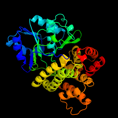
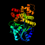
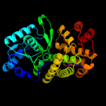
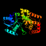
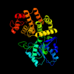
| 1 | c3bfjK_
|
|
 |
100.0 |
44 |
PDB header:oxidoreductase
Chain: K: PDB Molecule:1,3-propanediol oxidoreductase;
PDBTitle: crystal structure analysis of 1,3-propanediol oxidoreductase
|
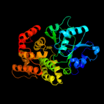
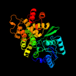
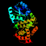
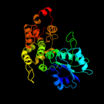
| 2 | d1rrma_
|
|
 |
100.0 |
97 |
Fold:Dehydroquinate synthase-like
Superfamily:Dehydroquinate synthase-like
Family:Iron-containing alcohol dehydrogenase |
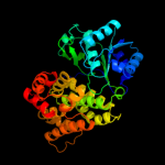
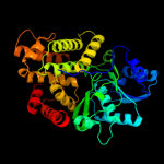
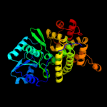
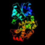
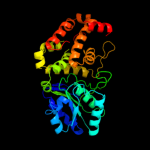
| 3 | c3ox4D_
|
|
 |
100.0 |
42 |
PDB header:oxidoreductase
Chain: D: PDB Molecule:alcohol dehydrogenase 2;
PDBTitle: structures of iron-dependent alcohol dehydrogenase 2 from zymomonas2 mobilis zm4 complexed with nad cofactor
|
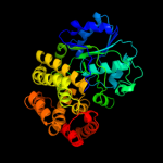
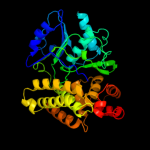
| 4 | d1vlja_
|
|
 |
100.0 |
25 |
Fold:Dehydroquinate synthase-like
Superfamily:Dehydroquinate synthase-like
Family:Iron-containing alcohol dehydrogenase |
| 5 | d1oj7a_
|
|
 |
100.0 |
23 |
Fold:Dehydroquinate synthase-like
Superfamily:Dehydroquinate synthase-like
Family:Iron-containing alcohol dehydrogenase |
| 6 | d1o2da_
|
|
 |
100.0 |
25 |
Fold:Dehydroquinate synthase-like
Superfamily:Dehydroquinate synthase-like
Family:Iron-containing alcohol dehydrogenase |
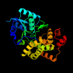
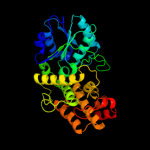
| 7 | c3hl0B_
|
|
 |
100.0 |
25 |
PDB header:oxidoreductase
Chain: B: PDB Molecule:maleylacetate reductase;
PDBTitle: crystal structure of maleylacetate reductase from agrobacterium2 tumefaciens
|
| 8 | c3iv7B_
|
|
 |
100.0 |
31 |
PDB header:oxidoreductase
Chain: B: PDB Molecule:alcohol dehydrogenase iv;
PDBTitle: crystal structure of iron-containing alcohol dehydrogenase2 (np_602249.1) from corynebacterium glutamicum atcc 13032 kitasato at3 2.07 a resolution
|
| 9 | c3jzdA_
|
|
 |
100.0 |
31 |
PDB header:oxidoreductase
Chain: A: PDB Molecule:iron-containing alcohol dehydrogenase;
PDBTitle: crystal structure of putative alcohol dehedrogenase (yp_298327.1) from2 ralstonia eutropha jmp134 at 2.10 a resolution
|
| 10 | c3rf7A_
|
|
 |
100.0 |
19 |
PDB header:oxidoreductase
Chain: A: PDB Molecule:iron-containing alcohol dehydrogenase;
PDBTitle: crystal structure of an iron-containing alcohol dehydrogenase2 (sden_2133) from shewanella denitrificans os-217 at 2.12 a resolution
|
| 11 | d1jq5a_
|
|
 |
100.0 |
22 |
Fold:Dehydroquinate synthase-like
Superfamily:Dehydroquinate synthase-like
Family:Iron-containing alcohol dehydrogenase |
| 12 | c1ta9A_
|
|
 |
100.0 |
21 |
PDB header:oxidoreductase
Chain: A: PDB Molecule:glycerol dehydrogenase;
PDBTitle: crystal structure of glycerol dehydrogenase from schizosaccharomyces2 pombe
|
| 13 | d1kq3a_
|
|
 |
100.0 |
24 |
Fold:Dehydroquinate synthase-like
Superfamily:Dehydroquinate synthase-like
Family:Iron-containing alcohol dehydrogenase |
| 14 | c3uhjE_
|
|
 |
100.0 |
22 |
PDB header:oxidoreductase
Chain: E: PDB Molecule:probable glycerol dehydrogenase;
PDBTitle: crystal structure of a probable glycerol dehydrogenase from2 sinorhizobium meliloti 1021
|
| 15 | c3ce9A_
|
|
 |
100.0 |
16 |
PDB header:oxidoreductase
Chain: A: PDB Molecule:glycerol dehydrogenase;
PDBTitle: crystal structure of glycerol dehydrogenase (np_348253.1) from2 clostridium acetobutylicum at 2.37 a resolution
|
| 16 | c3okfA_
|
|
 |
100.0 |
15 |
PDB header:lyase
Chain: A: PDB Molecule:3-dehydroquinate synthase;
PDBTitle: 2.5 angstrom resolution crystal structure of 3-dehydroquinate synthase2 (arob) from vibrio cholerae
|
| 17 | c1xahA_
|
|
 |
100.0 |
17 |
PDB header:lyase
Chain: A: PDB Molecule:3-dehydroquinate synthase;
PDBTitle: crystal structure of staphlyococcus aureus 3-dehydroquinate2 synthase (dhqs) in complex with zn2+ and nad+
|
| 18 | d1sg6a_
|
|
 |
100.0 |
14 |
Fold:Dehydroquinate synthase-like
Superfamily:Dehydroquinate synthase-like
Family:Dehydroquinate synthase, DHQS |
| 19 | c2gruB_
|
|
 |
100.0 |
14 |
PDB header:lyase
Chain: B: PDB Molecule:2-deoxy-scyllo-inosose synthase;
PDBTitle: crystal structure of 2-deoxy-scyllo-inosose synthase2 complexed with carbaglucose-6-phosphate, nad+ and co2+
|
| 20 | c3clhA_
|
|
 |
100.0 |
18 |
PDB header:lyase
Chain: A: PDB Molecule:3-dehydroquinate synthase;
PDBTitle: crystal structure of 3-dehydroquinate synthase (dhqs)from2 helicobacter pylori
|
| 21 | d1ujna_ |
|
not modelled |
100.0 |
19 |
Fold:Dehydroquinate synthase-like
Superfamily:Dehydroquinate synthase-like
Family:Dehydroquinate synthase, DHQS |
| 22 | c3orsD_ |
|
not modelled |
97.1 |
13 |
PDB header:isomerase,biosynthetic protein
Chain: D: PDB Molecule:n5-carboxyaminoimidazole ribonucleotide mutase;
PDBTitle: crystal structure of n5-carboxyaminoimidazole ribonucleotide mutase2 from staphylococcus aureus
|
| 23 | d1qcza_ |
|
not modelled |
97.1 |
16 |
Fold:Flavodoxin-like
Superfamily:N5-CAIR mutase (phosphoribosylaminoimidazole carboxylase, PurE)
Family:N5-CAIR mutase (phosphoribosylaminoimidazole carboxylase, PurE) |
| 24 | d1o4va_ |
|
not modelled |
97.0 |
18 |
Fold:Flavodoxin-like
Superfamily:N5-CAIR mutase (phosphoribosylaminoimidazole carboxylase, PurE)
Family:N5-CAIR mutase (phosphoribosylaminoimidazole carboxylase, PurE) |
| 25 | c3rggD_ |
|
not modelled |
97.0 |
19 |
PDB header:lyase
Chain: D: PDB Molecule:phosphoribosylaminoimidazole carboxylase, pure protein;
PDBTitle: crystal structure of treponema denticola pure bound to air
|
| 26 | d1u11a_ |
|
not modelled |
97.0 |
13 |
Fold:Flavodoxin-like
Superfamily:N5-CAIR mutase (phosphoribosylaminoimidazole carboxylase, PurE)
Family:N5-CAIR mutase (phosphoribosylaminoimidazole carboxylase, PurE) |
| 27 | c2fw9A_ |
|
not modelled |
97.0 |
13 |
PDB header:lyase
Chain: A: PDB Molecule:n5-carboxyaminoimidazole ribonucleotide mutase;
PDBTitle: structure of pure (n5-carboxyaminoimidazole ribonucleotide mutase)2 h59f from the acidophilic bacterium acetobacter aceti, at ph 8
|
| 28 | c3lp6D_ |
|
not modelled |
96.9 |
16 |
PDB header:lyase
Chain: D: PDB Molecule:phosphoribosylaminoimidazole carboxylase catalytic subunit;
PDBTitle: crystal structure of rv3275c-e60a from mycobacterium tuberculosis at2 1.7a resolution
|
| 29 | d2jgra1 |
|
not modelled |
96.8 |
12 |
Fold:NAD kinase/diacylglycerol kinase-like
Superfamily:NAD kinase/diacylglycerol kinase-like
Family:Diacylglycerol kinase-like |
| 30 | d2p1ra1 |
|
not modelled |
96.8 |
16 |
Fold:NAD kinase/diacylglycerol kinase-like
Superfamily:NAD kinase/diacylglycerol kinase-like
Family:Diacylglycerol kinase-like |
| 31 | d2bona1 |
|
not modelled |
96.7 |
12 |
Fold:NAD kinase/diacylglycerol kinase-like
Superfamily:NAD kinase/diacylglycerol kinase-like
Family:Diacylglycerol kinase-like |
| 32 | d1xmpa_ |
|
not modelled |
96.6 |
17 |
Fold:Flavodoxin-like
Superfamily:N5-CAIR mutase (phosphoribosylaminoimidazole carboxylase, PurE)
Family:N5-CAIR mutase (phosphoribosylaminoimidazole carboxylase, PurE) |
| 33 | c3trhI_ |
|
not modelled |
96.6 |
16 |
PDB header:lyase
Chain: I: PDB Molecule:phosphoribosylaminoimidazole carboxylase
PDBTitle: structure of a phosphoribosylaminoimidazole carboxylase catalytic2 subunit (pure) from coxiella burnetii
|
| 34 | c2bonB_ |
|
not modelled |
96.3 |
16 |
PDB header:transferase
Chain: B: PDB Molecule:lipid kinase;
PDBTitle: structure of an escherichia coli lipid kinase (yegs)
|
| 35 | c2h31A_ |
|
not modelled |
96.3 |
13 |
PDB header:ligase, lyase
Chain: A: PDB Molecule:multifunctional protein ade2;
PDBTitle: crystal structure of human paics, a bifunctional carboxylase and2 synthetase in purine biosynthesis
|
| 36 | d1u0ta_ |
|
not modelled |
96.1 |
24 |
Fold:NAD kinase/diacylglycerol kinase-like
Superfamily:NAD kinase/diacylglycerol kinase-like
Family:NAD kinase-like |
| 37 | d1qo0a_ |
|
not modelled |
95.5 |
9 |
Fold:Periplasmic binding protein-like I
Superfamily:Periplasmic binding protein-like I
Family:L-arabinose binding protein-like |
| 38 | c2qv7A_ |
|
not modelled |
95.3 |
22 |
PDB header:transferase
Chain: A: PDB Molecule:diacylglycerol kinase dgkb;
PDBTitle: crystal structure of diacylglycerol kinase dgkb in complex with adp2 and mg
|
| 39 | c3s40C_ |
|
not modelled |
95.2 |
22 |
PDB header:transferase
Chain: C: PDB Molecule:diacylglycerol kinase;
PDBTitle: the crystal structure of a diacylglycerol kinases from bacillus2 anthracis str. sterne
|
| 40 | c2ywxA_ |
|
not modelled |
95.0 |
22 |
PDB header:lyase
Chain: A: PDB Molecule:phosphoribosylaminoimidazole carboxylase catalytic subunit;
PDBTitle: crystal structure of phosphoribosylaminoimidazole carboxylase2 catalytic subunit from methanocaldococcus jannaschii
|
| 41 | d2qv7a1 |
|
not modelled |
94.7 |
22 |
Fold:NAD kinase/diacylglycerol kinase-like
Superfamily:NAD kinase/diacylglycerol kinase-like
Family:Diacylglycerol kinase-like |
| 42 | d1pfka_ |
|
not modelled |
92.8 |
18 |
Fold:Phosphofructokinase
Superfamily:Phosphofructokinase
Family:Phosphofructokinase |
| 43 | c3pfnB_ |
|
not modelled |
92.6 |
14 |
PDB header:transferase
Chain: B: PDB Molecule:nad kinase;
PDBTitle: crystal structure of human nad kinase
|
| 44 | c3snrA_ |
|
not modelled |
92.4 |
13 |
PDB header:transport protein
Chain: A: PDB Molecule:extracellular ligand-binding receptor;
PDBTitle: rpd_1889 protein, an extracellular ligand-binding receptor from2 rhodopseudomonas palustris.
|
| 45 | c1zxxA_ |
|
not modelled |
92.2 |
17 |
PDB header:transferase
Chain: A: PDB Molecule:6-phosphofructokinase;
PDBTitle: the crystal structure of phosphofructokinase from lactobacillus2 delbrueckii
|
| 46 | d1ewka_ |
|
not modelled |
92.1 |
13 |
Fold:Periplasmic binding protein-like I
Superfamily:Periplasmic binding protein-like I
Family:L-arabinose binding protein-like |
| 47 | c2j37W_ |
|
not modelled |
91.5 |
21 |
PDB header:ribosome
Chain: W: PDB Molecule:signal recognition particle 54 kda protein
PDBTitle: model of mammalian srp bound to 80s rncs
|
| 48 | d4pfka_ |
|
not modelled |
91.5 |
21 |
Fold:Phosphofructokinase
Superfamily:Phosphofructokinase
Family:Phosphofructokinase |
| 49 | c2an1D_ |
|
not modelled |
91.4 |
21 |
PDB header:transferase
Chain: D: PDB Molecule:putative kinase;
PDBTitle: structural genomics, the crystal structure of a putative kinase from2 salmonella typhimurim lt2
|
| 50 | c3sg0A_ |
|
not modelled |
91.3 |
10 |
PDB header:signaling protein
Chain: A: PDB Molecule:extracellular ligand-binding receptor;
PDBTitle: the crystal structure of an extracellular ligand-binding receptor from2 rhodopseudomonas palustris haa2
|
| 51 | c3q41B_ |
|
not modelled |
91.3 |
8 |
PDB header:transport protein
Chain: B: PDB Molecule:glutamate [nmda] receptor subunit zeta-1;
PDBTitle: crystal structure of the glun1 n-terminal domain (ntd)
|
| 52 | c3mdqA_ |
|
not modelled |
91.1 |
11 |
PDB header:hydrolase
Chain: A: PDB Molecule:exopolyphosphatase;
PDBTitle: crystal structure of an exopolyphosphatase (chu_0316) from cytophaga2 hutchinsonii atcc 33406 at 1.50 a resolution
|
| 53 | c3i09A_ |
|
not modelled |
91.0 |
15 |
PDB header:transport protein
Chain: A: PDB Molecule:periplasmic branched-chain amino acid-binding protein;
PDBTitle: crystal structure of a periplasmic binding protein (bma2936) from2 burkholderia mallei at 1.80 a resolution
|
| 54 | c3hi0B_ |
|
not modelled |
90.9 |
14 |
PDB header:hydrolase
Chain: B: PDB Molecule:putative exopolyphosphatase;
PDBTitle: crystal structure of putative exopolyphosphatase (17739545) from2 agrobacterium tumefaciens str. c58 (dupont) at 2.30 a resolution
|
| 55 | c2iy3A_ |
|
not modelled |
90.8 |
11 |
PDB header:rna-binding
Chain: A: PDB Molecule:signal recognition particle protein ffh;
PDBTitle: structure of the e. coli signal recognition particle2 bound to a translating ribosome
|
| 56 | c3i45A_ |
|
not modelled |
90.7 |
13 |
PDB header:signaling protein
Chain: A: PDB Molecule:twin-arginine translocation pathway signal protein;
PDBTitle: crystal structure of putative twin-arginine translocation pathway2 signal protein from rhodospirillum rubrum atcc 11170
|
| 57 | d2f48a1 |
|
not modelled |
90.6 |
17 |
Fold:Phosphofructokinase
Superfamily:Phosphofructokinase
Family:Phosphofructokinase |
| 58 | c3n0wA_ |
|
not modelled |
90.3 |
14 |
PDB header:transport protein
Chain: A: PDB Molecule:abc branched chain amino acid family transporter,
PDBTitle: crystal structure of a branched chain amino acid abc transporter2 periplasmic ligand-binding protein (bxe_c0949) from burkholderia3 xenovorans lb400 at 1.88 a resolution
|
| 59 | c1t6dB_ |
|
not modelled |
89.9 |
10 |
PDB header:hydrolase
Chain: B: PDB Molecule:exopolyphosphatase;
PDBTitle: miras phasing of the aquifex aeolicus ppx/gppa phosphatase: crystal2 structure of the type ii variant
|
| 60 | c3eafA_ |
|
not modelled |
89.8 |
13 |
PDB header:transport protein
Chain: A: PDB Molecule:abc transporter, substrate binding protein;
PDBTitle: crystal structure of abc transporter, substrate binding protein2 aeropyrum pernix
|
| 61 | d2ji7a1 |
|
not modelled |
89.8 |
12 |
Fold:DHS-like NAD/FAD-binding domain
Superfamily:DHS-like NAD/FAD-binding domain
Family:Pyruvate oxidase and decarboxylase, middle domain |
| 62 | c3opyH_ |
|
not modelled |
89.7 |
21 |
PDB header:transferase
Chain: H: PDB Molecule:6-phosphofructo-1-kinase beta-subunit;
PDBTitle: crystal structure of pichia pastoris phosphofructokinase in the t-2 state
|
| 63 | c3opyB_ |
|
not modelled |
89.7 |
21 |
PDB header:transferase
Chain: B: PDB Molecule:6-phosphofructo-1-kinase beta-subunit;
PDBTitle: crystal structure of pichia pastoris phosphofructokinase in the t-2 state
|
| 64 | c3rfqC_ |
|
not modelled |
89.0 |
20 |
PDB header:biosynthetic protein
Chain: C: PDB Molecule:pterin-4-alpha-carbinolamine dehydratase moab2;
PDBTitle: crystal structure of pterin-4-alpha-carbinolamine dehydratase moab22 from mycobacterium marinum
|
| 65 | d1ovma1 |
|
not modelled |
88.9 |
22 |
Fold:DHS-like NAD/FAD-binding domain
Superfamily:DHS-like NAD/FAD-binding domain
Family:Pyruvate oxidase and decarboxylase, middle domain |
| 66 | c3dm5A_ |
|
not modelled |
88.9 |
24 |
PDB header:rna binding protein, transport protein
Chain: A: PDB Molecule:signal recognition 54 kda protein;
PDBTitle: structures of srp54 and srp19, the two proteins assembling2 the ribonucleic core of the signal recognition particle3 from the archaeon pyrococcus furiosus.
|
| 67 | c2dwcB_ |
|
not modelled |
88.8 |
15 |
PDB header:transferase
Chain: B: PDB Molecule:433aa long hypothetical phosphoribosylglycinamide formyl
PDBTitle: crystal structure of probable phosphoribosylglycinamide formyl2 transferase from pyrococcus horikoshii ot3 complexed with adp
|
| 68 | c3lopA_ |
|
not modelled |
88.5 |
8 |
PDB header:substrate binding protein
Chain: A: PDB Molecule:substrate binding periplasmic protein;
PDBTitle: crystal structure of substrate-binding periplasmic protein2 (pbp) from ralstonia solanacearum
|
| 69 | c1kjjA_ |
|
not modelled |
88.3 |
11 |
PDB header:transferase
Chain: A: PDB Molecule:phosphoribosylglycinamide formyltransferase 2;
PDBTitle: crystal structure of glycniamide ribonucleotide2 transformylase in complex with mg-atp-gamma-s
|
| 70 | c3k2qA_ |
|
not modelled |
87.9 |
22 |
PDB header:transferase
Chain: A: PDB Molecule:pyrophosphate-dependent phosphofructokinase;
PDBTitle: crystal structure of pyrophosphate-dependent2 phosphofructokinase from marinobacter aquaeolei, northeast3 structural genomics consortium target mqr88
|
| 71 | d1v4va_ |
|
not modelled |
87.6 |
15 |
Fold:UDP-Glycosyltransferase/glycogen phosphorylase
Superfamily:UDP-Glycosyltransferase/glycogen phosphorylase
Family:UDP-N-acetylglucosamine 2-epimerase |
| 72 | c1qzwC_ |
|
not modelled |
87.5 |
22 |
PDB header:signaling protein/rna
Chain: C: PDB Molecule:signal recognition 54 kda protein;
PDBTitle: crystal structure of the complete core of archaeal srp and2 implications for inter-domain communication
|
| 73 | d1xi8a3 |
|
not modelled |
87.4 |
15 |
Fold:Molybdenum cofactor biosynthesis proteins
Superfamily:Molybdenum cofactor biosynthesis proteins
Family:MoeA central domain-like |
| 74 | c2e4wA_ |
|
not modelled |
87.4 |
8 |
PDB header:signaling protein
Chain: A: PDB Molecule:metabotropic glutamate receptor 3;
PDBTitle: crystal structure of the extracellular region of the group ii2 metabotropic glutamate receptor complexed with 1s,3s-acpd
|
| 75 | c2j289_ |
|
not modelled |
87.3 |
16 |
PDB header:ribosome
Chain: 9: PDB Molecule:signal recognition particle 54;
PDBTitle: model of e. coli srp bound to 70s rncs
|
| 76 | c2i2aA_ |
|
not modelled |
87.2 |
18 |
PDB header:transferase
Chain: A: PDB Molecule:probable inorganic polyphosphate/atp-nad kinase 1;
PDBTitle: crystal structure of lmnadk1 from listeria monocytogenes
|
| 77 | c2higA_ |
|
not modelled |
87.0 |
18 |
PDB header:transferase
Chain: A: PDB Molecule:6-phospho-1-fructokinase;
PDBTitle: crystal structure of phosphofructokinase apoenzyme from trypanosoma2 brucei.
|
| 78 | c2floA_ |
|
not modelled |
87.0 |
17 |
PDB header:hydrolase
Chain: A: PDB Molecule:exopolyphosphatase;
PDBTitle: crystal structure of exopolyphosphatase (ppx) from e. coli o157:h7
|
| 79 | c3tqrA_ |
|
not modelled |
87.0 |
7 |
PDB header:transferase
Chain: A: PDB Molecule:phosphoribosylglycinamide formyltransferase;
PDBTitle: structure of the phosphoribosylglycinamide formyltransferase (purn) in2 complex with ches from coxiella burnetii
|
| 80 | d1zpda1 |
|
not modelled |
86.9 |
23 |
Fold:DHS-like NAD/FAD-binding domain
Superfamily:DHS-like NAD/FAD-binding domain
Family:Pyruvate oxidase and decarboxylase, middle domain |
| 81 | c3h5lB_ |
|
not modelled |
86.6 |
15 |
PDB header:transport protein
Chain: B: PDB Molecule:putative branched-chain amino acid abc
PDBTitle: crystal structure of a putative branched-chain amino acid2 abc transporter from silicibacter pomeroyi
|
| 82 | c3o8oB_ |
|
not modelled |
86.6 |
18 |
PDB header:transferase
Chain: B: PDB Molecule:6-phosphofructokinase subunit beta;
PDBTitle: structure of phosphofructokinase from saccharomyces cerevisiae
|
| 83 | c3hutA_ |
|
not modelled |
86.4 |
7 |
PDB header:transport protein
Chain: A: PDB Molecule:putative branched-chain amino acid abc
PDBTitle: crystal structure of a putative branched-chain amino acid2 abc transporter from rhodospirillum rubrum
|
| 84 | c3cf4G_ |
|
not modelled |
86.2 |
14 |
PDB header:oxidoreductase
Chain: G: PDB Molecule:acetyl-coa decarboxylase/synthase epsilon subunit;
PDBTitle: structure of the codh component of the m. barkeri acds complex
|
| 85 | d1vh3a_ |
|
not modelled |
86.0 |
13 |
Fold:Nucleotide-diphospho-sugar transferases
Superfamily:Nucleotide-diphospho-sugar transferases
Family:Cytidylytransferase |
| 86 | c3opyE_ |
|
not modelled |
85.7 |
23 |
PDB header:transferase
Chain: E: PDB Molecule:6-phosphofructo-1-kinase alpha-subunit;
PDBTitle: crystal structure of pichia pastoris phosphofructokinase in the t-2 state
|
| 87 | d2ihta1 |
|
not modelled |
85.6 |
9 |
Fold:DHS-like NAD/FAD-binding domain
Superfamily:DHS-like NAD/FAD-binding domain
Family:Pyruvate oxidase and decarboxylase, middle domain |
| 88 | c2q5cA_ |
|
not modelled |
85.6 |
13 |
PDB header:transcription
Chain: A: PDB Molecule:ntrc family transcriptional regulator;
PDBTitle: crystal structure of ntrc family transcriptional regulator from2 clostridium acetobutylicum
|
| 89 | d2ez9a1 |
|
not modelled |
85.5 |
23 |
Fold:DHS-like NAD/FAD-binding domain
Superfamily:DHS-like NAD/FAD-binding domain
Family:Pyruvate oxidase and decarboxylase, middle domain |
| 90 | c3cerD_ |
|
not modelled |
85.5 |
18 |
PDB header:structural genomics, unknown function
Chain: D: PDB Molecule:possible exopolyphosphatase-like protein;
PDBTitle: crystal structure of the exopolyphosphatase-like protein2 q8g5j2. northeast structural genomics consortium target3 blr13
|
| 91 | c3kg2A_ |
|
not modelled |
85.4 |
10 |
PDB header:membrane protein, transport protein
Chain: A: PDB Molecule:glutamate receptor 2;
PDBTitle: ampa subtype ionotropic glutamate receptor in complex with competitive2 antagonist zk 200775
|
| 92 | d1zl0a2 |
|
not modelled |
84.5 |
9 |
Fold:Flavodoxin-like
Superfamily:Class I glutamine amidotransferase-like
Family:LD-carboxypeptidase A N-terminal domain-like |
| 93 | d1pvda1 |
|
not modelled |
84.4 |
25 |
Fold:DHS-like NAD/FAD-binding domain
Superfamily:DHS-like NAD/FAD-binding domain
Family:Pyruvate oxidase and decarboxylase, middle domain |
| 94 | c3lkbB_ |
|
not modelled |
84.0 |
15 |
PDB header:transport protein
Chain: B: PDB Molecule:probable branched-chain amino acid abc
PDBTitle: crystal structure of a branched chain amino acid abc2 transporter from thermus thermophilus with bound valine
|
| 95 | d1t9ba1 |
|
not modelled |
84.0 |
16 |
Fold:DHS-like NAD/FAD-binding domain
Superfamily:DHS-like NAD/FAD-binding domain
Family:Pyruvate oxidase and decarboxylase, middle domain |
| 96 | c3t0nA_ |
|
not modelled |
83.6 |
12 |
PDB header:signaling protein
Chain: A: PDB Molecule:twin-arginine translocation pathway signal;
PDBTitle: crystal structure of twin-arginine translocation pathway signal from2 rhodopseudomonas palustris bisb5
|
| 97 | c1zrsB_ |
|
not modelled |
83.4 |
9 |
PDB header:hydrolase
Chain: B: PDB Molecule:hypothetical protein;
PDBTitle: wild-type ld-carboxypeptidase
|
| 98 | c3dcjA_ |
|
not modelled |
83.1 |
7 |
PDB header:transferase
Chain: A: PDB Molecule:probable 5'-phosphoribosylglycinamide
PDBTitle: crystal structure of glycinamide formyltransferase (purn)2 from mycobacterium tuberculosis in complex with 5-methyl-5,3 6,7,8-tetrahydrofolic acid derivative
|
| 99 | d2auna2 |
|
not modelled |
82.8 |
9 |
Fold:Flavodoxin-like
Superfamily:Class I glutamine amidotransferase-like
Family:LD-carboxypeptidase A N-terminal domain-like |
| 100 | d1q6za1 |
|
not modelled |
82.5 |
13 |
Fold:DHS-like NAD/FAD-binding domain
Superfamily:DHS-like NAD/FAD-binding domain
Family:Pyruvate oxidase and decarboxylase, middle domain |
| 101 | c2xecD_ |
|
not modelled |
82.1 |
17 |
PDB header:isomerase
Chain: D: PDB Molecule:putative maleate isomerase;
PDBTitle: nocardia farcinica maleate cis-trans isomerase bound to2 tris
|
| 102 | c2g4rB_ |
|
not modelled |
81.9 |
14 |
PDB header:biosynthetic protein
Chain: B: PDB Molecule:molybdopterin biosynthesis mog protein;
PDBTitle: anomalous substructure of moga
|
| 103 | c3opyG_ |
|
not modelled |
81.8 |
18 |
PDB header:transferase
Chain: G: PDB Molecule:6-phosphofructo-1-kinase alpha-subunit;
PDBTitle: crystal structure of pichia pastoris phosphofructokinase in the t-2 state
|
| 104 | d1y5ea1 |
|
not modelled |
81.5 |
15 |
Fold:Molybdenum cofactor biosynthesis proteins
Superfamily:Molybdenum cofactor biosynthesis proteins
Family:MogA-like |
| 105 | d2pjua1 |
|
not modelled |
81.4 |
11 |
Fold:Chelatase-like
Superfamily:PrpR receptor domain-like
Family:PrpR receptor domain-like |
| 106 | c3ip5A_ |
|
not modelled |
81.1 |
18 |
PDB header:transport protein
Chain: A: PDB Molecule:abc transporter, substrate binding protein (amino acid);
PDBTitle: structure of atu2422-gaba receptor in complex with alanine
|
| 107 | c2pjkA_ |
|
not modelled |
80.7 |
16 |
PDB header:structural genomics, unknown function
Chain: A: PDB Molecule:178aa long hypothetical molybdenum cofactor
PDBTitle: structure of hypothetical molybdenum cofactor biosynthesis2 protein b from sulfolobus tokodaii
|
| 108 | c3o8oC_ |
|
not modelled |
80.3 |
19 |
PDB header:transferase
Chain: C: PDB Molecule:6-phosphofructokinase subunit alpha;
PDBTitle: structure of phosphofructokinase from saccharomyces cerevisiae
|
| 109 | c1m6vE_ |
|
not modelled |
79.9 |
13 |
PDB header:ligase
Chain: E: PDB Molecule:carbamoyl phosphate synthetase large chain;
PDBTitle: crystal structure of the g359f (small subunit) point mutant of2 carbamoyl phosphate synthetase
|
| 110 | c3dnfB_ |
|
not modelled |
79.8 |
20 |
PDB header:oxidoreductase
Chain: B: PDB Molecule:4-hydroxy-3-methylbut-2-enyl diphosphate reductase;
PDBTitle: structure of (e)-4-hydroxy-3-methyl-but-2-enyl diphosphate reductase,2 the terminal enzyme of the non-mevalonate pathway
|
| 111 | c3o8nA_ |
|
not modelled |
79.3 |
15 |
PDB header:transferase
Chain: A: PDB Molecule:6-phosphofructokinase, muscle type;
PDBTitle: structure of phosphofructokinase from rabbit skeletal muscle
|
| 112 | c2ywrA_ |
|
not modelled |
79.2 |
8 |
PDB header:transferase
Chain: A: PDB Molecule:phosphoribosylglycinamide formyltransferase;
PDBTitle: crystal structure of gar transformylase from aquifex2 aeolicus
|
| 113 | c3jpyA_ |
|
not modelled |
79.2 |
9 |
PDB header:transport protein
Chain: A: PDB Molecule:glutamate [nmda] receptor subunit epsilon-2;
PDBTitle: crystal structure of the zinc-bound amino terminal domain of the nmda2 receptor subunit nr2b
|
| 114 | d1a9xa4 |
|
not modelled |
79.1 |
12 |
Fold:PreATP-grasp domain
Superfamily:PreATP-grasp domain
Family:BC N-terminal domain-like |
| 115 | c2is8A_ |
|
not modelled |
79.1 |
13 |
PDB header:structural protein
Chain: A: PDB Molecule:molybdopterin biosynthesis enzyme, moab;
PDBTitle: crystal structure of the molybdopterin biosynthesis enzyme moab2 (ttha0341) from thermus theromophilus hb8
|
| 116 | c3p9xB_ |
|
not modelled |
79.1 |
18 |
PDB header:transferase
Chain: B: PDB Molecule:phosphoribosylglycinamide formyltransferase;
PDBTitle: crystal structure of phosphoribosylglycinamide formyltransferase from2 bacillus halodurans
|
| 117 | c2yhsA_ |
|
not modelled |
79.0 |
15 |
PDB header:cell cycle
Chain: A: PDB Molecule:cell division protein ftsy;
PDBTitle: structure of the e. coli srp receptor ftsy
|
| 118 | c2jkzB_ |
|
not modelled |
78.6 |
25 |
PDB header:transferase
Chain: B: PDB Molecule:hypoxanthine-guanine phosphoribosyltransferase;
PDBTitle: saccharomyces cerevisiae hypoxanthine-guanine2 phosphoribosyltransferase in complex with gmp (guanosine 5'3 -monophosphate) (orthorhombic crystal form)
|
| 119 | d2g2ca1 |
|
not modelled |
78.6 |
16 |
Fold:Molybdenum cofactor biosynthesis proteins
Superfamily:Molybdenum cofactor biosynthesis proteins
Family:MogA-like |
| 120 | d1mkza_ |
|
not modelled |
78.3 |
15 |
Fold:Molybdenum cofactor biosynthesis proteins
Superfamily:Molybdenum cofactor biosynthesis proteins
Family:MogA-like |