| 1 | d1s5pa_
|
|
 |
100.0 |
97 |
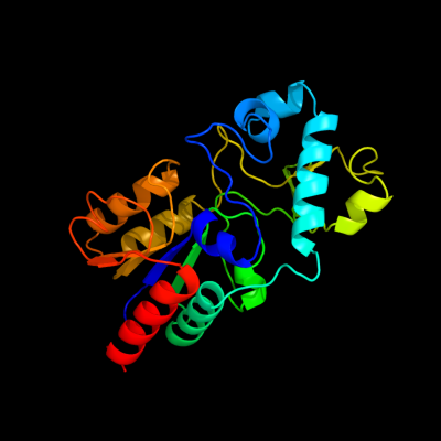
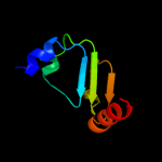
Fold:DHS-like NAD/FAD-binding domain
Superfamily:DHS-like NAD/FAD-binding domain
Family:Sir2 family of transcriptional regulators |
| 2 | d1m2ka_
|
|
 |
100.0 |
40 |
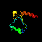
Fold:DHS-like NAD/FAD-binding domain
Superfamily:DHS-like NAD/FAD-binding domain
Family:Sir2 family of transcriptional regulators |
| 3 | c3jwpA_
|
|
 |
100.0 |
28 |
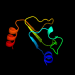
PDB header:transcription
Chain: A: PDB Molecule:transcriptional regulatory protein sir2 homologue;
PDBTitle: crystal structure of plasmodium falciparum sir2a (pf13_0152) in2 complex with amp
|
| 4 | d1j8fa_
|
|
 |
100.0 |
26 |
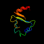
Fold:DHS-like NAD/FAD-binding domain
Superfamily:DHS-like NAD/FAD-binding domain
Family:Sir2 family of transcriptional regulators |
| 5 | c2hjhB_
|
|
 |
100.0 |
28 |
PDB header:hydrolase
Chain: B: PDB Molecule:nad-dependent histone deacetylase sir2;
PDBTitle: crystal structure of the sir2 deacetylase
|
| 6 | d1yc5a1
|
|
 |
100.0 |
31 |
Fold:DHS-like NAD/FAD-binding domain
Superfamily:DHS-like NAD/FAD-binding domain
Family:Sir2 family of transcriptional regulators |
| 7 | c3glsC_
|
|
 |
100.0 |
26 |
PDB header:hydrolase
Chain: C: PDB Molecule:nad-dependent deacetylase sirtuin-3,
PDBTitle: crystal structure of human sirt3
|
| 8 | d2b4ya1
|
|
 |
100.0 |
37 |
Fold:DHS-like NAD/FAD-binding domain
Superfamily:DHS-like NAD/FAD-binding domain
Family:Sir2 family of transcriptional regulators |
| 9 | d1q1aa_
|
|
 |
100.0 |
27 |
Fold:DHS-like NAD/FAD-binding domain
Superfamily:DHS-like NAD/FAD-binding domain
Family:Sir2 family of transcriptional regulators |
| 10 | c1q14A_
|
|
 |
100.0 |
27 |
PDB header:hydrolase
Chain: A: PDB Molecule:hst2 protein;
PDBTitle: structure and autoregulation of the yeast hst2 homolog of sir2
|
| 11 | c3k35D_
|
|
 |
100.0 |
27 |
PDB header:hydrolase
Chain: D: PDB Molecule:nad-dependent deacetylase sirtuin-6;
PDBTitle: crystal structure of human sirt6
|
| 12 | c3pkiF_
|
|
 |
100.0 |
26 |
PDB header:hydrolase
Chain: F: PDB Molecule:nad-dependent deacetylase sirtuin-6;
PDBTitle: human sirt6 crystal structure in complex with adp ribose
|
| 13 | d1ma3a_
|
|
 |
100.0 |
35 |
Fold:DHS-like NAD/FAD-binding domain
Superfamily:DHS-like NAD/FAD-binding domain
Family:Sir2 family of transcriptional regulators |
| 14 | d2ji7a1
|
|
 |
97.2 |
16 |
Fold:DHS-like NAD/FAD-binding domain
Superfamily:DHS-like NAD/FAD-binding domain
Family:Pyruvate oxidase and decarboxylase, middle domain |
| 15 | d2djia1
|
|
 |
97.2 |
15 |
Fold:DHS-like NAD/FAD-binding domain
Superfamily:DHS-like NAD/FAD-binding domain
Family:Pyruvate oxidase and decarboxylase, middle domain |
| 16 | d2ez9a1
|
|
 |
97.0 |
13 |
Fold:DHS-like NAD/FAD-binding domain
Superfamily:DHS-like NAD/FAD-binding domain
Family:Pyruvate oxidase and decarboxylase, middle domain |
| 17 | d1ybha1
|
|
 |
97.0 |
14 |
Fold:DHS-like NAD/FAD-binding domain
Superfamily:DHS-like NAD/FAD-binding domain
Family:Pyruvate oxidase and decarboxylase, middle domain |
| 18 | d1ozha1
|
|
 |
96.8 |
19 |
Fold:DHS-like NAD/FAD-binding domain
Superfamily:DHS-like NAD/FAD-binding domain
Family:Pyruvate oxidase and decarboxylase, middle domain |
| 19 | d1t9ba1
|
|
 |
96.8 |
15 |
Fold:DHS-like NAD/FAD-binding domain
Superfamily:DHS-like NAD/FAD-binding domain
Family:Pyruvate oxidase and decarboxylase, middle domain |
| 20 | d2ihta1
|
|
 |
96.7 |
9 |
Fold:DHS-like NAD/FAD-binding domain
Superfamily:DHS-like NAD/FAD-binding domain
Family:Pyruvate oxidase and decarboxylase, middle domain |
| 21 | c2ivfA_ |
|
not modelled |
96.4 |
13 |
PDB header:oxidoreductase
Chain: A: PDB Molecule:ethylbenzene dehydrogenase alpha-subunit;
PDBTitle: ethylbenzene dehydrogenase from aromatoleum aromaticum
|
| 22 | d1h0ha2 |
|
not modelled |
96.4 |
14 |
Fold:Formate dehydrogenase/DMSO reductase, domains 1-3
Superfamily:Formate dehydrogenase/DMSO reductase, domains 1-3
Family:Formate dehydrogenase/DMSO reductase, domains 1-3 |
| 23 | c2vpyE_ |
|
not modelled |
96.4 |
14 |
PDB header:oxidoreductase
Chain: E: PDB Molecule:thiosulfate reductase;
PDBTitle: polysulfide reductase with bound quinone inhibitor,2 pentachlorophenol (pcp)
|
| 24 | c1powA_ |
|
not modelled |
96.3 |
13 |
PDB header:oxidoreductase(oxygen as acceptor)
Chain: A: PDB Molecule:pyruvate oxidase;
PDBTitle: the refined structures of a stabilized mutant and of wild-type2 pyruvate oxidase from lactobacillus plantarum
|
| 25 | c3lq1A_ |
|
not modelled |
96.0 |
6 |
PDB header:transferase
Chain: A: PDB Molecule:2-succinyl-5-enolpyruvyl-6-hydroxy-3-cyclohexene-
PDBTitle: crystal structure of 2-succinyl-6-hydroxy-2,4-cyclohexadiene2 1-carboxylic acid synthase/2-oxoglutarate decarboxylase3 from listeria monocytogenes str. 4b f2365
|
| 26 | d1q6za1 |
|
not modelled |
96.0 |
13 |
Fold:DHS-like NAD/FAD-binding domain
Superfamily:DHS-like NAD/FAD-binding domain
Family:Pyruvate oxidase and decarboxylase, middle domain |
| 27 | c2djiA_ |
|
not modelled |
96.0 |
15 |
PDB header:oxidoreductase
Chain: A: PDB Molecule:pyruvate oxidase;
PDBTitle: crystal structure of pyruvate oxidase from aerococcus2 viridans containing fad
|
| 28 | c2ji6B_ |
|
not modelled |
96.0 |
16 |
PDB header:lyase
Chain: B: PDB Molecule:oxalyl-coa decarboxylase;
PDBTitle: x-ray structure of oxalyl-coa decarboxylase in complex with2 3-deaza-thdp and oxalyl-coa
|
| 29 | c2x7jA_ |
|
not modelled |
95.9 |
13 |
PDB header:transferase
Chain: A: PDB Molecule:2-succinyl-5-enolpyruvyl-6-hydroxy-3-cyclohexene
PDBTitle: structure of the menaquinone biosynthesis protein mend from2 bacillus subtilis
|
| 30 | c1h0hA_ |
|
not modelled |
95.9 |
14 |
PDB header:dehydrogenase
Chain: A: PDB Molecule:formate dehydrogenase (large subunit);
PDBTitle: tungsten containing formate dehydrogenase from2 desulfovibrio gigas
|
| 31 | c1h5nC_ |
|
not modelled |
95.8 |
10 |
PDB header:oxidoreductase
Chain: C: PDB Molecule:dmso reductase;
PDBTitle: dmso reductase modified by the presence of dms and air
|
| 32 | c1yi1A_ |
|
not modelled |
95.6 |
9 |
PDB header:transferase
Chain: A: PDB Molecule:acetolactate synthase;
PDBTitle: crystal structure of arabidopsis thaliana acetohydroxyacid synthase in2 complex with a sulfonylurea herbicide, tribenuron methyl
|
| 33 | c2e7zA_ |
|
not modelled |
95.6 |
12 |
PDB header:lyase
Chain: A: PDB Molecule:acetylene hydratase ahy;
PDBTitle: acetylene hydratase from pelobacter acetylenicus
|
| 34 | c2q27B_ |
|
not modelled |
95.6 |
18 |
PDB header:lyase
Chain: B: PDB Molecule:oxalyl-coa decarboxylase;
PDBTitle: crystal structure of oxalyl-coa decarboxylase from escherichia coli
|
| 35 | c1ozhD_ |
|
not modelled |
95.4 |
19 |
PDB header:lyase
Chain: D: PDB Molecule:acetolactate synthase, catabolic;
PDBTitle: the crystal structure of klebsiella pneumoniae acetolactate2 synthase with enzyme-bound cofactor and with an unusual3 intermediate.
|
| 36 | c2panF_ |
|
not modelled |
95.4 |
20 |
PDB header:lyase
Chain: F: PDB Molecule:glyoxylate carboligase;
PDBTitle: crystal structure of e. coli glyoxylate carboligase
|
| 37 | c3eyaE_ |
|
not modelled |
95.4 |
14 |
PDB header:oxidoreductase
Chain: E: PDB Molecule:pyruvate dehydrogenase [cytochrome];
PDBTitle: structural basis for membrane binding and catalytic2 activation of the peripheral membrane enzyme pyruvate3 oxidase from escherichia coli
|
| 38 | d3clsd2 |
|
not modelled |
95.3 |
19 |
Fold:DHS-like NAD/FAD-binding domain
Superfamily:DHS-like NAD/FAD-binding domain
Family:C-terminal domain of the electron transfer flavoprotein alpha subunit |
| 39 | c2iv2X_ |
|
not modelled |
95.3 |
13 |
PDB header:oxidoreductase
Chain: X: PDB Molecule:formate dehydrogenase h;
PDBTitle: reinterpretation of reduced form of formate dehydrogenase h2 from e. coli
|
| 40 | d1y5ia2 |
|
not modelled |
95.3 |
17 |
Fold:Formate dehydrogenase/DMSO reductase, domains 1-3
Superfamily:Formate dehydrogenase/DMSO reductase, domains 1-3
Family:Formate dehydrogenase/DMSO reductase, domains 1-3 |
| 41 | c1efpC_ |
|
not modelled |
95.2 |
25 |
PDB header:electron transport
Chain: C: PDB Molecule:protein (electron transfer flavoprotein);
PDBTitle: electron transfer flavoprotein (etf) from paracoccus2 denitrificans
|
| 42 | d1kqfa2 |
|
not modelled |
95.2 |
7 |
Fold:Formate dehydrogenase/DMSO reductase, domains 1-3
Superfamily:Formate dehydrogenase/DMSO reductase, domains 1-3
Family:Formate dehydrogenase/DMSO reductase, domains 1-3 |
| 43 | c1t9dB_ |
|
not modelled |
95.2 |
17 |
PDB header:transferase
Chain: B: PDB Molecule:acetolactate synthase, mitochondrial;
PDBTitle: crystal structure of yeast acetohydroxyacid synthase in2 complex with a sulfonylurea herbicide, metsulfuron methyl
|
| 44 | d2iv2x2 |
|
not modelled |
95.1 |
13 |
Fold:Formate dehydrogenase/DMSO reductase, domains 1-3
Superfamily:Formate dehydrogenase/DMSO reductase, domains 1-3
Family:Formate dehydrogenase/DMSO reductase, domains 1-3 |
| 45 | d1vlfm2 |
|
not modelled |
95.1 |
12 |
Fold:Formate dehydrogenase/DMSO reductase, domains 1-3
Superfamily:Formate dehydrogenase/DMSO reductase, domains 1-3
Family:Formate dehydrogenase/DMSO reductase, domains 1-3 |
| 46 | c2pgnA_ |
|
not modelled |
95.1 |
16 |
PDB header:hydrolase
Chain: A: PDB Molecule:cyclohexane-1,2-dione hydrolase (cdh);
PDBTitle: the crystal structure of fad and thdp-dependent cyclohexane-1,2-dione2 hydrolase in complex with cyclohexane-1,2-dione
|
| 47 | c1vlfQ_ |
|
not modelled |
94.9 |
11 |
PDB header:oxidoreductase
Chain: Q: PDB Molecule:pyrogallol hydroxytransferase large subunit;
PDBTitle: crystal structure of pyrogallol-phloroglucinol2 transhydroxylase from pelobacter acidigallici complexed3 with inhibitor 1,2,4,5-tetrahydroxy-benzene
|
| 48 | d1dmra2 |
|
not modelled |
94.9 |
10 |
Fold:Formate dehydrogenase/DMSO reductase, domains 1-3
Superfamily:Formate dehydrogenase/DMSO reductase, domains 1-3
Family:Formate dehydrogenase/DMSO reductase, domains 1-3 |
| 49 | c2v45A_ |
|
not modelled |
94.7 |
14 |
PDB header:oxidoreductase
Chain: A: PDB Molecule:periplasmic nitrate reductase;
PDBTitle: a new catalytic mechanism of periplasmic nitrate reductase2 from desulfovibrio desulfuricans atcc 27774 from3 crystallographic and epr data and based on detailed4 analysis of the sixth ligand
|
| 50 | c3shoA_ |
|
not modelled |
94.7 |
14 |
PDB header:transcription regulator
Chain: A: PDB Molecule:transcriptional regulator, rpir family;
PDBTitle: crystal structure of rpir transcription factor from sphaerobacter2 thermophilus (sugar isomerase domain)
|
| 51 | d1efpa2 |
|
not modelled |
94.6 |
25 |
Fold:DHS-like NAD/FAD-binding domain
Superfamily:DHS-like NAD/FAD-binding domain
Family:C-terminal domain of the electron transfer flavoprotein alpha subunit |
| 52 | d1efva2 |
|
not modelled |
94.6 |
27 |
Fold:DHS-like NAD/FAD-binding domain
Superfamily:DHS-like NAD/FAD-binding domain
Family:C-terminal domain of the electron transfer flavoprotein alpha subunit |
| 53 | c1y5iA_ |
|
not modelled |
94.6 |
17 |
PDB header:oxidoreductase
Chain: A: PDB Molecule:respiratory nitrate reductase 1 alpha chain;
PDBTitle: the crystal structure of the narghi mutant nari-k86a
|
| 54 | c2nyaF_ |
|
not modelled |
94.5 |
7 |
PDB header:oxidoreductase
Chain: F: PDB Molecule:periplasmic nitrate reductase;
PDBTitle: crystal structure of the periplasmic nitrate reductase2 (nap) from escherichia coli
|
| 55 | d1ogya2 |
|
not modelled |
94.5 |
9 |
Fold:Formate dehydrogenase/DMSO reductase, domains 1-3
Superfamily:Formate dehydrogenase/DMSO reductase, domains 1-3
Family:Formate dehydrogenase/DMSO reductase, domains 1-3 |
| 56 | c1kqgA_ |
|
not modelled |
94.3 |
7 |
PDB header:oxidoreductase
Chain: A: PDB Molecule:formate dehydrogenase, nitrate-inducible, major subunit;
PDBTitle: formate dehydrogenase n from e. coli
|
| 57 | d1ovma1 |
|
not modelled |
94.3 |
26 |
Fold:DHS-like NAD/FAD-binding domain
Superfamily:DHS-like NAD/FAD-binding domain
Family:Pyruvate oxidase and decarboxylase, middle domain |
| 58 | c2jlaD_ |
|
not modelled |
94.3 |
8 |
PDB header:transferase
Chain: D: PDB Molecule:2-succinyl-5-enolpyruvyl-6-hydroxy-3-cyclohexene
PDBTitle: crystal structure of e.coli mend, 2-succinyl-5-enolpyruvyl-2 6-hydroxy-3-cyclohexadiene-1-carboxylate synthase - semet3 protein
|
| 59 | c1ogyA_ |
|
not modelled |
94.2 |
9 |
PDB header:oxidoreductase
Chain: A: PDB Molecule:periplasmic nitrate reductase;
PDBTitle: crystal structure of the heterodimeric nitrate reductase2 from rhodobacter sphaeroides
|
| 60 | c2khzB_ |
|
not modelled |
94.2 |
16 |
PDB header:nuclear protein
Chain: B: PDB Molecule:c-myc-responsive protein rcl;
PDBTitle: solution structure of rcl
|
| 61 | d2jioa2 |
|
not modelled |
94.2 |
11 |
Fold:Formate dehydrogenase/DMSO reductase, domains 1-3
Superfamily:Formate dehydrogenase/DMSO reductase, domains 1-3
Family:Formate dehydrogenase/DMSO reductase, domains 1-3 |
| 62 | d1d4oa_ |
|
not modelled |
94.1 |
19 |
Fold:DHS-like NAD/FAD-binding domain
Superfamily:DHS-like NAD/FAD-binding domain
Family:Transhydrogenase domain III (dIII) |
| 63 | c1tmoA_ |
|
not modelled |
94.1 |
11 |
PDB header:oxidoreductase
Chain: A: PDB Molecule:trimethylamine n-oxide reductase;
PDBTitle: trimethylamine n-oxide reductase from shewanella massilia
|
| 64 | c1pt9B_ |
|
not modelled |
93.8 |
19 |
PDB header:oxidoreductase
Chain: B: PDB Molecule:nad(p) transhydrogenase, mitochondrial;
PDBTitle: crystal structure analysis of the diii component of transhydrogenase2 with a thio-nicotinamide nucleotide analogue
|
| 65 | c1jscA_ |
|
not modelled |
93.6 |
17 |
PDB header:lyase
Chain: A: PDB Molecule:acetohydroxy-acid synthase;
PDBTitle: crystal structure of the catalytic subunit of yeast2 acetohydroxyacid synthase: a target for herbicidal3 inhibitors
|
| 66 | d1zpda1 |
|
not modelled |
93.6 |
14 |
Fold:DHS-like NAD/FAD-binding domain
Superfamily:DHS-like NAD/FAD-binding domain
Family:Pyruvate oxidase and decarboxylase, middle domain |
| 67 | c1upaC_ |
|
not modelled |
93.2 |
7 |
PDB header:synthase
Chain: C: PDB Molecule:carboxyethylarginine synthase;
PDBTitle: carboxyethylarginine synthase from streptomyces2 clavuligerus (semet structure)
|
| 68 | d1pvda1 |
|
not modelled |
93.2 |
17 |
Fold:DHS-like NAD/FAD-binding domain
Superfamily:DHS-like NAD/FAD-binding domain
Family:Pyruvate oxidase and decarboxylase, middle domain |
| 69 | c2ag1A_ |
|
not modelled |
93.0 |
12 |
PDB header:lyase
Chain: A: PDB Molecule:benzaldehyde lyase;
PDBTitle: crystal structure of benzaldehyde lyase (bal)- semet
|
| 70 | d1tmoa2 |
|
not modelled |
92.9 |
11 |
Fold:Formate dehydrogenase/DMSO reductase, domains 1-3
Superfamily:Formate dehydrogenase/DMSO reductase, domains 1-3
Family:Formate dehydrogenase/DMSO reductase, domains 1-3 |
| 71 | d1tk9a_ |
|
not modelled |
92.0 |
15 |
Fold:SIS domain
Superfamily:SIS domain
Family:mono-SIS domain |
| 72 | d1pnoa_ |
|
not modelled |
91.9 |
17 |
Fold:DHS-like NAD/FAD-binding domain
Superfamily:DHS-like NAD/FAD-binding domain
Family:Transhydrogenase domain III (dIII) |
| 73 | c1ovmC_ |
|
not modelled |
90.9 |
27 |
PDB header:lyase
Chain: C: PDB Molecule:indole-3-pyruvate decarboxylase;
PDBTitle: crystal structure of indolepyruvate decarboxylase from2 enterobacter cloacae
|
| 74 | c2v3wC_ |
|
not modelled |
90.4 |
13 |
PDB header:lyase
Chain: C: PDB Molecule:benzoylformate decarboxylase;
PDBTitle: crystal structure of the benzoylformate decarboxylase2 variant l461a from pseudomonas putida
|
| 75 | c3dnfB_ |
|
not modelled |
89.7 |
13 |
PDB header:oxidoreductase
Chain: B: PDB Molecule:4-hydroxy-3-methylbut-2-enyl diphosphate reductase;
PDBTitle: structure of (e)-4-hydroxy-3-methyl-but-2-enyl diphosphate reductase,2 the terminal enzyme of the non-mevalonate pathway
|
| 76 | c2x3yA_ |
|
not modelled |
89.7 |
17 |
PDB header:isomerase
Chain: A: PDB Molecule:phosphoheptose isomerase;
PDBTitle: crystal structure of gmha from burkholderia pseudomallei
|
| 77 | d1g8ka2 |
|
not modelled |
89.5 |
13 |
Fold:Formate dehydrogenase/DMSO reductase, domains 1-3
Superfamily:Formate dehydrogenase/DMSO reductase, domains 1-3
Family:Formate dehydrogenase/DMSO reductase, domains 1-3 |
| 78 | c2vbiF_ |
|
not modelled |
89.2 |
12 |
PDB header:lyase
Chain: F: PDB Molecule:pyruvate decarboxylase;
PDBTitle: holostructure of pyruvate decarboxylase from acetobacter2 pasteurianus
|
| 79 | d1m3sa_ |
|
not modelled |
88.8 |
19 |
Fold:SIS domain
Superfamily:SIS domain
Family:mono-SIS domain |
| 80 | d1vima_ |
|
not modelled |
88.5 |
17 |
Fold:SIS domain
Superfamily:SIS domain
Family:mono-SIS domain |
| 81 | d1x94a_ |
|
not modelled |
88.4 |
15 |
Fold:SIS domain
Superfamily:SIS domain
Family:mono-SIS domain |
| 82 | d1x92a_ |
|
not modelled |
88.3 |
16 |
Fold:SIS domain
Superfamily:SIS domain
Family:mono-SIS domain |
| 83 | c2xhzC_ |
|
not modelled |
87.9 |
15 |
PDB header:isomerase
Chain: C: PDB Molecule:arabinose 5-phosphate isomerase;
PDBTitle: probing the active site of the sugar isomerase domain from e. coli2 arabinose-5-phosphate isomerase via x-ray crystallography
|
| 84 | c3trjC_ |
|
not modelled |
87.7 |
20 |
PDB header:isomerase
Chain: C: PDB Molecule:phosphoheptose isomerase;
PDBTitle: structure of a phosphoheptose isomerase from francisella tularensis
|
| 85 | c1zpdA_ |
|
not modelled |
87.6 |
14 |
PDB header:alcohol fermentation
Chain: A: PDB Molecule:pyruvate decarboxylase;
PDBTitle: pyruvate decarboxylase from zymomonas mobilis
|
| 86 | d1eu1a2 |
|
not modelled |
87.5 |
10 |
Fold:Formate dehydrogenase/DMSO reductase, domains 1-3
Superfamily:Formate dehydrogenase/DMSO reductase, domains 1-3
Family:Formate dehydrogenase/DMSO reductase, domains 1-3 |
| 87 | c3cf4G_ |
|
not modelled |
86.6 |
15 |
PDB header:oxidoreductase
Chain: G: PDB Molecule:acetyl-coa decarboxylase/synthase epsilon subunit;
PDBTitle: structure of the codh component of the m. barkeri acds complex
|
| 88 | c1g8jC_ |
|
not modelled |
86.2 |
11 |
PDB header:oxidoreductase
Chain: C: PDB Molecule:arsenite oxidase;
PDBTitle: crystal structure analysis of arsenite oxidase from2 alcaligenes faecalis
|
| 89 | d1jeoa_ |
|
not modelled |
85.8 |
16 |
Fold:SIS domain
Superfamily:SIS domain
Family:mono-SIS domain |
| 90 | c2yvaB_ |
|
not modelled |
85.6 |
18 |
PDB header:dna binding protein
Chain: B: PDB Molecule:dnaa initiator-associating protein diaa;
PDBTitle: crystal structure of escherichia coli diaa
|
| 91 | c2bruC_ |
|
not modelled |
85.4 |
24 |
PDB header:oxidoreductase
Chain: C: PDB Molecule:nad(p) transhydrogenase subunit beta;
PDBTitle: complex of the domain i and domain iii of escherichia coli2 transhydrogenase
|
| 92 | c1s24A_ |
|
not modelled |
85.2 |
23 |
PDB header:electron transport
Chain: A: PDB Molecule:rubredoxin 2;
PDBTitle: rubredoxin domain ii from pseudomonas oleovorans
|
| 93 | d1s24a_ |
|
not modelled |
85.2 |
23 |
Fold:Rubredoxin-like
Superfamily:Rubredoxin-like
Family:Rubredoxin |
| 94 | c3cvjB_ |
|
not modelled |
84.7 |
21 |
PDB header:isomerase
Chain: B: PDB Molecule:putative phosphoheptose isomerase;
PDBTitle: crystal structure of a putative phosphoheptose isomerase (bh3325) from2 bacillus halodurans c-125 at 2.00 a resolution
|
| 95 | c2lcqA_ |
|
not modelled |
84.6 |
10 |
PDB header:metal binding protein
Chain: A: PDB Molecule:putative toxin vapc6;
PDBTitle: solution structure of the endonuclease nob1 from p.horikoshii
|
| 96 | c3ke8A_ |
|
not modelled |
84.1 |
12 |
PDB header:oxidoreductase
Chain: A: PDB Molecule:4-hydroxy-3-methylbut-2-enyl diphosphate
PDBTitle: crystal structure of isph:hmbpp-complex
|
| 97 | c3fxaA_ |
|
not modelled |
83.8 |
18 |
PDB header:sugar binding protein
Chain: A: PDB Molecule:sis domain protein;
PDBTitle: crystal structure of a putative sugar-phosphate isomerase2 (lmof2365_0531) from listeria monocytogenes str. 4b f2365 at 1.60 a3 resolution
|
| 98 | c2vbgB_ |
|
not modelled |
83.0 |
18 |
PDB header:lyase
Chain: B: PDB Molecule:branched-chain alpha-ketoacid decarboxylase;
PDBTitle: the complex structure of the branched-chain keto acid2 decarboxylase (kdca) from lactococcus lactis with 2r-1-3 hydroxyethyl-deazathdp
|
| 99 | d4rxna_ |
|
not modelled |
81.8 |
14 |
Fold:Rubredoxin-like
Superfamily:Rubredoxin-like
Family:Rubredoxin |
| 100 | c3hbaA_ |
|
not modelled |
81.7 |
23 |
PDB header:isomerase
Chain: A: PDB Molecule:putative phosphosugar isomerase;
PDBTitle: crystal structure of a putative phosphosugar isomerase (sden_2705)2 from shewanella denitrificans os217 at 2.00 a resolution
|
| 101 | c2w93A_ |
|
not modelled |
81.2 |
20 |
PDB header:lyase
Chain: A: PDB Molecule:pyruvate decarboxylase isozyme 1;
PDBTitle: crystal structure of the saccharomyces cerevisiae pyruvate2 decarboxylase variant e477q in complex with the surrogate3 pyruvamide
|
| 102 | c1eu1A_ |
|
not modelled |
81.1 |
10 |
PDB header:oxidoreductase
Chain: A: PDB Molecule:dimethyl sulfoxide reductase;
PDBTitle: the crystal structure of rhodobacter sphaeroides dimethylsulfoxide2 reductase reveals two distinct molybdenum coordination environments.
|
| 103 | c1nriA_ |
|
not modelled |
80.6 |
21 |
PDB header:structural genomics, unknown function
Chain: A: PDB Molecule:hypothetical protein hi0754;
PDBTitle: crystal structure of putative phosphosugar isomerase hi0754 from2 haemophilus influenzae
|
| 104 | d1nria_ |
|
not modelled |
80.6 |
21 |
Fold:SIS domain
Superfamily:SIS domain
Family:mono-SIS domain |
| 105 | c1yuzB_ |
|
not modelled |
80.2 |
24 |
PDB header:oxidoreductase
Chain: B: PDB Molecule:nigerythrin;
PDBTitle: partially reduced state of nigerythrin
|
| 106 | c3a44D_ |
|
not modelled |
79.5 |
6 |
PDB header:metal binding protein
Chain: D: PDB Molecule:hydrogenase nickel incorporation protein hypa;
PDBTitle: crystal structure of hypa in the dimeric form
|
| 107 | d1brfa_ |
|
not modelled |
78.7 |
17 |
Fold:Rubredoxin-like
Superfamily:Rubredoxin-like
Family:Rubredoxin |
| 108 | d1ltla_ |
|
not modelled |
78.5 |
18 |
Fold:OB-fold
Superfamily:Nucleic acid-binding proteins
Family:DNA replication initiator (cdc21/cdc54) N-terminal domain |
| 109 | c2kdxA_ |
|
not modelled |
78.3 |
15 |
PDB header:metal-binding protein
Chain: A: PDB Molecule:hydrogenase/urease nickel incorporation protein
PDBTitle: solution structure of hypa protein
|
| 110 | c3etnD_ |
|
not modelled |
75.4 |
19 |
PDB header:isomerase
Chain: D: PDB Molecule:putative phosphosugar isomerase involved in capsule
PDBTitle: crystal structure of putative phosphosugar isomerase involved in2 capsule formation (yp_209877.1) from bacteroides fragilis nctc 93433 at 1.70 a resolution
|
| 111 | d1qcva_ |
|
not modelled |
75.2 |
12 |
Fold:Rubredoxin-like
Superfamily:Rubredoxin-like
Family:Rubredoxin |
| 112 | c2zj3A_ |
|
not modelled |
75.2 |
21 |
PDB header:transferase
Chain: A: PDB Molecule:glucosamine--fructose-6-phosphate
PDBTitle: isomerase domain of human glucose:fructose-6-phosphate2 amidotransferase
|
| 113 | c3c2qA_ |
|
not modelled |
73.7 |
16 |
PDB header:structural genomics, unknown function
Chain: A: PDB Molecule:uncharacterized conserved protein;
PDBTitle: crystal structure of conserved putative lor/sdh protein2 from methanococcus maripaludis s2
|
| 114 | c2puwA_ |
|
not modelled |
73.6 |
18 |
PDB header:transferase
Chain: A: PDB Molecule:isomerase domain of glutamine-fructose-6-phosphate
PDBTitle: the crystal structure of isomerase domain of glucosamine-6-phosphate2 synthase from candida albicans
|
| 115 | c3euaD_ |
|
not modelled |
73.0 |
18 |
PDB header:isomerase
Chain: D: PDB Molecule:putative fructose-aminoacid-6-phosphate deglycase;
PDBTitle: crystal structure of a putative phosphosugar isomerase (bsu32610) from2 bacillus subtilis at 1.90 a resolution
|
| 116 | c2a3nA_ |
|
not modelled |
72.5 |
14 |
PDB header:sugar binding protein
Chain: A: PDB Molecule:putative glucosamine-fructose-6-phosphate aminotransferase;
PDBTitle: crystal structure of a putative glucosamine-fructose-6-phosphate2 aminotransferase (stm4540.s) from salmonella typhimurium lt2 at 1.353 a resolution
|
| 117 | d2dsxa1 |
|
not modelled |
71.8 |
15 |
Fold:Rubredoxin-like
Superfamily:Rubredoxin-like
Family:Rubredoxin |
| 118 | d1x9ia_ |
|
not modelled |
71.6 |
17 |
Fold:SIS domain
Superfamily:SIS domain
Family:double-SIS domain |
| 119 | d1dx8a_ |
|
not modelled |
70.7 |
16 |
Fold:Rubredoxin-like
Superfamily:Rubredoxin-like
Family:Rubredoxin |
| 120 | c1ltlE_ |
|
not modelled |
70.1 |
18 |
PDB header:replication
Chain: E: PDB Molecule:dna replication initiator (cdc21/cdc54);
PDBTitle: the dodecamer structure of mcm from archaeal m.2 thermoautotrophicum
|