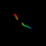
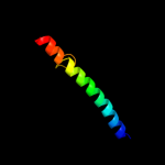
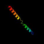
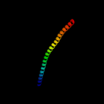
1 c1gk4A_
89.0
13
PDB header: vimentinChain: A: PDB Molecule: vimentin;PDBTitle: human vimentin coil 2b fragment (cys2)
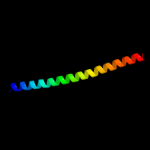
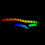
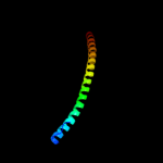
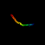
2 c1x8yA_
84.0
7
PDB header: structural proteinChain: A: PDB Molecule: lamin a/c;PDBTitle: human lamin coil 2b
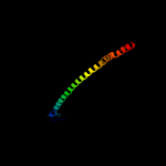
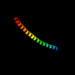
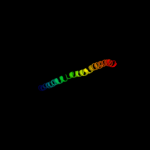
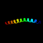
3 d1ucua_
80.9
11
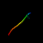
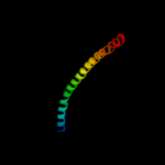
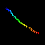
Fold: Phase 1 flagellinSuperfamily: Phase 1 flagellinFamily: Phase 1 flagellin4 c3movB_
78.0
9
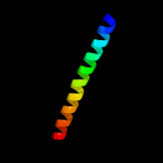
PDB header: structural proteinChain: B: PDB Molecule: lamin-b1;PDBTitle: crystal structure of human lamin-b1 coil 2 segment
5 c1ei3E_
77.7
12
PDB header: PDB COMPND: 6 c1ik9B_
73.2
11
PDB header: gene regulation/ligaseChain: B: PDB Molecule: dna repair protein xrcc4;PDBTitle: crystal structure of a xrcc4-dna ligase iv complex
7 c2pnvA_
69.8
19
PDB header: membrane proteinChain: A: PDB Molecule: small conductance calcium-activated potassiumPDBTitle: crystal structure of the leucine zipper domain of small-2 conductance ca2+-activated k+ (skca) channel from rattus3 norvegicus
8 c1ei3C_
63.0
13
PDB header: PDB COMPND: 9 c2d4yA_
61.7
10
PDB header: structural proteinChain: A: PDB Molecule: flagellar hook-associated protein 1;PDBTitle: crystal structure of a 49k fragment of hap1 (flgk)
10 c2zdiA_
55.6
11
PDB header: chaperoneChain: A: PDB Molecule: prefoldin subunit beta;PDBTitle: crystal structure of prefoldin from pyrococcus horikoshii2 ot3
11 d1fxka_
52.8
15
Fold: Long alpha-hairpinSuperfamily: PrefoldinFamily: Prefoldin12 c1sfcD_
52.4
8
PDB header: transport proteinChain: D: PDB Molecule: protein (snap-25b);PDBTitle: neuronal synaptic fusion complex
13 c1deqO_
48.8
12
PDB header: PDB COMPND: 14 c1l4aD_
47.5
10
PDB header: endocytosis/exocytosisChain: D: PDB Molecule: s-snap25 fusion protein;PDBTitle: x-ray structure of the neuronal complexin/snare complex2 from the squid loligo pealei
15 c3ojaA_
45.0
7
PDB header: protein bindingChain: A: PDB Molecule: leucine-rich immune molecule 1;PDBTitle: crystal structure of lrim1/apl1c complex
16 c3ghgD_
44.2
11
PDB header: blood clottingChain: D: PDB Molecule: fibrinogen alpha chain;PDBTitle: crystal structure of human fibrinogen
17 c2gr7C_
43.4
18
PDB header: membrane proteinChain: C: PDB Molecule: adhesin;PDBTitle: hia 992-1098
18 d2gr7a1
43.4
18
Fold: Pili subunitsSuperfamily: Pili subunitsFamily: YadA C-terminal domain-like19 c3ghgK_
43.0
9
PDB header: blood clottingChain: K: PDB Molecule: fibrinogen beta chain;PDBTitle: crystal structure of human fibrinogen
20 c3swyB_
42.9
14
PDB header: transport proteinChain: B: PDB Molecule: cyclic nucleotide-gated cation channel alpha-3;PDBTitle: cnga3 626-672 containing clz domain
21 c1deqF_
not modelled
42.3
8
PDB header: PDB COMPND: 22 c1kzzA_
not modelled
37.0
12
PDB header: signaling proteinChain: A: PDB Molecule: tnf receptor associated factor 3;PDBTitle: downstream regulator tank binds to the cd40 recognition2 site on traf3
23 c2npsD_
not modelled
36.3
13
PDB header: transport proteinChain: D: PDB Molecule: syntaxin-6;PDBTitle: crystal structure of the early endosomal snare complex
24 c2zdiC_
not modelled
30.9
11
PDB header: chaperoneChain: C: PDB Molecule: prefoldin subunit alpha;PDBTitle: crystal structure of prefoldin from pyrococcus horikoshii2 ot3
25 c1fllA_
not modelled
29.4
13
PDB header: apoptosisChain: A: PDB Molecule: tnf receptor associated factor 3;PDBTitle: molecular basis for cd40 signaling mediated by traf3
26 c3k8vB_
not modelled
27.3
12
PDB header: structural proteinChain: B: PDB Molecule: flagellin homolog;PDBTitle: crysatl structure of a bacterial cell-surface flagellin n20c20
27 c1m1jA_
not modelled
26.7
9
PDB header: blood clottingChain: A: PDB Molecule: fibrinogen alpha subunit;PDBTitle: crystal structure of native chicken fibrinogen with two different2 bound ligands
28 c1zxaB_
not modelled
24.8
10
PDB header: transferaseChain: B: PDB Molecule: cgmp-dependent protein kinase 1, alpha isozyme;PDBTitle: solution structure of the coiled-coil domain of cgmp-2 dependent protein kinase ia
29 c1r48A_
not modelled
22.4
7
PDB header: transport proteinChain: A: PDB Molecule: proline/betaine transporter;PDBTitle: solution structure of the c-terminal cytoplasmic domain2 residues 468-497 of escherichia coli protein prop
30 c3b5nL_
not modelled
22.2
17
PDB header: membrane proteinChain: L: PDB Molecule: protein transport protein sec9;PDBTitle: structure of the yeast plasma membrane snare complex
31 c2j69D_
not modelled
22.2
7
PDB header: hydrolaseChain: D: PDB Molecule: bacterial dynamin-like protein;PDBTitle: bacterial dynamin-like protein bdlp
32 c2ayuA_
not modelled
21.9
13
PDB header: chaperoneChain: A: PDB Molecule: nucleosome assembly protein;PDBTitle: the structure of nucleosome assembly protein suggests a mechanism for2 histone binding and shuttling
33 d2ayua1
not modelled
21.9
13
Fold: NAP-likeSuperfamily: NAP-likeFamily: NAP-like34 c1aq5C_
not modelled
20.8
24
PDB header: coiled-coilChain: C: PDB Molecule: cartilage matrix protein;PDBTitle: high-resolution solution nmr structure of the trimeric coiled-coil2 domain of chicken cartilage matrix protein, 20 structures
35 d2j0na1
not modelled
20.4
13
Fold: IpaD-likeSuperfamily: IpaD-likeFamily: IpaD-like36 c2nrjA_
not modelled
20.4
12
PDB header: toxinChain: A: PDB Molecule: hbl b protein;PDBTitle: crystal structure of hemolysin binding component from2 bacillus cereus
37 c1ca9D_
not modelled
19.0
3
PDB header: tnf signalingChain: D: PDB Molecule: protein (tnf receptor associated factor 2);PDBTitle: structure of tnf receptor associated factor 2 in complex2 with a peptide from tnf-r2
38 d1fxkc_
not modelled
18.3
5
Fold: Long alpha-hairpinSuperfamily: PrefoldinFamily: Prefoldin39 c1gl2D_
not modelled
17.3
8
PDB header: membrane proteinChain: D: PDB Molecule: syntaxin 8;PDBTitle: crystal structure of an endosomal snare core complex
40 c2zvfG_
not modelled
16.8
7
PDB header: ligaseChain: G: PDB Molecule: alanyl-trna synthetase;PDBTitle: crystal structure of archaeoglobus fulgidus alanyl-trna2 synthetase c-terminal dimerization domain
41 c2fxmB_
not modelled
16.3
13
PDB header: contractile proteinChain: B: PDB Molecule: myosin heavy chain, cardiac muscle beta isoform;PDBTitle: structure of the human beta-myosin s2 fragment
42 d1z0pa1
not modelled
15.6
16
Fold: Long alpha-hairpinSuperfamily: SPy1572-likeFamily: SPy1572-like43 c3bvhE_
not modelled
14.7
15
PDB header: blood clottingChain: E: PDB Molecule: fibrinogen beta chain;PDBTitle: crystal structure of recombinant gammad364a fibrinogen fragment d with2 the peptide ligand gly-pro-arg-pro-amide
44 c2oqqB_
not modelled
14.4
16
PDB header: transcriptionChain: B: PDB Molecule: transcription factor hy5;PDBTitle: crystal structure of hy5 leucine zipper homodimer from2 arabidopsis thaliana
45 c3ci9B_
not modelled
14.2
14
PDB header: transcriptionChain: B: PDB Molecule: heat shock factor-binding protein 1;PDBTitle: crystal structure of the human hsbp1
46 d1ivsa1
not modelled
14.2
13
Fold: Long alpha-hairpinSuperfamily: tRNA-binding armFamily: Valyl-tRNA synthetase (ValRS) C-terminal domain47 c2z5hB_
not modelled
13.8
20
PDB header: contractile proteinChain: B: PDB Molecule: general control protein gcn4 and tropomyosinPDBTitle: crystal structure of the head-to-tail junction of2 tropomyosin complexed with a fragment of tnt
48 c1l8dB_
not modelled
11.4
12
PDB header: replicationChain: B: PDB Molecule: dna double-strand break repair rad50 atpase;PDBTitle: rad50 coiled-coil zn hook
49 c1htmB_
not modelled
11.3
7
PDB header: viral proteinChain: B: PDB Molecule: hemagglutinin ha2 chain;PDBTitle: structure of influenza haemagglutinin at the ph of membrane2 fusion
50 c2oszA_
not modelled
11.1
12
PDB header: structural proteinChain: A: PDB Molecule: nucleoporin p58/p45;PDBTitle: structure of nup58/45 suggests flexible nuclear pore diameter by2 intermolecular sliding
51 d1owaa_
not modelled
11.1
7
Fold: Spectrin repeat-likeSuperfamily: Spectrin repeatFamily: Spectrin repeat52 c3ibpA_
not modelled
10.9
23
PDB header: cell cycleChain: A: PDB Molecule: chromosome partition protein mukb;PDBTitle: the crystal structure of the dimerization domain of escherichia coli2 structural maintenance of chromosomes protein mukb
53 c3swfA_
not modelled
10.7
13
PDB header: transport proteinChain: A: PDB Molecule: cgmp-gated cation channel alpha-1;PDBTitle: cnga1 621-690 containing clz domain
54 d1wa8b1
not modelled
10.5
18
Fold: Ferritin-likeSuperfamily: EsxAB dimer-likeFamily: ESAT-6 like55 c2akfB_
not modelled
9.4
10
PDB header: protein bindingChain: B: PDB Molecule: coronin-1a;PDBTitle: crystal structure of the coiled-coil domain of coronin 1
56 c2akfA_
not modelled
9.4
10
PDB header: protein bindingChain: A: PDB Molecule: coronin-1a;PDBTitle: crystal structure of the coiled-coil domain of coronin 1
57 c2akfC_
not modelled
9.4
10
PDB header: protein bindingChain: C: PDB Molecule: coronin-1a;PDBTitle: crystal structure of the coiled-coil domain of coronin 1
58 c3f6hA_
not modelled
8.8
13
PDB header: transferaseChain: A: PDB Molecule: alpha-isopropylmalate synthase;PDBTitle: crystal structure of the regulatory domain of licms in2 complexed with isoleucine - type iii
59 c2xdjF_
not modelled
8.7
13
PDB header: unknown functionChain: F: PDB Molecule: uncharacterized protein ybgf;PDBTitle: crystal structure of the n-terminal domain of e.coli ybgf
60 c2xzrA_
not modelled
7.6
13
PDB header: cell adhesionChain: A: PDB Molecule: immunoglobulin-binding protein eibd;PDBTitle: escherichia coli immunoglobulin-binding protein eibd 391-438 fused2 to gcn4 adaptors
61 c3ipkA_
not modelled
7.4
11
PDB header: cell adhesionChain: A: PDB Molecule: agi/ii;PDBTitle: crystal structure of a3vp1 of agi/ii of streptococcus mutans
62 c3bvhC_
not modelled
7.4
6
PDB header: blood clottingChain: C: PDB Molecule: fibrinogen gamma chain;PDBTitle: crystal structure of recombinant gammad364a fibrinogen fragment d with2 the peptide ligand gly-pro-arg-pro-amide
63 c3l9oA_
not modelled
7.0
14
PDB header: hydrolaseChain: A: PDB Molecule: atp-dependent rna helicase dob1;PDBTitle: crystal structure of mtr4, a co-factor of the nuclear exosome
64 d1hq1a_
not modelled
6.9
22
Fold: Signal peptide-binding domainSuperfamily: Signal peptide-binding domainFamily: Signal peptide-binding domain65 c2dq3A_
not modelled
6.9
13
PDB header: ligaseChain: A: PDB Molecule: seryl-trna synthetase;PDBTitle: crystal structure of aq_298
66 c2hpcH_
not modelled
6.8
11
PDB header: blood clottingChain: H: PDB Molecule: fibrinogen beta chain;PDBTitle: crystal structure of fragment d from human fibrinogen complexed with2 gly-pro-arg-pro-amide.
67 c2d3eD_
not modelled
6.8
11
PDB header: contractile proteinChain: D: PDB Molecule: general control protein gcn4 and tropomyosin 1PDBTitle: crystal structure of the c-terminal fragment of rabbit2 skeletal alpha-tropomyosin
68 c3na7A_
not modelled
6.7
5
PDB header: gene regulation, chaperoneChain: A: PDB Molecule: hp0958;PDBTitle: 2.2 angstrom structure of the hp0958 protein from helicobacter pylori2 ccug 17874
69 d2azeb1
not modelled
6.7
9
Fold: E2F-DP heterodimerization regionSuperfamily: E2F-DP heterodimerization regionFamily: E2F dimerization segment70 c3dtpA_
not modelled
6.6
13
PDB header: contractile proteinChain: A: PDB Molecule: myosin 2 heavy chain chimera of smooth andPDBTitle: tarantula heavy meromyosin obtained by flexible docking to2 tarantula muscle thick filament cryo-em 3d-map
71 c1x59A_
not modelled
6.5
20
PDB header: protein bindingChain: A: PDB Molecule: histidyl-trna synthetase;PDBTitle: solution structures of the whep-trs domain of human2 histidyl-trna synthetase
72 c2gtlO_
not modelled
6.5
15
PDB header: oxygen storage/transportChain: O: PDB Molecule: extracellular hemoglobin linker l3 subunit;PDBTitle: lumbricus erythrocruorin at 3.5a resolution
73 d1e52a_
not modelled
6.5
15
Fold: Long alpha-hairpinSuperfamily: C-terminal UvrC-binding domain of UvrBFamily: C-terminal UvrC-binding domain of UvrB74 d1jyoa_
not modelled
6.4
18
Fold: Secretion chaperone-likeSuperfamily: Type III secretory system chaperone-likeFamily: Type III secretory system chaperone75 c1dlcA_
not modelled
6.4
11
PDB header: toxinChain: A: PDB Molecule: delta-endotoxin cryiiia;PDBTitle: crystal structure of insecticidal delta-endotoxin from2 bacillus thuringiensis at 2.5 angstroms resolution
76 c2h3sB_
not modelled
6.4
25
PDB header: de novo proteinChain: B: PDB Molecule: pancreatic hormone;PDBTitle: cis-azobenzene-avian pancreatic polypeptide bound to dpc2 micelles
77 c2h4bC_
not modelled
6.4
25
PDB header: de novo proteinChain: C: PDB Molecule: pancreatic hormone;PDBTitle: cis-4-aminomethylphenylazobenzoic acid-avian pancreatic2 polypeptide
78 c2h3tB_
not modelled
6.4
25
PDB header: de novo proteinChain: B: PDB Molecule: pancreatic hormone;PDBTitle: trans-(4-aminomethyl)phenylazobenzoic acid-app bound to dpc2 micelles
79 c2h4bD_
not modelled
6.4
25
PDB header: de novo proteinChain: D: PDB Molecule: pancreatic hormone;PDBTitle: cis-4-aminomethylphenylazobenzoic acid-avian pancreatic2 polypeptide
80 d1r6fa_
not modelled
6.3
15
Fold: Virulence-associated V antigenSuperfamily: Virulence-associated V antigenFamily: Virulence-associated V antigen81 c2ym9C_
not modelled
6.3
11
PDB header: cell invasionChain: C: PDB Molecule: cell invasion protein sipd;PDBTitle: sipd from salmonella typhimurium
82 c2k48A_
not modelled
6.2
24
PDB header: viral proteinChain: A: PDB Molecule: nucleoprotein;PDBTitle: nmr structure of the n-terminal coiled coil domain of the2 andes hantavirus nucleocapsid protein
83 c3rrkA_
not modelled
6.2
5
PDB header: proton transportChain: A: PDB Molecule: v-type atpase 116 kda subunit;PDBTitle: crystal structure of the cytoplasmic n-terminal domain of subunit i,2 homolog of subunit a, of v-atpase
84 d1w8oa1
not modelled
6.1
21
Fold: Immunoglobulin-like beta-sandwichSuperfamily: E set domainsFamily: E-set domains of sugar-utilizing enzymes85 c3g9rF_
not modelled
6.1
0
PDB header: viral proteinChain: F: PDB Molecule: fusion complex of hiv-1 envelope glycoproteinPDBTitle: structure of the hiv-1 gp41 membrane-proximal ectodomain2 region in a putative prefusion conformation
86 c1jsdB_
not modelled
6.1
17
PDB header: viral proteinChain: B: PDB Molecule: haemagglutinin (ha2 chain);PDBTitle: crystal structure of swine h9 haemagglutinin
87 d2okua1
not modelled
6.0
10
Fold: N-cbl likeSuperfamily: PG0775 C-terminal domain-likeFamily: PG0775 C-terminal domain-like88 c2v71A_
not modelled
6.0
13
PDB header: nuclear proteinChain: A: PDB Molecule: nuclear distribution protein nude-like 1;PDBTitle: coiled-coil region of nudel
89 c2hpcF_
not modelled
5.9
14
PDB header: blood clottingChain: F: PDB Molecule: fibrinogen, gamma polypeptide;PDBTitle: crystal structure of fragment d from human fibrinogen complexed with2 gly-pro-arg-pro-amide.
90 c1bf5A_
not modelled
5.9
8
PDB header: gene regulation/dnaChain: A: PDB Molecule: signal transducer and activator of transcriptionPDBTitle: tyrosine phosphorylated stat-1/dna complex
91 c2ykqC_
not modelled
5.8
11
PDB header: rna-binding proteinChain: C: PDB Molecule: line-1 orf1p;PDBTitle: structure of the human line-1 orf1p trimer
92 c3bj4B_
not modelled
5.8
9
PDB header: signaling proteinChain: B: PDB Molecule: potassium voltage-gated channel subfamily kqtPDBTitle: the kcnq1 (kv7.1) c-terminus, a multi-tiered scaffold for2 subunit assembly and protein interaction
93 c3mtuE_
not modelled
5.8
16
PDB header: contractile proteinChain: E: PDB Molecule: head morphogenesis protein, tropomyosin alpha-1 chain;PDBTitle: structure of the tropomyosin overlap complex from chicken smooth2 muscle
94 c2ic6B_
not modelled
5.7
22
PDB header: viral proteinChain: B: PDB Molecule: nucleocapsid protein;PDBTitle: the coiled-coil domain (residues 1-75) structure of the sin2 nombre virus nucleocapsid protein
95 c1ezjA_
not modelled
5.6
13
PDB header: viral protein, transferaseChain: A: PDB Molecule: nucleocapsid phosphoprotein;PDBTitle: crystal structure of the multimerization domain of the phosphoprotein2 from sendai virus
96 c2ym0B_
not modelled
5.6
12
PDB header: cell invasionChain: B: PDB Molecule: cell invasion protein sipd;PDBTitle: truncated sipd from salmonella typhimurium
97 c1t6zB_
not modelled
5.5
17
PDB header: transferaseChain: B: PDB Molecule: riboflavin kinase/fmn adenylyltransferase;PDBTitle: crystal structure of riboflavin bound tm379
98 c1ic2B_
not modelled
5.4
18
PDB header: contractile proteinChain: B: PDB Molecule: tropomyosin alpha chain, skeletal muscle;PDBTitle: deciphering the design of the tropomyosin molecule
99 c3hfeC_
not modelled
5.4
14
PDB header: transport proteinChain: C: PDB Molecule: potassium voltage-gated channel subfamily kqt member 1;PDBTitle: a trimeric form of the kv7.1 a domain tail