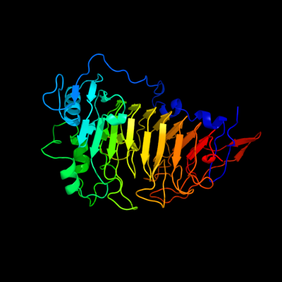
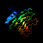
1 c3grhA_
100.0
100
PDB header: hydrolaseChain: A: PDB Molecule: acyl-coa thioester hydrolase ybgc;PDBTitle: crystal structure of escherichia coli ybhc
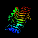
2 d1gq8a_
100.0
30
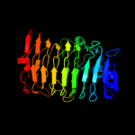
Fold: Single-stranded right-handed beta-helixSuperfamily: Pectin lyase-likeFamily: Pectin methylesterase3 c1xg2A_
100.0
29
PDB header: hydrolase/hydrolase inhibitorChain: A: PDB Molecule: pectinesterase 1;PDBTitle: crystal structure of the complex between pectin2 methylesterase and its inhibitor protein
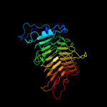
4 d1qjva_
100.0
26
Fold: Single-stranded right-handed beta-helixSuperfamily: Pectin lyase-likeFamily: Pectin methylesterase5 d1ofla_
99.0
13
Fold: Single-stranded right-handed beta-helixSuperfamily: Pectin lyase-likeFamily: Chondroitinase B6 c1dbgA_
98.9
13
PDB header: lyaseChain: A: PDB Molecule: chondroitinase b;PDBTitle: crystal structure of chondroitinase b
7 d1ru4a_
98.9
16
Fold: Single-stranded right-handed beta-helixSuperfamily: Pectin lyase-likeFamily: Pectate transeliminase8 d1bhea_
98.7
11
Fold: Single-stranded right-handed beta-helixSuperfamily: Pectin lyase-likeFamily: Galacturonase9 c3eqnB_
98.7
15
PDB header: hydrolaseChain: B: PDB Molecule: glucan 1,3-beta-glucosidase;PDBTitle: crystal structure of beta-1,3-glucanase from phanerochaete2 chrysosporium (lam55a)
10 d1czfa_
98.5
14
Fold: Single-stranded right-handed beta-helixSuperfamily: Pectin lyase-likeFamily: Galacturonase11 c1czfB_
98.5
13
PDB header: hydrolaseChain: B: PDB Molecule: polygalacturonase ii;PDBTitle: endo-polygalacturonase ii from aspergillus niger
12 c2pyhB_
98.4
13
PDB header: isomeraseChain: B: PDB Molecule: poly(beta-d-mannuronate) c5 epimerase 4;PDBTitle: azotobacter vinelandii mannuronan c-5 epimerase alge4 a-module2 complexed with mannuronan trisaccharide
13 c2iq7D_
98.2
15
PDB header: hydrolaseChain: D: PDB Molecule: endopolygalacturonase;PDBTitle: crystal structure of the polygalacturonase from colletotrichum lupini2 and its implications for the interaction with polygalacturonase-3 inhibiting proteins
14 d1nhca_
98.0
13
Fold: Single-stranded right-handed beta-helixSuperfamily: Pectin lyase-likeFamily: Galacturonase15 c2uveA_
98.0
16
PDB header: hydrolaseChain: A: PDB Molecule: exopolygalacturonase;PDBTitle: structure of yersinia enterocolitica family 282 exopolygalacturonase
16 c2inuC_
98.0
14
PDB header: lyaseChain: C: PDB Molecule: insulin fructotransferase;PDBTitle: crystal structure of insulin fructotransferase in the absence of2 substrate
17 d1hg8a_
98.0
15
Fold: Single-stranded right-handed beta-helixSuperfamily: Pectin lyase-likeFamily: Galacturonase18 d1ia5a_
97.9
12
Fold: Single-stranded right-handed beta-helixSuperfamily: Pectin lyase-likeFamily: Galacturonase19 c3jurA_
97.7
14
PDB header: hydrolaseChain: A: PDB Molecule: exo-poly-alpha-d-galacturonosidase;PDBTitle: the crystal structure of a hyperthermoactive exopolygalacturonase from2 thermotoga maritima
20 d1rmga_
97.2
10
Fold: Single-stranded right-handed beta-helixSuperfamily: Pectin lyase-likeFamily: Galacturonase21 c3gqnA_
not modelled
96.9
15
PDB header: viral proteinChain: A: PDB Molecule: preneck appendage protein;PDBTitle: crystal structure of the pre-mature bacteriophage phi29 gene product2 12
22 c3gq8A_
not modelled
96.5
12
PDB header: viral proteinChain: A: PDB Molecule: preneck appendage protein;PDBTitle: crystal structure of the bacteriophage phi29 gene product2 12 n-terminal fragment in complex with 2-(n-3 cyclohexylamino)ethane sulfonic acid (ches)
23 c2qy1B_
not modelled
96.4
21
PDB header: lyaseChain: B: PDB Molecule: pectate lyase ii;PDBTitle: pectate lyase a31g/r236f from xanthomonas campestris
24 d1idka_
not modelled
96.0
28
Fold: Single-stranded right-handed beta-helixSuperfamily: Pectin lyase-likeFamily: Pectin lyase25 d1pcla_
not modelled
95.6
21
Fold: Single-stranded right-handed beta-helixSuperfamily: Pectin lyase-likeFamily: Pectate lyase-like26 d1qcxa_
not modelled
95.2
25
Fold: Single-stranded right-handed beta-helixSuperfamily: Pectin lyase-likeFamily: Pectin lyase27 d1bn8a_
not modelled
95.0
18
Fold: Single-stranded right-handed beta-helixSuperfamily: Pectin lyase-likeFamily: Pectate lyase-like28 d1k5ca_
not modelled
94.9
12
Fold: Single-stranded right-handed beta-helixSuperfamily: Pectin lyase-likeFamily: Galacturonase29 d1pe9a_
not modelled
93.9
27
Fold: Single-stranded right-handed beta-helixSuperfamily: Pectin lyase-likeFamily: Pectate lyase-like30 d1ogmx2
not modelled
93.4
11
Fold: Single-stranded right-handed beta-helixSuperfamily: Pectin lyase-likeFamily: Dextranase, catalytic domain31 d1o88a_
not modelled
93.4
21
Fold: Single-stranded right-handed beta-helixSuperfamily: Pectin lyase-likeFamily: Pectate lyase-like32 c2vjjA_
not modelled
92.8
19
PDB header: viral proteinChain: A: PDB Molecule: tailspike protein;PDBTitle: tailspike protein of e.coli bacteriophage hk620 in complex2 with hexasaccharide
33 c1vblA_
not modelled
92.4
23
PDB header: lyaseChain: A: PDB Molecule: pectate lyase 47;PDBTitle: structure of the thermostable pectate lyase pl 47
34 c2z8gB_
not modelled
85.1
15
PDB header: hydrolaseChain: B: PDB Molecule: isopullulanase;PDBTitle: aspergillus niger atcc9642 isopullulanase complexed with isopanose
35 d1pxza_
not modelled
85.0
19
Fold: Single-stranded right-handed beta-helixSuperfamily: Pectin lyase-likeFamily: Pectate lyase-like36 c1ogoX_
not modelled
41.2
13
PDB header: hydrolaseChain: X: PDB Molecule: dextranase;PDBTitle: dex49a from penicillium minioluteum complex with isomaltose
37 d1j83a_
not modelled
18.8
23
Fold: Galactose-binding domain-likeSuperfamily: Galactose-binding domain-likeFamily: Family 17 carbohydrate binding module, CBM1738 c3cegA_
not modelled
18.1
26
PDB header: ligaseChain: A: PDB Molecule: baculoviral iap repeat-containing protein 6;PDBTitle: crystal structure of the ubc domain of baculoviral iap2 repeat-containing protein 6
39 d1acoa1
not modelled
16.2
20
Fold: The "swivelling" beta/beta/alpha domainSuperfamily: LeuD/IlvD-likeFamily: LeuD-like40 c2l02B_
not modelled
12.3
45
PDB header: structural genomics, unknown functionChain: B: PDB Molecule: uncharacterized protein;PDBTitle: solution nmr structure of protein bt2368 from bacteroides2 thetaiotaomicron, northeast structural genomics consortium target3 btr375
41 c2p4gA_
not modelled
10.9
7
PDB header: oxidoreductaseChain: A: PDB Molecule: hypothetical protein;PDBTitle: crystal structure of a pyrimidine reductase-like protein (dip1392)2 from corynebacterium diphtheriae nctc at 2.30 a resolution
42 c3owvA_
not modelled
10.0
7
PDB header: hydrolaseChain: A: PDB Molecule: dna-entry nuclease;PDBTitle: structural insights into catalytic and substrate binding mechanisms of2 the strategic enda nuclease from streptococcus pneumoniae
43 c2vbeA_
not modelled
9.9
29
PDB header: viral proteinChain: A: PDB Molecule: tailspike-protein;PDBTitle: tailspike protein of bacteriophage sf6
44 d1dmla1
not modelled
9.8
20
Fold: DNA clampSuperfamily: DNA clampFamily: DNA polymerase processivity factor45 c2xz8A_
not modelled
9.1
33
PDB header: immune systemChain: A: PDB Molecule: peptidoglycan-recognition protein lf;PDBTitle: crystal structure of the lfw ectodomain of the2 peptidoglycan recognition protein lf
46 c1dmlG_
not modelled
8.9
20
PDB header: dna binding protein/transferaseChain: G: PDB Molecule: dna polymerase processivity factor;PDBTitle: crystal structure of herpes simplex ul42 bound to the c-2 terminus of hsv pol
47 c2rfyB_
not modelled
8.9
36
PDB header: hydrolaseChain: B: PDB Molecule: cellulose 1,4-beta-cellobiosidase;PDBTitle: crystal structure of cellobiohydrolase from melanocarpus2 albomyces complexed with cellobiose
48 d2nn6h3
not modelled
8.4
17
Fold: Eukaryotic type KH-domain (KH-domain type I)Superfamily: Eukaryotic type KH-domain (KH-domain type I)Family: Eukaryotic type KH-domain (KH-domain type I)49 c2qyvB_
not modelled
7.8
19
PDB header: hydrolaseChain: B: PDB Molecule: xaa-his dipeptidase;PDBTitle: crystal structure of putative xaa-his dipeptidase (yp_718209.1) from2 haemophilus somnus 129pt at 2.11 a resolution
50 d1autl2
not modelled
7.7
40
Fold: Knottins (small inhibitors, toxins, lectins)Superfamily: EGF/LamininFamily: EGF-type module51 d1ckva_
not modelled
7.6
50
Fold: Monooxygenase (hydroxylase) regulatory proteinSuperfamily: Monooxygenase (hydroxylase) regulatory proteinFamily: Monooxygenase (hydroxylase) regulatory protein52 c3b4qA_
not modelled
7.5
29
PDB header: structural genomics, unknown functionChain: A: PDB Molecule: uncharacterized protein;PDBTitle: crystal structure of a conserved protein domain (unknown2 function) from corynebacterium diphtheriae
53 c3ep1B_
not modelled
7.1
38
PDB header: immune systemChain: B: PDB Molecule: pgrp-hd - peptidoglycan recognition proteinPDBTitle: structure of the pgrp-hd from alvinella pompejana
54 d2cu8a1
not modelled
7.0
21
Fold: Glucocorticoid receptor-like (DNA-binding domain)Superfamily: Glucocorticoid receptor-like (DNA-binding domain)Family: LIM domain55 c3gw6F_
not modelled
6.6
21
PDB header: chaperoneChain: F: PDB Molecule: endo-n-acetylneuraminidase;PDBTitle: intramolecular chaperone
56 d1hqia_
not modelled
6.0
67
Fold: Monooxygenase (hydroxylase) regulatory proteinSuperfamily: Monooxygenase (hydroxylase) regulatory proteinFamily: Monooxygenase (hydroxylase) regulatory protein57 d1ex4a1
not modelled
5.8
37
Fold: SH3-like barrelSuperfamily: DNA-binding domain of retroviral integraseFamily: DNA-binding domain of retroviral integrase58 d1el6a_
not modelled
5.8
25
Fold: Baseplate structural protein gp11Superfamily: Baseplate structural protein gp11Family: Baseplate structural protein gp1159 d2moba_
not modelled
5.6
40
Fold: Monooxygenase (hydroxylase) regulatory proteinSuperfamily: Monooxygenase (hydroxylase) regulatory proteinFamily: Monooxygenase (hydroxylase) regulatory protein60 c2mobA_
not modelled
5.6
40
PDB header: oxidoreductaseChain: A: PDB Molecule: protein (methane monooxygenase regulatoryPDBTitle: methane monooxygenase component b
61 c3rlcA_
not modelled
5.2
21
PDB header: structural proteinChain: A: PDB Molecule: a1 protein;PDBTitle: crystal structure of the read-through domain from bacteriophage qbeta2 a1 protein, hexagonal crystal form
62 d1k4ia_
not modelled
5.1
10
Fold: YrdC/RibBSuperfamily: YrdC/RibBFamily: 3,4-dihydroxy-2-butanone 4-phosphate synthase, DHBP synthase, RibB63 d1xpaa2
not modelled
5.1
27
Fold: Glucocorticoid receptor-like (DNA-binding domain)Superfamily: Glucocorticoid receptor-like (DNA-binding domain)Family: DNA repair factor XPA DNA- and RPA-binding domain, N-terminal subdomain64 c1s2jA_
not modelled
5.0
25
PDB header: hydrolaseChain: A: PDB Molecule: peptidoglycan recognition protein sa cg11709-pa;PDBTitle: crystal structure of the drosophila pattern-recognition2 receptor pgrp-sa
65 d1jb0d_
not modelled
5.0
14
Fold: Photosystem I subunit PsaDSuperfamily: Photosystem I subunit PsaDFamily: Photosystem I subunit PsaD