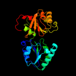
1 c1pjtB_
100.0
90
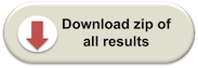
PDB header: transferase/oxidoreductase/lyaseChain: B: PDB Molecule: siroheme synthase;PDBTitle: the structure of the ser128ala point-mutant variant of cysg,2 the multifunctional3 methyltransferase/dehydrogenase/ferrochelatase for4 siroheme synthesis
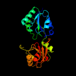
2 d1s4da_
100.0
44
Fold: Tetrapyrrole methylaseSuperfamily: Tetrapyrrole methylaseFamily: Tetrapyrrole methylase3 d1pjqa2
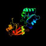
100.0
91
Fold: Tetrapyrrole methylaseSuperfamily: Tetrapyrrole methylaseFamily: Tetrapyrrole methylase4 c2yboA_
100.0
55
PDB header: transferaseChain: A: PDB Molecule: methyltransferase;PDBTitle: the x-ray structure of the sam-dependent uroporphyrinogen2 iii methyltransferase nire from pseudomonas aeruginosa in3 complex with sah
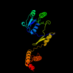
5 c1kyqC_
100.0
21
PDB header: oxidoreductase, lyaseChain: C: PDB Molecule: siroheme biosynthesis protein met8;PDBTitle: met8p: a bifunctional nad-dependent dehydrogenase and2 ferrochelatase involved in siroheme synthesis.
6 d1cbfa_
100.0
30
Fold: Tetrapyrrole methylaseSuperfamily: Tetrapyrrole methylaseFamily: Tetrapyrrole methylase7 c1cbfA_
100.0
30
PDB header: methyltransferaseChain: A: PDB Molecule: cobalt-precorrin-4 transmethylase;PDBTitle: the x-ray structure of a cobalamin biosynthetic enzyme, cobalt2 precorrin-4 methyltransferase, cbif
8 c3dfzB_
100.0
21
PDB header: oxidoreductaseChain: B: PDB Molecule: precorrin-2 dehydrogenase;PDBTitle: sirc, precorrin-2 dehydrogenase
9 d1ve2a1
100.0
42
Fold: Tetrapyrrole methylaseSuperfamily: Tetrapyrrole methylaseFamily: Tetrapyrrole methylase10 c3ndcB_
100.0
32
PDB header: transferaseChain: B: PDB Molecule: precorrin-4 c(11)-methyltransferase;PDBTitle: crystal structure of precorrin-4 c11-methyltransferase from2 rhodobacter capsulatus
11 c3kwpA_
100.0
19
PDB header: transferaseChain: A: PDB Molecule: predicted methyltransferase;PDBTitle: crystal structure of putative methyltransferase from lactobacillus2 brevis
12 d1va0a1
100.0
39
Fold: Tetrapyrrole methylaseSuperfamily: Tetrapyrrole methylaseFamily: Tetrapyrrole methylase13 c2zvbA_
100.0
25
PDB header: transferaseChain: A: PDB Molecule: precorrin-3 c17-methyltransferase;PDBTitle: crystal structure of tt0207 from thermus thermophilus hb8
14 d1wdea_
100.0
18
Fold: Tetrapyrrole methylaseSuperfamily: Tetrapyrrole methylaseFamily: Tetrapyrrole methylase15 c3nutC_
100.0
21
PDB header: transferaseChain: C: PDB Molecule: precorrin-3 methylase;PDBTitle: crystal structure of the methyltransferase cobj
16 c2e0kA_
100.0
20
PDB header: transferaseChain: A: PDB Molecule: precorrin-2 c20-methyltransferase;PDBTitle: crystal structure of cbil, a methyltransferase involved in anaerobic2 vitamin b12 biosynthesis
17 d1wyza1
100.0
14
Fold: Tetrapyrrole methylaseSuperfamily: Tetrapyrrole methylaseFamily: Tetrapyrrole methylase18 c3i4tA_
100.0
18
PDB header: structural genomics, unknown functionChain: A: PDB Molecule: diphthine synthase;PDBTitle: crystal structure of putative diphthine synthase from2 entamoeba histolytica
19 d2deka1
100.0
18
Fold: Tetrapyrrole methylaseSuperfamily: Tetrapyrrole methylaseFamily: Tetrapyrrole methylase20 d1vhva_
100.0
18
Fold: Tetrapyrrole methylaseSuperfamily: Tetrapyrrole methylaseFamily: Tetrapyrrole methylase21 c2qbuA_
not modelled
100.0
21
PDB header: transferaseChain: A: PDB Molecule: precorrin-2 methyltransferase;PDBTitle: crystal structure of methanothermobacter thermautotrophicus cbil
22 c2npnA_
not modelled
100.0
19
PDB header: transferaseChain: A: PDB Molecule: putative cobalamin synthesis related protein;PDBTitle: crystal structure of putative cobalamin synthesis related protein2 (cobf) from corynebacterium diphtheriae
23 c3nd1B_
not modelled
100.0
21
PDB header: transferaseChain: B: PDB Molecule: precorrin-6a synthase/cobf protein;PDBTitle: crystal structure of precorrin-6a synthase from rhodobacter capsulatus
24 c2bb3B_
not modelled
100.0
20
PDB header: transferaseChain: B: PDB Molecule: cobalamin biosynthesis precorrin-6y methylase (cbie);PDBTitle: crystal structure of cobalamin biosynthesis precorrin-6y methylase2 (cbie) from archaeoglobus fulgidus
25 d2bb3a1
not modelled
100.0
18
Fold: Tetrapyrrole methylaseSuperfamily: Tetrapyrrole methylaseFamily: Tetrapyrrole methylase26 d1kyqa1
not modelled
100.0
21
Fold: NAD(P)-binding Rossmann-fold domainsSuperfamily: NAD(P)-binding Rossmann-fold domainsFamily: Siroheme synthase N-terminal domain-like27 d1pjqa1
not modelled
100.0
90
Fold: NAD(P)-binding Rossmann-fold domainsSuperfamily: NAD(P)-binding Rossmann-fold domainsFamily: Siroheme synthase N-terminal domain-like28 c3hh1D_
not modelled
99.9
19
PDB header: transferaseChain: D: PDB Molecule: tetrapyrrole methylase family protein;PDBTitle: the structure of a tetrapyrrole methylase family protein domain from2 chlorobium tepidum tls
29 d1pjqa3
not modelled
99.8
87
Fold: Siroheme synthase middle domains-likeSuperfamily: Siroheme synthase middle domains-likeFamily: Siroheme synthase middle domains-like30 d1kyqa2
not modelled
99.5
20
Fold: Siroheme synthase middle domains-likeSuperfamily: Siroheme synthase middle domains-likeFamily: Siroheme synthase middle domains-like31 c3fq6A_
not modelled
99.0
19
PDB header: transferaseChain: A: PDB Molecule: methyltransferase;PDBTitle: the crystal structure of a methyltransferase domain from bacteroides2 thetaiotaomicron vpi
32 c3d4oA_
not modelled
98.4
15
PDB header: oxidoreductaseChain: A: PDB Molecule: dipicolinate synthase subunit a;PDBTitle: crystal structure of dipicolinate synthase subunit a (np_243269.1)2 from bacillus halodurans at 2.10 a resolution
33 c2rirA_
not modelled
98.2
17
PDB header: oxidoreductaseChain: A: PDB Molecule: dipicolinate synthase, a chain;PDBTitle: crystal structure of dipicolinate synthase, a chain, from bacillus2 subtilis
34 d2jfga1
not modelled
98.1
16
Fold: MurCD N-terminal domainSuperfamily: MurCD N-terminal domainFamily: MurCD N-terminal domain35 d1lssa_
not modelled
97.8
19
Fold: NAD(P)-binding Rossmann-fold domainsSuperfamily: NAD(P)-binding Rossmann-fold domainsFamily: Potassium channel NAD-binding domain36 d1e5qa1
not modelled
97.7
18
Fold: NAD(P)-binding Rossmann-fold domainsSuperfamily: NAD(P)-binding Rossmann-fold domainsFamily: Glyceraldehyde-3-phosphate dehydrogenase-like, N-terminal domain37 c2g1uA_
not modelled
97.6
22
PDB header: transport proteinChain: A: PDB Molecule: hypothetical protein tm1088a;PDBTitle: crystal structure of a putative transport protein (tm1088a) from2 thermotoga maritima at 1.50 a resolution
38 d1li4a1
not modelled
97.6
18
Fold: NAD(P)-binding Rossmann-fold domainsSuperfamily: NAD(P)-binding Rossmann-fold domainsFamily: Formate/glycerate dehydrogenases, NAD-domain39 c3gvpB_
not modelled
97.6
18
PDB header: hydrolaseChain: B: PDB Molecule: adenosylhomocysteinase 3;PDBTitle: human sahh-like domain of human adenosylhomocysteinase 3
40 c1gpjA_
not modelled
97.6
19
PDB header: reductaseChain: A: PDB Molecule: glutamyl-trna reductase;PDBTitle: glutamyl-trna reductase from methanopyrus kandleri
41 c1y8qA_
not modelled
97.5
16
PDB header: ligaseChain: A: PDB Molecule: ubiquitin-like 1 activating enzyme e1a;PDBTitle: sumo e1 activating enzyme sae1-sae2-mg-atp complex
42 c1d4fD_
not modelled
97.5
18
PDB header: hydrolaseChain: D: PDB Molecule: s-adenosylhomocysteine hydrolase;PDBTitle: crystal structure of recombinant rat-liver d244e mutant s-2 adenosylhomocysteine hydrolase
43 c3fwzA_
not modelled
97.5
17
PDB header: membrane proteinChain: A: PDB Molecule: inner membrane protein ybal;PDBTitle: crystal structure of trka-n domain of inner membrane protein ybal from2 escherichia coli
44 d1p3da1
not modelled
97.5
17
Fold: MurCD N-terminal domainSuperfamily: MurCD N-terminal domainFamily: MurCD N-terminal domain45 c3llvA_
not modelled
97.5
20
PDB header: nad(p) binding proteinChain: A: PDB Molecule: exopolyphosphatase-related protein;PDBTitle: the crystal structure of the nad(p)-binding domain of an2 exopolyphosphatase-related protein from archaeoglobus fulgidus to3 1.7a
46 c3dhyC_
not modelled
97.5
22
PDB header: hydrolaseChain: C: PDB Molecule: adenosylhomocysteinase;PDBTitle: crystal structures of mycobacterium tuberculosis s-adenosyl-l-2 homocysteine hydrolase in ternary complex with substrate and3 inhibitors
47 d1jw9b_
not modelled
97.5
29
Fold: Activating enzymes of the ubiquitin-like proteinsSuperfamily: Activating enzymes of the ubiquitin-like proteinsFamily: Molybdenum cofactor biosynthesis protein MoeB48 c3l4bG_
not modelled
97.4
17
PDB header: transport proteinChain: G: PDB Molecule: trka k+ channel protien tm1088b;PDBTitle: crystal structure of an octomeric two-subunit trka k+ channel ring2 gating assembly, tm1088a:tm1088b, from thermotoga maritima
49 c3n58D_
not modelled
97.4
21
PDB header: hydrolaseChain: D: PDB Molecule: adenosylhomocysteinase;PDBTitle: crystal structure of s-adenosyl-l-homocysteine hydrolase from brucella2 melitensis in ternary complex with nad and adenosine, orthorhombic3 form
50 c3gucB_
not modelled
97.4
14
PDB header: transferaseChain: B: PDB Molecule: ubiquitin-like modifier-activating enzyme 5;PDBTitle: human ubiquitin-activating enzyme 5 in complex with amppnp
51 c3d64A_
not modelled
97.4
21
PDB header: hydrolaseChain: A: PDB Molecule: adenosylhomocysteinase;PDBTitle: crystal structure of s-adenosyl-l-homocysteine hydrolase from2 burkholderia pseudomallei
52 c3eywA_
not modelled
97.4
23
PDB header: transport proteinChain: A: PDB Molecule: c-terminal domain of glutathione-regulated potassium-effluxPDBTitle: crystal structure of the c-terminal domain of e. coli kefc in complex2 with keff
53 d1gpja2
not modelled
97.4
20
Fold: NAD(P)-binding Rossmann-fold domainsSuperfamily: NAD(P)-binding Rossmann-fold domainsFamily: Aminoacid dehydrogenase-like, C-terminal domain54 c1zfnA_
not modelled
97.4
29
PDB header: transferaseChain: A: PDB Molecule: adenylyltransferase thif;PDBTitle: structural analysis of escherichia coli thif
55 c1v8bA_
not modelled
97.3
18
PDB header: hydrolaseChain: A: PDB Molecule: adenosylhomocysteinase;PDBTitle: crystal structure of a hydrolase
56 c3oneA_
not modelled
97.3
20
PDB header: hydrolase/hydrolase substrateChain: A: PDB Molecule: adenosylhomocysteinase;PDBTitle: crystal structure of lupinus luteus s-adenosyl-l-homocysteine2 hydrolase in complex with adenine
57 d1v8ba1
not modelled
97.3
17
Fold: NAD(P)-binding Rossmann-fold domainsSuperfamily: NAD(P)-binding Rossmann-fold domainsFamily: Formate/glycerate dehydrogenases, NAD-domain58 c3ic5A_
not modelled
97.2
18
PDB header: structural genomics, unknown functionChain: A: PDB Molecule: putative saccharopine dehydrogenase;PDBTitle: n-terminal domain of putative saccharopine dehydrogenase from ruegeria2 pomeroyi.
59 d1j6ua1
not modelled
97.2
15
Fold: MurCD N-terminal domainSuperfamily: MurCD N-terminal domainFamily: MurCD N-terminal domain60 c2f00A_
not modelled
97.1
22
PDB header: ligaseChain: A: PDB Molecule: udp-n-acetylmuramate--l-alanine ligase;PDBTitle: escherichia coli murc
61 c2axqA_
not modelled
97.1
15
PDB header: oxidoreductaseChain: A: PDB Molecule: saccharopine dehydrogenase;PDBTitle: apo histidine-tagged saccharopine dehydrogenase (l-glu2 forming) from saccharomyces cerevisiae
62 d2hmva1
not modelled
97.1
14
Fold: NAD(P)-binding Rossmann-fold domainsSuperfamily: NAD(P)-binding Rossmann-fold domainsFamily: Potassium channel NAD-binding domain63 c1e5lA_
not modelled
97.1
17
PDB header: oxidoreductaseChain: A: PDB Molecule: saccharopine reductase;PDBTitle: apo saccharopine reductase from magnaporthe grisea
64 d1f0ya2
not modelled
97.1
18
Fold: NAD(P)-binding Rossmann-fold domainsSuperfamily: NAD(P)-binding Rossmann-fold domainsFamily: 6-phosphogluconate dehydrogenase-like, N-terminal domain65 d1gdha1
not modelled
97.1
12
Fold: NAD(P)-binding Rossmann-fold domainsSuperfamily: NAD(P)-binding Rossmann-fold domainsFamily: Formate/glycerate dehydrogenases, NAD-domain66 d1np3a2
not modelled
97.0
14
Fold: NAD(P)-binding Rossmann-fold domainsSuperfamily: NAD(P)-binding Rossmann-fold domainsFamily: 6-phosphogluconate dehydrogenase-like, N-terminal domain67 c3gznB_
not modelled
97.0
12
PDB header: protein binding/ligaseChain: B: PDB Molecule: nedd8-activating enzyme e1 catalytic subunit;PDBTitle: structure of nedd8-activating enzyme in complex with nedd82 and mln4924
68 c3c85A_
not modelled
97.0
16
PDB header: transport proteinChain: A: PDB Molecule: putative glutathione-regulated potassium-efflux systemPDBTitle: crystal structure of trka domain of putative glutathione-regulated2 potassium-efflux kefb from vibrio parahaemolyticus
69 d1ygya1
not modelled
97.0
15
Fold: NAD(P)-binding Rossmann-fold domainsSuperfamily: NAD(P)-binding Rossmann-fold domainsFamily: Formate/glycerate dehydrogenases, NAD-domain70 c3uagA_
not modelled
97.0
16
PDB header: ligaseChain: A: PDB Molecule: protein (udp-n-acetylmuramoyl-l-alanine:d-PDBTitle: udp-n-acetylmuramoyl-l-alanine:d-glutamate ligase
71 c1y8qD_
not modelled
97.0
16
PDB header: ligaseChain: D: PDB Molecule: ubiquitin-like 2 activating enzyme e1b;PDBTitle: sumo e1 activating enzyme sae1-sae2-mg-atp complex
72 c3hn7A_
not modelled
97.0
15
PDB header: ligaseChain: A: PDB Molecule: udp-n-acetylmuramate-l-alanine ligase;PDBTitle: crystal structure of a murein peptide ligase mpl (psyc_0032) from2 psychrobacter arcticus 273-4 at 1.65 a resolution
73 c1wwkA_
not modelled
96.9
15
PDB header: oxidoreductaseChain: A: PDB Molecule: phosphoglycerate dehydrogenase;PDBTitle: crystal structure of phosphoglycerate dehydrogenase from pyrococcus2 horikoshii ot3
74 c3pgjB_
not modelled
96.9
19
PDB header: oxidoreductaseChain: B: PDB Molecule: shikimate dehydrogenase;PDBTitle: 2.49 angstrom resolution crystal structure of shikimate 5-2 dehydrogenase (aroe) from vibrio cholerae o1 biovar eltor str. n169613 in complex with shikimate
75 c3kydB_
not modelled
96.9
15
PDB header: ligaseChain: B: PDB Molecule: sumo-activating enzyme subunit 2;PDBTitle: human sumo e1~sumo1-amp tetrahedral intermediate mimic
76 c2eezG_
not modelled
96.9
17
PDB header: oxidoreductaseChain: G: PDB Molecule: alanine dehydrogenase;PDBTitle: crystal structure of alanine dehydrogenase from themus thermophilus
77 c2nvuB_
not modelled
96.9
12
PDB header: protein turnover, ligaseChain: B: PDB Molecule: maltose binding protein/nedd8-activating enzymePDBTitle: structure of appbp1-uba3~nedd8-nedd8-mgatp-ubc12(c111a), a2 trapped ubiquitin-like protein activation complex
78 d1id1a_
not modelled
96.9
22
Fold: NAD(P)-binding Rossmann-fold domainsSuperfamily: NAD(P)-binding Rossmann-fold domainsFamily: Potassium channel NAD-binding domain79 d2naca1
not modelled
96.9
13
Fold: NAD(P)-binding Rossmann-fold domainsSuperfamily: NAD(P)-binding Rossmann-fold domainsFamily: Formate/glycerate dehydrogenases, NAD-domain80 c2hk8B_
not modelled
96.9
17
PDB header: oxidoreductaseChain: B: PDB Molecule: shikimate dehydrogenase;PDBTitle: crystal structure of shikimate dehydrogenase from aquifex2 aeolicus at 2.35 angstrom resolution
81 c1np3B_
not modelled
96.8
16
PDB header: oxidoreductaseChain: B: PDB Molecule: ketol-acid reductoisomerase;PDBTitle: crystal structure of class i acetohydroxy acid isomeroreductase from2 pseudomonas aeruginosa
82 c1gdhA_
not modelled
96.8
12
PDB header: oxidoreductase(choh (d)-nad(p)+ (a))Chain: A: PDB Molecule: d-glycerate dehydrogenase;PDBTitle: crystal structure of a nad-dependent d-glycerate2 dehydrogenase at 2.4 angstroms resolution
83 c3h9gA_
not modelled
96.8
12
PDB header: transferase/antibioticChain: A: PDB Molecule: mccb protein;PDBTitle: crystal structure of e. coli mccb + mcca-n7isoasn
84 c2eggA_
not modelled
96.8
19
PDB header: oxidoreductaseChain: A: PDB Molecule: shikimate 5-dehydrogenase;PDBTitle: crystal structure of shikimate 5-dehydrogenase (aroe) from2 geobacillus kaustophilus
85 d1j4aa1
not modelled
96.7
15
Fold: NAD(P)-binding Rossmann-fold domainsSuperfamily: NAD(P)-binding Rossmann-fold domainsFamily: Formate/glycerate dehydrogenases, NAD-domain86 c2g76A_
not modelled
96.7
18
PDB header: oxidoreductaseChain: A: PDB Molecule: d-3-phosphoglycerate dehydrogenase;PDBTitle: crystal structure of human 3-phosphoglycerate dehydrogenase
87 d1l7da1
not modelled
96.7
11
Fold: NAD(P)-binding Rossmann-fold domainsSuperfamily: NAD(P)-binding Rossmann-fold domainsFamily: Formate/glycerate dehydrogenases, NAD-domain88 c2g5cD_
not modelled
96.7
13
PDB header: oxidoreductaseChain: D: PDB Molecule: prephenate dehydrogenase;PDBTitle: crystal structure of prephenate dehydrogenase from aquifex aeolicus
89 d1pjca1
not modelled
96.6
17
Fold: NAD(P)-binding Rossmann-fold domainsSuperfamily: NAD(P)-binding Rossmann-fold domainsFamily: Formate/glycerate dehydrogenases, NAD-domain90 c1nytC_
not modelled
96.6
17
PDB header: oxidoreductaseChain: C: PDB Molecule: shikimate 5-dehydrogenase;PDBTitle: shikimate dehydrogenase aroe complexed with nadp+
91 d2fy8a1
not modelled
96.6
15
Fold: NAD(P)-binding Rossmann-fold domainsSuperfamily: NAD(P)-binding Rossmann-fold domainsFamily: Potassium channel NAD-binding domain92 d1mx3a1
not modelled
96.6
14
Fold: NAD(P)-binding Rossmann-fold domainsSuperfamily: NAD(P)-binding Rossmann-fold domainsFamily: Formate/glycerate dehydrogenases, NAD-domain93 c3pwzA_
not modelled
96.6
18
PDB header: oxidoreductaseChain: A: PDB Molecule: shikimate dehydrogenase 3;PDBTitle: crystal structure of an ael1 enzyme from pseudomonas putida
94 c3vh3A_
not modelled
96.6
18
PDB header: metal binding protein/protein transportChain: A: PDB Molecule: ubiquitin-like modifier-activating enzyme atg7;PDBTitle: crystal structure of atg7ctd-atg8 complex
95 c1j4aA_
not modelled
96.6
16
PDB header: oxidoreductaseChain: A: PDB Molecule: d-lactate dehydrogenase;PDBTitle: insights into domain closure, substrate specificity and2 catalysis of d-lactate dehydrogenase from lactobacillus3 bulgaricus
96 c2f1kD_
not modelled
96.5
16
PDB header: oxidoreductaseChain: D: PDB Molecule: prephenate dehydrogenase;PDBTitle: crystal structure of synechocystis arogenate dehydrogenase
97 c2dbqA_
not modelled
96.5
13
PDB header: oxidoreductaseChain: A: PDB Molecule: glyoxylate reductase;PDBTitle: crystal structure of glyoxylate reductase (ph0597) from pyrococcus2 horikoshii ot3, complexed with nadp (i41)
98 d1nyta1
not modelled
96.5
19
Fold: NAD(P)-binding Rossmann-fold domainsSuperfamily: NAD(P)-binding Rossmann-fold domainsFamily: Aminoacid dehydrogenase-like, C-terminal domain99 d2f1ka2
not modelled
96.5
17
Fold: NAD(P)-binding Rossmann-fold domainsSuperfamily: NAD(P)-binding Rossmann-fold domainsFamily: 6-phosphogluconate dehydrogenase-like, N-terminal domain100 c3g0oA_
not modelled
96.5
20
PDB header: oxidoreductaseChain: A: PDB Molecule: 3-hydroxyisobutyrate dehydrogenase;PDBTitle: crystal structure of 3-hydroxyisobutyrate dehydrogenase2 (ygbj) from salmonella typhimurium
101 c3evtA_
not modelled
96.5
15
PDB header: oxidoreductaseChain: A: PDB Molecule: phosphoglycerate dehydrogenase;PDBTitle: crystal structure of phosphoglycerate dehydrogenase from2 lactobacillus plantarum
102 d1yovb1
not modelled
96.5
11
Fold: Activating enzymes of the ubiquitin-like proteinsSuperfamily: Activating enzymes of the ubiquitin-like proteinsFamily: Ubiquitin activating enzymes (UBA)103 c3d1lB_
not modelled
96.4
17
PDB header: oxidoreductaseChain: B: PDB Molecule: putative nadp oxidoreductase bf3122;PDBTitle: crystal structure of putative nadp oxidoreductase bf3122 from2 bacteroides fragilis
104 c3vh1A_
not modelled
96.4
16
PDB header: metal binding proteinChain: A: PDB Molecule: ubiquitin-like modifier-activating enzyme atg7;PDBTitle: crystal structure of saccharomyces cerevisiae atg7 (1-595)
105 c3b1fA_
not modelled
96.4
10
PDB header: oxidoreductaseChain: A: PDB Molecule: putative prephenate dehydrogenase;PDBTitle: crystal structure of prephenate dehydrogenase from streptococcus2 mutans
106 c3cmmA_
not modelled
96.4
14
PDB header: ligase/protein bindingChain: A: PDB Molecule: ubiquitin-activating enzyme e1 1;PDBTitle: crystal structure of the uba1-ubiquitin complex
107 c3hwrA_
not modelled
96.4
22
PDB header: oxidoreductaseChain: A: PDB Molecule: 2-dehydropantoate 2-reductase;PDBTitle: crystal structure of pane/apba family ketopantoate reductase2 (yp_299159.1) from ralstonia eutropha jmp134 at 2.15 a resolution
108 c3o8qB_
not modelled
96.4
23
PDB header: oxidoreductaseChain: B: PDB Molecule: shikimate 5-dehydrogenase i alpha;PDBTitle: 1.45 angstrom resolution crystal structure of shikimate 5-2 dehydrogenase (aroe) from vibrio cholerae
109 c2pv7B_
not modelled
96.4
14
PDB header: isomerase, oxidoreductaseChain: B: PDB Molecule: t-protein [includes: chorismate mutase (ec 5.4.99.5) (cm)PDBTitle: crystal structure of chorismate mutase / prephenate dehydrogenase2 (tyra) (1574749) from haemophilus influenzae rd at 2.00 a resolution
110 c3cumA_
not modelled
96.3
19
PDB header: oxidoreductaseChain: A: PDB Molecule: probable 3-hydroxyisobutyrate dehydrogenase;PDBTitle: crystal structure of a possible 3-hydroxyisobutyrate dehydrogenase2 from pseudomonas aeruginosa pao1
111 d1nvta1
not modelled
96.3
18
Fold: NAD(P)-binding Rossmann-fold domainsSuperfamily: NAD(P)-binding Rossmann-fold domainsFamily: Aminoacid dehydrogenase-like, C-terminal domain112 c3oj0A_
not modelled
96.3
25
PDB header: oxidoreductaseChain: A: PDB Molecule: glutamyl-trna reductase;PDBTitle: crystal structure of glutamyl-trna reductase from thermoplasma2 volcanium (nucleotide binding domain)
113 c3n7uD_
not modelled
96.3
13
PDB header: oxidoreductaseChain: D: PDB Molecule: formate dehydrogenase;PDBTitle: nad-dependent formate dehydrogenase from higher-plant arabidopsis2 thaliana in complex with nad and azide
114 c1l7eC_
not modelled
96.3
8
PDB header: oxidoreductaseChain: C: PDB Molecule: nicotinamide nucleotide transhydrogenase,PDBTitle: crystal structure of r. rubrum transhydrogenase domain i2 with bound nadh
115 d1leha1
not modelled
96.3
20
Fold: NAD(P)-binding Rossmann-fold domainsSuperfamily: NAD(P)-binding Rossmann-fold domainsFamily: Aminoacid dehydrogenase-like, C-terminal domain116 d1txga2
not modelled
96.3
18
Fold: NAD(P)-binding Rossmann-fold domainsSuperfamily: NAD(P)-binding Rossmann-fold domainsFamily: 6-phosphogluconate dehydrogenase-like, N-terminal domain117 c3kboB_
not modelled
96.2
16
PDB header: oxidoreductaseChain: B: PDB Molecule: glyoxylate/hydroxypyruvate reductase a;PDBTitle: 2.14 angstrom crystal structure of putative oxidoreductase (ycdw) from2 salmonella typhimurium in complex with nadp
118 c3bazA_
not modelled
96.2
12
PDB header: oxidoreductaseChain: A: PDB Molecule: hydroxyphenylpyruvate reductase;PDBTitle: structure of hydroxyphenylpyruvate reductase from coleus blumei in2 complex with nadp+
119 d2gz1a1
not modelled
96.2
11
Fold: NAD(P)-binding Rossmann-fold domainsSuperfamily: NAD(P)-binding Rossmann-fold domainsFamily: Glyceraldehyde-3-phosphate dehydrogenase-like, N-terminal domain120 c2nloA_
not modelled
96.2
19
PDB header: oxidoreductaseChain: A: PDB Molecule: shikimate dehydrogenase;PDBTitle: crystal structure of the quinate dehydrogenase from corynebacterium2 glutamicum