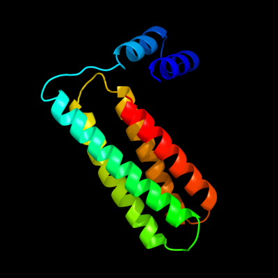
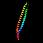
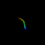
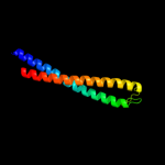
1 c1y4cA_
75.4
9
PDB header: de novo proteinChain: A: PDB Molecule: maltose binding protein fused with designedPDBTitle: designed helical protein fusion mbp
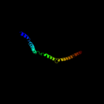
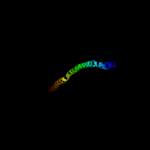
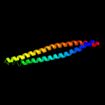
2 c3ojaB_
72.9
14
PDB header: protein bindingChain: B: PDB Molecule: anopheles plasmodium-responsive leucine-rich repeat proteinPDBTitle: crystal structure of lrim1/apl1c complex
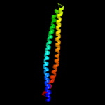
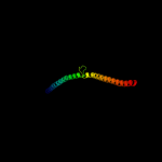
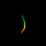
3 c2fxmB_
70.7
12
PDB header: contractile proteinChain: B: PDB Molecule: myosin heavy chain, cardiac muscle beta isoform;PDBTitle: structure of the human beta-myosin s2 fragment
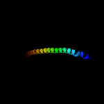
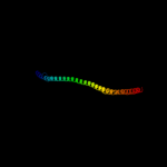
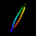
4 c3u59C_
66.9
12
PDB header: contractile proteinChain: C: PDB Molecule: tropomyosin beta chain;PDBTitle: n-terminal 98-aa fragment of smooth muscle tropomyosin beta
5 c2efrB_
66.3
11
PDB header: contractile proteinChain: B: PDB Molecule: general control protein gcn4 and tropomyosin 1 alpha chain;PDBTitle: crystal structure of the c-terminal tropomyosin fragment with n- and2 c-terminal extensions of the leucine zipper at 1.8 angstroms3 resolution
6 c2d3eD_
53.6
16
PDB header: contractile proteinChain: D: PDB Molecule: general control protein gcn4 and tropomyosin 1PDBTitle: crystal structure of the c-terminal fragment of rabbit2 skeletal alpha-tropomyosin
7 c2y3aB_
40.2
13
PDB header: transferaseChain: B: PDB Molecule: phosphatidylinositol 3-kinase regulatory subunit beta;PDBTitle: crystal structure of p110beta in complex with icsh2 of p85beta and2 the drug gdc-0941
8 c1deqF_
37.3
11
PDB header: PDB COMPND: 9 c4a55B_
35.6
13
PDB header: transferaseChain: B: PDB Molecule: phosphatidylinositol 3-kinase regulatory subunit alpha;PDBTitle: crystal structure of p110alpha in complex with ish2 of p85alpha and2 the inhibitor pik-108
10 c3cvfA_
34.4
15
PDB header: signaling proteinChain: A: PDB Molecule: homer protein homolog 3;PDBTitle: crystal structure of the carboxy terminus of homer3
11 c2b9cA_
30.8
12
PDB header: contractile proteinChain: A: PDB Molecule: striated-muscle alpha tropomyosin;PDBTitle: structure of tropomyosin's mid-region: bending and binding2 sites for actin
12 c3ghgK_
27.3
8
PDB header: blood clottingChain: K: PDB Molecule: fibrinogen beta chain;PDBTitle: crystal structure of human fibrinogen
13 c3ol1A_
25.4
12
PDB header: structural proteinChain: A: PDB Molecule: vimentin;PDBTitle: crystal structure of vimentin (fragment 144-251) from homo sapiens,2 northeast structural genomics consortium target hr4796b
14 c1ei3E_
25.1
10
PDB header: PDB COMPND: 15 c1l8dB_
23.6
11
PDB header: replicationChain: B: PDB Molecule: dna double-strand break repair rad50 atpase;PDBTitle: rad50 coiled-coil zn hook
16 c3hizB_
22.8
13
PDB header: transferase/oncoproteinChain: B: PDB Molecule: phosphatidylinositol 3-kinase regulatory subunitPDBTitle: crystal structure of p110alpha h1047r mutant in complex with2 nish2 of p85alpha
17 c3ipkA_
20.3
11
PDB header: cell adhesionChain: A: PDB Molecule: agi/ii;PDBTitle: crystal structure of a3vp1 of agi/ii of streptococcus mutans
18 c2rd0B_
19.4
9
PDB header: transferase/oncoproteinChain: B: PDB Molecule: phosphatidylinositol 3-kinase regulatory subunit alpha;PDBTitle: structure of a human p110alpha/p85alpha complex
19 c3cveC_
19.3
15
PDB header: signaling proteinChain: C: PDB Molecule: homer protein homolog 1;PDBTitle: crystal structure of the carboxy terminus of homer1
20 c3o0zD_
16.8
11
PDB header: transferaseChain: D: PDB Molecule: rho-associated protein kinase 1;PDBTitle: crystal structure of a coiled-coil domain from human rock i
21 c2gl2B_
not modelled
16.2
18
PDB header: cell adhesionChain: B: PDB Molecule: adhesion a;PDBTitle: crystal structure of the tetra muntant (t66g,r67g,f68g,2 y69g) of bacterial adhesin fada
22 c2v71A_
not modelled
16.1
13
PDB header: nuclear proteinChain: A: PDB Molecule: nuclear distribution protein nude-like 1;PDBTitle: coiled-coil region of nudel
23 c3dtpA_
not modelled
15.8
13
PDB header: contractile proteinChain: A: PDB Molecule: myosin 2 heavy chain chimera of smooth andPDBTitle: tarantula heavy meromyosin obtained by flexible docking to2 tarantula muscle thick filament cryo-em 3d-map
24 c3na7A_
not modelled
15.5
8
PDB header: gene regulation, chaperoneChain: A: PDB Molecule: hp0958;PDBTitle: 2.2 angstrom structure of the hp0958 protein from helicobacter pylori2 ccug 17874
25 c3ojaA_
not modelled
14.6
9
PDB header: protein bindingChain: A: PDB Molecule: leucine-rich immune molecule 1;PDBTitle: crystal structure of lrim1/apl1c complex
26 c3a5tB_
not modelled
14.5
15
PDB header: transcription regulator/dnaChain: B: PDB Molecule: transcription factor mafg;PDBTitle: crystal structure of mafg-dna complex
27 c3cwgA_
not modelled
13.6
11
PDB header: transcriptionChain: A: PDB Molecule: signal transducer and activator of transcriptionPDBTitle: unphosphorylated mouse stat3 core fragment
28 c1deqO_
not modelled
13.0
8
PDB header: PDB COMPND: 29 c3u1aC_
not modelled
11.8
19
PDB header: contractile proteinChain: C: PDB Molecule: smooth muscle tropomyosin alpha;PDBTitle: n-terminal 81-aa fragment of smooth muscle tropomyosin alpha
30 c3ajwA_
not modelled
11.5
15
PDB header: protein transportChain: A: PDB Molecule: flagellar flij protein;PDBTitle: structure of flij, a soluble component of flagellar type iii export2 apparatus
31 c1c1gA_
not modelled
11.5
16
PDB header: contractile proteinChain: A: PDB Molecule: tropomyosin;PDBTitle: crystal structure of tropomyosin at 7 angstroms resolution2 in the spermine-induced crystal form
32 d2obpa1
not modelled
11.4
18
Fold: DNA/RNA-binding 3-helical bundleSuperfamily: "Winged helix" DNA-binding domainFamily: ReutB4095-like33 c1ei3C_
not modelled
11.0
21
PDB header: PDB COMPND: 34 c2jziB_
not modelled
11.0
18
PDB header: metal binding proteinChain: B: PDB Molecule: serine/threonine-protein phosphatase 2bPDBTitle: structure of calmodulin complexed with the calmodulin2 binding domain of calcineurin
35 c2v66C_
not modelled
10.8
13
PDB header: structural proteinChain: C: PDB Molecule: nuclear distribution protein nude-like 1;PDBTitle: crystal structure of the coiled-coil domain of ndel1 (a.a.2 58 to 169)c
36 c1jchC_
not modelled
10.6
10
PDB header: ribosome inhibitor, hydrolaseChain: C: PDB Molecule: colicin e3;PDBTitle: crystal structure of colicin e3 in complex with its immunity protein
37 c1ic2B_
not modelled
10.5
18
PDB header: contractile proteinChain: B: PDB Molecule: tropomyosin alpha chain, skeletal muscle;PDBTitle: deciphering the design of the tropomyosin molecule
38 c1g8xB_
not modelled
10.4
13
PDB header: structural proteinChain: B: PDB Molecule: myosin ii heavy chain fused to alpha-actinin 3;PDBTitle: structure of a genetically engineered molecular motor
39 c3m9bK_
not modelled
10.2
24
PDB header: chaperoneChain: K: PDB Molecule: proteasome-associated atpase;PDBTitle: crystal structure of the amino terminal coiled coil domain and the2 inter domain of the mycobacterium tuberculosis proteasomal atpase mpa
40 c2wt7B_
not modelled
10.2
14
PDB header: transcriptionChain: B: PDB Molecule: transcription factor mafb;PDBTitle: crystal structure of the bzip heterodimeric complex2 mafb:cfos bound to dna
41 d1vmaa1
not modelled
9.9
21
Fold: Four-helical up-and-down bundleSuperfamily: Domain of the SRP/SRP receptor G-proteinsFamily: Domain of the SRP/SRP receptor G-proteins42 c2z2qF_
not modelled
9.8
18
PDB header: virus/rnaChain: F: PDB Molecule: coat protein gamma;PDBTitle: crystal structure of flock house virus
43 c1deqD_
not modelled
9.5
13
PDB header: PDB COMPND: 44 d1kbha_
not modelled
9.5
24
Fold: Nuclear receptor coactivator interlocking domainSuperfamily: Nuclear receptor coactivator interlocking domainFamily: Nuclear receptor coactivator interlocking domain45 c1fosE_
not modelled
9.3
12
PDB header: transcription/dnaChain: E: PDB Molecule: p55-c-fos proto-oncogene protein;PDBTitle: two human c-fos:c-jun:dna complexes
46 c3hnwB_
not modelled
9.3
10
PDB header: structural genomics, unknown functionChain: B: PDB Molecule: uncharacterized protein;PDBTitle: crystal structure of a basic coiled-coil protein of unknown function2 from eubacterium eligens atcc 27750
47 d1xvha1
not modelled
9.3
16
Fold: immunoglobulin/albumin-binding domain-likeSuperfamily: Bacterial immunoglobulin/albumin-binding domainsFamily: GA module, an albumin-binding domain48 c1fosF_
not modelled
9.1
15
PDB header: transcription/dnaChain: F: PDB Molecule: c-jun proto-oncogene protein;PDBTitle: two human c-fos:c-jun:dna complexes
49 c2xdjF_
not modelled
9.0
7
PDB header: unknown functionChain: F: PDB Molecule: uncharacterized protein ybgf;PDBTitle: crystal structure of the n-terminal domain of e.coli ybgf
50 d1z0kb1
not modelled
8.8
31
Fold: Long alpha-hairpinSuperfamily: Rabenosyn-5 Rab-binding domain-likeFamily: Rabenosyn-5 Rab-binding domain-like51 c2oevA_
not modelled
8.8
13
PDB header: protein transportChain: A: PDB Molecule: programmed cell death 6-interacting protein;PDBTitle: crystal structure of alix/aip1
52 c2v1yB_
not modelled
8.6
9
PDB header: transferaseChain: B: PDB Molecule: phosphatidylinositol 3-kinase regulatory subunit alpha;PDBTitle: structure of a phosphoinositide 3-kinase alpha adaptor-2 binding domain (abd) in a complex with the ish2 domain3 from p85 alpha
53 c2k10A_
not modelled
8.5
26
PDB header: antimicrobial proteinChain: A: PDB Molecule: ranatuerin-2csa;PDBTitle: confirmational analysis of the broad-spectrum antibacterial2 peptide, rantuerin-2csa: identification of a full length3 helix-turn-helix motif
54 d1yzma1
not modelled
8.4
31
Fold: Long alpha-hairpinSuperfamily: Rabenosyn-5 Rab-binding domain-likeFamily: Rabenosyn-5 Rab-binding domain-like55 c2x7aB_
not modelled
8.4
11
PDB header: immune systemChain: B: PDB Molecule: bone marrow stromal antigen 2;PDBTitle: structural basis of hiv-1 tethering to membranes by the2 bst2-tetherin ectodomain
56 c3n4xB_
not modelled
8.4
14
PDB header: replicationChain: B: PDB Molecule: monopolin complex subunit csm1;PDBTitle: structure of csm1 full-length
57 c2dohC_
not modelled
8.3
40
PDB header: hydrolaseChain: C: PDB Molecule: plasminogen-binding group a streptococcal m-like proteinPDBTitle: the x-ray crystallographic structure of the angiogenesis inhibitor,2 angiostatin, bound a to a peptide from the group a streptococcal3 surface protein pam
58 c1t2kD_
not modelled
8.3
15
PDB header: transcription/dnaChain: D: PDB Molecule: cyclic-amp-dependent transcription factor atf-2;PDBTitle: structure of the dna binding domains of irf3, atf-2 and jun2 bound to dna
59 d2c52b1
not modelled
8.2
15
Fold: Nuclear receptor coactivator interlocking domainSuperfamily: Nuclear receptor coactivator interlocking domainFamily: Nuclear receptor coactivator interlocking domain60 c2doiB_
not modelled
8.2
40
PDB header: hydrolaseChain: B: PDB Molecule: plasminogen-binding group a streptococcal m-like proteinPDBTitle: the x-ray crystallographic structure of the angiogenesis inhibitor,2 angiostatin, bound to a peptide from the group a streptococcus3 protein pam
61 c2doiC_
not modelled
8.2
40
PDB header: hydrolaseChain: C: PDB Molecule: plasminogen-binding group a streptococcal m-like proteinPDBTitle: the x-ray crystallographic structure of the angiogenesis inhibitor,2 angiostatin, bound to a peptide from the group a streptococcus3 protein pam
62 c1i5kD_
not modelled
8.1
40
PDB header: blood clottingChain: D: PDB Molecule: m protein;PDBTitle: structure and binding determinants of the recombinant kringle-2 domain2 of human plasminogen to an internal peptide from a group a3 streptococcal surface protein
63 c1i5kC_
not modelled
8.1
40
PDB header: blood clottingChain: C: PDB Molecule: m protein;PDBTitle: structure and binding determinants of the recombinant kringle-2 domain2 of human plasminogen to an internal peptide from a group a3 streptococcal surface protein
64 c1bg1A_
not modelled
8.1
15
PDB header: transcription/dnaChain: A: PDB Molecule: protein (transcription factor stat3b);PDBTitle: transcription factor stat3b/dna complex
65 d1k8ib2
not modelled
8.0
19
Fold: MHC antigen-recognition domainSuperfamily: MHC antigen-recognition domainFamily: MHC antigen-recognition domain66 c1ci6A_
not modelled
7.9
6
PDB header: transcriptionChain: A: PDB Molecule: transcription factor atf-4;PDBTitle: transcription factor atf4-c/ebp beta bzip heterodimer
67 c2kj4B_
not modelled
7.5
40
PDB header: blood clottingChain: B: PDB Molecule: vek-30;PDBTitle: solution structure of the complex of vek-30 and plasminogen2 kringle 2
68 c1junB_
not modelled
7.4
24
PDB header: transcription regulationChain: B: PDB Molecule: c-jun homodimer;PDBTitle: nmr study of c-jun homodimer
69 d2gqba1
not modelled
7.4
18
Fold: RPA2825-likeSuperfamily: RPA2825-likeFamily: RPA2825-like70 c1uo3B_
not modelled
7.4
36
PDB header: four helix bundleChain: B: PDB Molecule: general control protein gcn4;PDBTitle: structure based engineering of internal molecular surfaces2 of four helix bundles
71 c1u9fC_
not modelled
7.3
36
PDB header: transcriptionChain: C: PDB Molecule: general control protein gcn4;PDBTitle: heterocyclic peptide backbone modification in gcn4-pli based coiled2 coils: replacement of k(15)l(16)
72 c1u9fB_
not modelled
7.3
36
PDB header: transcriptionChain: B: PDB Molecule: general control protein gcn4;PDBTitle: heterocyclic peptide backbone modification in gcn4-pli based coiled2 coils: replacement of k(15)l(16)
73 c1vmaA_
not modelled
7.3
13
PDB header: protein transportChain: A: PDB Molecule: cell division protein ftsy;PDBTitle: crystal structure of cell division protein ftsy (tm0570) from2 thermotoga maritima at 1.60 a resolution
74 c1o8tA_
not modelled
7.3
19
PDB header: lipid transportChain: A: PDB Molecule: apolipoprotein c-ii;PDBTitle: global structure and dynamics of human apolipoprotein cii2 in complex with micelles: evidence for increased mobility3 of the helix involvved in the activation of lipoprotein4 lipase.
75 c1unuA_
not modelled
7.3
36
PDB header: four helix bundleChain: A: PDB Molecule: general control protein gcn4;PDBTitle: structure based engineering of internal molecular surfaces2 of four helix bundles
76 c1unuB_
not modelled
7.3
36
PDB header: four helix bundleChain: B: PDB Molecule: general control protein gcn4;PDBTitle: structure based engineering of internal molecular surfaces2 of four helix bundles
77 c1uo5A_
not modelled
7.2
36
PDB header: four helix bundleChain: A: PDB Molecule: general control protein gcn4;PDBTitle: structure based engineering of internal molecular surfaces2 of four helix bundles
78 c1uo5B_
not modelled
7.2
36
PDB header: four helix bundleChain: B: PDB Molecule: general control protein gcn4;PDBTitle: structure based engineering of internal molecular surfaces2 of four helix bundles
79 c2kxoA_
not modelled
7.2
38
PDB header: cell cycleChain: A: PDB Molecule: cell division topological specificity factor;PDBTitle: solution nmr structure of the cell division regulator mine protein2 from neisseria gonorrhoeae
80 c1u9fD_
not modelled
7.1
36
PDB header: transcriptionChain: D: PDB Molecule: general control protein gcn4;PDBTitle: heterocyclic peptide backbone modification in gcn4-pli based coiled2 coils: replacement of k(15)l(16)
81 c1yybA_
not modelled
7.1
50
PDB header: apoptosisChain: A: PDB Molecule: programmed cell death protein 5;PDBTitle: solution structure of 1-26 fragment of human programmed2 cell death 5 protein
82 c1gd2G_
not modelled
7.1
13
PDB header: transcription/dnaChain: G: PDB Molecule: transcription factor pap1;PDBTitle: crystal structure of bzip transcription factor pap1 bound2 to dna
83 c1untA_
not modelled
7.0
36
PDB header: four helix bundleChain: A: PDB Molecule: general control protein gcn4;PDBTitle: structure based engineering of internal molecular surfaces2 of four helix bundles
84 c2kesA_
not modelled
7.0
16
PDB header: protein bindingChain: A: PDB Molecule: synphilin-1;PDBTitle: solution structure of the coiled-coil domain of synphilin-1
85 c1m1jA_
not modelled
7.0
10
PDB header: blood clottingChain: A: PDB Molecule: fibrinogen alpha subunit;PDBTitle: crystal structure of native chicken fibrinogen with two different2 bound ligands
86 c1untB_
not modelled
6.9
36
PDB header: four helix bundleChain: B: PDB Molecule: general control protein gcn4;PDBTitle: structure based engineering of internal molecular surfaces2 of four helix bundles
87 d1fxkc_
not modelled
6.8
9
Fold: Long alpha-hairpinSuperfamily: PrefoldinFamily: Prefoldin88 c3a7pB_
not modelled
6.7
15
PDB header: protein transportChain: B: PDB Molecule: autophagy protein 16;PDBTitle: the crystal structure of saccharomyces cerevisiae atg16
89 c3jqhA_
not modelled
6.7
45
PDB header: sugar binding proteinChain: A: PDB Molecule: c-type lectin domain family 4 member m;PDBTitle: structure of the neck region of the glycan-binding receptor2 dc-signr
90 c2cceB_
not modelled
6.5
36
PDB header: four helix bundleChain: B: PDB Molecule: general control protein gcn4;PDBTitle: parallel configuration of pli e20s
91 c2cceA_
not modelled
6.5
36
PDB header: four helix bundleChain: A: PDB Molecule: general control protein gcn4;PDBTitle: parallel configuration of pli e20s
92 c1gk4A_
not modelled
6.5
18
PDB header: vimentinChain: A: PDB Molecule: vimentin;PDBTitle: human vimentin coil 2b fragment (cys2)
93 c3kltB_
not modelled
6.5
11
PDB header: structural proteinChain: B: PDB Molecule: vimentin;PDBTitle: crystal structure of a vimentin fragment
94 d2auwa1
not modelled
6.4
25
Fold: lambda repressor-like DNA-binding domainsSuperfamily: lambda repressor-like DNA-binding domainsFamily: NE0471 C-terminal domain-like95 c2eqbC_
not modelled
6.4
17
PDB header: endocytosis/exocytosisChain: C: PDB Molecule: rab guanine nucleotide exchange factor sec2;PDBTitle: crystal structure of the rab gtpase sec4p, the sec2p gef2 domain, and phosphate complex
96 d1fxka_
not modelled
6.4
11
Fold: Long alpha-hairpinSuperfamily: PrefoldinFamily: Prefoldin97 c3qh9A_
not modelled
6.1
13
PDB header: structural proteinChain: A: PDB Molecule: liprin-beta-2;PDBTitle: human liprin-beta2 coiled-coil
98 c1unvA_
not modelled
6.1
38
PDB header: four helix bundleChain: A: PDB Molecule: general control protein gcn4;PDBTitle: structure based engineering of internal molecular surfaces2 of four helix bundles
99 c2e7sM_
not modelled
6.1
15
PDB header: endocytosis/exocytosisChain: M: PDB Molecule: rab guanine nucleotide exchange factor sec2;PDBTitle: crystal structure of the yeast sec2p gef domain