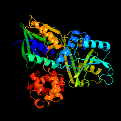
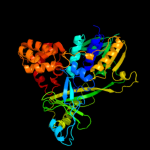
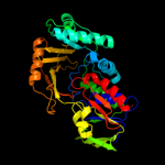
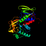
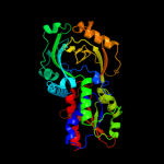
| 1 | c2r4jA_
|
|
 |
100.0 |
99 |
PDB header:oxidoreductase
Chain: A: PDB Molecule:aerobic glycerol-3-phosphate dehydrogenase;
PDBTitle: crystal structure of escherichia coli semet substituted2 glycerol-3-phosphate dehydrogenase in complex with dhap
|
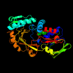
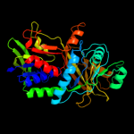
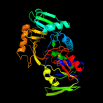
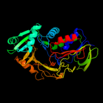
| 2 | c2rgoA_
|
|
 |
100.0 |
30 |
PDB header:oxidoreductase
Chain: A: PDB Molecule:alpha-glycerophosphate oxidase;
PDBTitle: structure of alpha-glycerophosphate oxidase from2 streptococcus sp.: a template for the mitochondrial alpha-3 glycerophosphate dehydrogenase
|
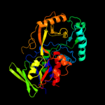
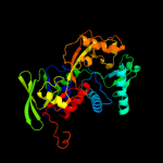
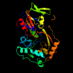
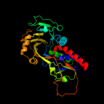
| 3 | c2rghA_
|
|
 |
100.0 |
30 |
PDB header:oxidoreductase
Chain: A: PDB Molecule:alpha-glycerophosphate oxidase;
PDBTitle: structure of alpha-glycerophosphate oxidase from2 streptococcus sp.: a template for the mitochondrial alpha-3 glycerophosphate dehydrogenase
|
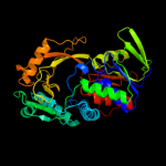
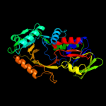
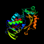
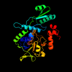
| 4 | c3da1A_
|
|
 |
100.0 |
31 |
PDB header:oxidoreductase
Chain: A: PDB Molecule:glycerol-3-phosphate dehydrogenase;
PDBTitle: x-ray structure of the glycerol-3-phosphate dehydrogenase2 from bacillus halodurans complexed with fad. northeast3 structural genomics consortium target bhr167.
|
| 5 | c3dmeB_
|
|
 |
100.0 |
19 |
PDB header:structural genomics, unknown function
Chain: B: PDB Molecule:conserved exported protein;
PDBTitle: crystal structure of conserved exported protein from2 bordetella pertussis. northeast structural genomics target3 ber141
|
| 6 | c1pj6A_
|
|
 |
100.0 |
15 |
PDB header:oxidoreductase
Chain: A: PDB Molecule:n,n-dimethylglycine oxidase;
PDBTitle: crystal structure of dimethylglycine oxidase of arthrobacter2 globiformis in complex with folic acid
|
| 7 | c1y56B_
|
|
 |
100.0 |
14 |
PDB header:oxidoreductase
Chain: B: PDB Molecule:sarcosine oxidase;
PDBTitle: crystal structure of l-proline dehydrogenase from p.horikoshii
|
| 8 | c2gahB_
|
|
 |
100.0 |
14 |
PDB header:oxidoreductase
Chain: B: PDB Molecule:heterotetrameric sarcosine oxidase beta-subunit;
PDBTitle: heterotetrameric sarcosine: structure of a diflavin2 metaloenzyme at 1.85 a resolution
|
| 9 | c2olnA_
|
|
 |
100.0 |
16 |
PDB header:oxidoreductase
Chain: A: PDB Molecule:nikd protein;
PDBTitle: nikd, an unusual amino acid oxidase essential for2 nikkomycin biosynthesis: closed form at 1.15 a resolution
|
| 10 | c3ps9A_
|
|
 |
100.0 |
13 |
PDB header:transferase
Chain: A: PDB Molecule:trna 5-methylaminomethyl-2-thiouridine biosynthesis
PDBTitle: crystal structure of mnmc from e. coli
|
| 11 | c3nyeA_
|
|
 |
100.0 |
17 |
PDB header:oxidoreductase
Chain: A: PDB Molecule:d-arginine dehydrogenase;
PDBTitle: crystal structure of pseudomonas aeruginosa d-arginine dehydrogenase2 in complex with imino-arginine
|
| 12 | c3pvcA_
|
|
 |
100.0 |
15 |
PDB header:oxidoreductase, transferase
Chain: A: PDB Molecule:trna 5-methylaminomethyl-2-thiouridine biosynthesis
PDBTitle: crystal structure of apo mnmc from yersinia pestis
|
| 13 | c3bhkA_
|
|
 |
100.0 |
14 |
PDB header:oxidoreductase
Chain: A: PDB Molecule:monomeric sarcosine oxidase;
PDBTitle: crystal structure of r49k mutant of monomeric sarcosine oxidase2 crystallized in phosphate as precipitant
|
| 14 | c3djeA_
|
|
 |
100.0 |
16 |
PDB header:oxidoreductase
Chain: A: PDB Molecule:fructosyl amine: oxygen oxidoreductase;
PDBTitle: crystal structure of the deglycating enzyme fructosamine2 oxidase from aspergillus fumigatus (amadoriase ii) in3 complex with fsa
|
| 15 | c1ryiB_
|
|
 |
100.0 |
14 |
PDB header:oxidoreductase
Chain: B: PDB Molecule:glycine oxidase;
PDBTitle: structure of glycine oxidase with bound inhibitor glycolate
|
| 16 | c1kifE_
|
|
 |
100.0 |
15 |
PDB header:flavoprotein
Chain: E: PDB Molecule:d-amino acid oxidase;
PDBTitle: d-amino acid oxidase from pig kidney
|
| 17 | c2uzzD_
|
|
 |
100.0 |
17 |
PDB header:oxidoreductase
Chain: D: PDB Molecule:n-methyl-l-tryptophan oxidase;
PDBTitle: x-ray structure of n-methyl-l-tryptophan oxidase (mtox)
|
| 18 | c1c0iA_
|
|
 |
100.0 |
17 |
PDB header:oxidoreductase
Chain: A: PDB Molecule:d-amino acid oxidase;
PDBTitle: crystal structure of d-amino acid oxidase in complex with2 two anthranylate molecules
|
| 19 | d1pj5a2
|
|
 |
99.9 |
15 |
Fold:FAD/NAD(P)-binding domain
Superfamily:FAD/NAD(P)-binding domain
Family:FAD-linked reductases, N-terminal domain |
| 20 | d1ryia1
|
|
 |
99.9 |
16 |
Fold:FAD/NAD(P)-binding domain
Superfamily:FAD/NAD(P)-binding domain
Family:FAD-linked reductases, N-terminal domain |
| 21 | d2gf3a1 |
|
not modelled |
99.9 |
19 |
Fold:FAD/NAD(P)-binding domain
Superfamily:FAD/NAD(P)-binding domain
Family:FAD-linked reductases, N-terminal domain |
| 22 | c1x31A_ |
|
not modelled |
99.9 |
15 |
PDB header:oxidoreductase
Chain: A: PDB Molecule:sarcosine oxidase alpha subunit;
PDBTitle: crystal structure of heterotetrameric sarcosine oxidase from2 corynebacterium sp. u-96
|
| 23 | d1c0pa1 |
|
not modelled |
99.8 |
18 |
Fold:Nucleotide-binding domain
Superfamily:Nucleotide-binding domain
Family:D-aminoacid oxidase, N-terminal domain |
| 24 | c2aczA_ |
|
not modelled |
99.8 |
16 |
PDB header:oxidoreductase/electron transport
Chain: A: PDB Molecule:succinate dehydrogenase flavoprotein subunit;
PDBTitle: complex ii (succinate dehydrogenase) from e. coli with atpenin a52 inhibitor co-crystallized at the ubiquinone binding site
|
| 25 | c1yq4A_ |
|
not modelled |
99.8 |
17 |
PDB header:oxidoreductase
Chain: A: PDB Molecule:succinate dehydrogenase flavoprotein subunit;
PDBTitle: avian respiratory complex ii with 3-nitropropionate and ubiquinone
|
| 26 | d1kifa1 |
|
not modelled |
99.8 |
19 |
Fold:Nucleotide-binding domain
Superfamily:Nucleotide-binding domain
Family:D-aminoacid oxidase, N-terminal domain |
| 27 | c1y56A_ |
|
not modelled |
99.8 |
14 |
PDB header:oxidoreductase
Chain: A: PDB Molecule:hypothetical protein ph1363;
PDBTitle: crystal structure of l-proline dehydrogenase from p.horikoshii
|
| 28 | d1neka2 |
|
not modelled |
99.8 |
15 |
Fold:FAD/NAD(P)-binding domain
Superfamily:FAD/NAD(P)-binding domain
Family:Succinate dehydrogenase/fumarate reductase flavoprotein N-terminal domain |
| 29 | c2bs3A_ |
|
not modelled |
99.8 |
16 |
PDB header:oxidoreductase
Chain: A: PDB Molecule:quinol-fumarate reductase flavoprotein subunit a;
PDBTitle: glu c180 -> gln variant quinol:fumarate reductase from2 wolinella succinogenes
|
| 30 | c3atrA_ |
|
not modelled |
99.8 |
12 |
PDB header:oxidoreductase
Chain: A: PDB Molecule:conserved archaeal protein;
PDBTitle: geranylgeranyl reductase (ggr) from sulfolobus acidocaldarius co-2 crystallized with its ligand
|
| 31 | c1jrxA_ |
|
not modelled |
99.8 |
16 |
PDB header:oxidoreductase
Chain: A: PDB Molecule:flavocytochrome c;
PDBTitle: crystal structure of arg402ala mutant flavocytochrome c32 from shewanella frigidimarina
|
| 32 | c1qo8A_ |
|
not modelled |
99.8 |
13 |
PDB header:oxidoreductase
Chain: A: PDB Molecule:flavocytochrome c3 fumarate reductase;
PDBTitle: the structure of the open conformation of a flavocytochrome2 c3 fumarate reductase
|
| 33 | c3ka7A_ |
|
not modelled |
99.8 |
16 |
PDB header:oxidoreductase
Chain: A: PDB Molecule:oxidoreductase;
PDBTitle: crystal structure of an oxidoreductase from methanosarcina2 mazei. northeast structural genomics consortium target id3 mar208
|
| 34 | d2bs2a2 |
|
not modelled |
99.7 |
16 |
Fold:FAD/NAD(P)-binding domain
Superfamily:FAD/NAD(P)-binding domain
Family:Succinate dehydrogenase/fumarate reductase flavoprotein N-terminal domain |
| 35 | c3p4rM_ |
|
not modelled |
99.7 |
14 |
PDB header:oxidoreductase
Chain: M: PDB Molecule:fumarate reductase flavoprotein subunit;
PDBTitle: crystal structure of menaquinol:fumarate oxidoreductase in complex2 with glutarate
|
| 36 | d1kf6a2 |
|
not modelled |
99.7 |
11 |
Fold:FAD/NAD(P)-binding domain
Superfamily:FAD/NAD(P)-binding domain
Family:Succinate dehydrogenase/fumarate reductase flavoprotein N-terminal domain |
| 37 | c1d4cB_ |
|
not modelled |
99.7 |
14 |
PDB header:oxidoreductase
Chain: B: PDB Molecule:flavocytochrome c fumarate reductase;
PDBTitle: crystal structure of the uncomplexed form of the2 flavocytochrome c fumarate reductase of shewanella3 putrefaciens strain mr-1
|
| 38 | c3i3lA_ |
|
not modelled |
99.7 |
13 |
PDB header:hydrolase
Chain: A: PDB Molecule:alkylhalidase cmls;
PDBTitle: crystal structure of cmls, a flavin-dependent halogenase
|
| 39 | c1kf6A_ |
|
not modelled |
99.7 |
14 |
PDB header:oxidoreductase
Chain: A: PDB Molecule:fumarate reductase flavoprotein;
PDBTitle: e. coli quinol-fumarate reductase with bound inhibitor hqno
|
| 40 | d1y0pa2 |
|
not modelled |
99.7 |
15 |
Fold:FAD/NAD(P)-binding domain
Superfamily:FAD/NAD(P)-binding domain
Family:Succinate dehydrogenase/fumarate reductase flavoprotein N-terminal domain |
| 41 | d1qo8a2 |
|
not modelled |
99.7 |
13 |
Fold:FAD/NAD(P)-binding domain
Superfamily:FAD/NAD(P)-binding domain
Family:Succinate dehydrogenase/fumarate reductase flavoprotein N-terminal domain |
| 42 | c3e1tA_ |
|
not modelled |
99.7 |
17 |
PDB header:flavoprotein
Chain: A: PDB Molecule:halogenase;
PDBTitle: structure and action of the myxobacterial chondrochloren2 halogenase cndh, a new variant of fad-dependent halogenases
|
| 43 | c3nixF_ |
|
not modelled |
99.7 |
16 |
PDB header:oxidoreductase
Chain: F: PDB Molecule:flavoprotein/dehydrogenase;
PDBTitle: crystal structure of flavoprotein/dehydrogenase from cytophaga2 hutchinsonii. northeast structural genomics consortium target chr43.
|
| 44 | c2weuD_ |
|
not modelled |
99.7 |
15 |
PDB header:antifungal protein
Chain: D: PDB Molecule:tryptophan 5-halogenase;
PDBTitle: crystal structure of tryptophan 5-halogenase (pyrh) complex2 with substrate tryptophan
|
| 45 | c3i6dA_ |
|
not modelled |
99.7 |
13 |
PDB header:oxidoreductase
Chain: A: PDB Molecule:protoporphyrinogen oxidase;
PDBTitle: crystal structure of ppo from bacillus subtilis with af
|
| 46 | d2gqfa1 |
|
not modelled |
99.6 |
19 |
Fold:FAD/NAD(P)-binding domain
Superfamily:FAD/NAD(P)-binding domain
Family:HI0933 N-terminal domain-like |
| 47 | c3cgvA_ |
|
not modelled |
99.6 |
16 |
PDB header:structural genomics, unknown function
Chain: A: PDB Molecule:geranylgeranyl reductase related protein;
PDBTitle: crystal structure of geranylgeranyl bacteriochlorophyll reductase-like2 fixc homolog (np_393992.1) from thermoplasma acidophilum at 1.60 a3 resolution
|
| 48 | c2gmhA_ |
|
not modelled |
99.6 |
14 |
PDB header:oxidoreductase
Chain: A: PDB Molecule:electron transfer flavoprotein-ubiquinone
PDBTitle: structure of porcine electron transfer flavoprotein-2 ubiquinone oxidoreductase in complexed with ubiquinone
|
| 49 | d1d5ta1 |
|
not modelled |
99.6 |
14 |
Fold:FAD/NAD(P)-binding domain
Superfamily:FAD/NAD(P)-binding domain
Family:GDI-like N domain |
| 50 | c2ardA_ |
|
not modelled |
99.6 |
10 |
PDB header:biosynthetic protein
Chain: A: PDB Molecule:tryptophan halogenase prna;
PDBTitle: the structure of tryptophan 7-halogenase (prna) suggests a mechanism2 for regioselective chlorination
|
| 51 | c2zxiC_ |
|
not modelled |
99.6 |
14 |
PDB header:fad-binding protein
Chain: C: PDB Molecule:trna uridine 5-carboxymethylaminomethyl
PDBTitle: structure of aquifex aeolicus gida in the form ii crystal
|
| 52 | c1ltxR_ |
|
not modelled |
99.6 |
12 |
PDB header:transferase/protein binding
Chain: R: PDB Molecule:rab escort protein 1;
PDBTitle: structure of rab escort protein-1 in complex with rab2 geranylgeranyl transferase and isoprenoid
|
| 53 | c2eq8E_ |
|
not modelled |
99.6 |
23 |
PDB header:oxidoreductase
Chain: E: PDB Molecule:pyruvate dehydrogenase complex, dihydrolipoamide
PDBTitle: crystal structure of lipoamide dehydrogenase from thermus thermophilus2 hb8 with psbdp
|
| 54 | d1o5wa1 |
|
not modelled |
99.6 |
18 |
Fold:FAD/NAD(P)-binding domain
Superfamily:FAD/NAD(P)-binding domain
Family:FAD-linked reductases, N-terminal domain |
| 55 | d1d4ca2 |
|
not modelled |
99.6 |
14 |
Fold:FAD/NAD(P)-binding domain
Superfamily:FAD/NAD(P)-binding domain
Family:Succinate dehydrogenase/fumarate reductase flavoprotein N-terminal domain |
| 56 | c2qa2A_ |
|
not modelled |
99.6 |
15 |
PDB header:oxidoreductase
Chain: A: PDB Molecule:polyketide oxygenase cabe;
PDBTitle: crystal structure of cabe, an aromatic hydroxylase from angucycline2 biosynthesis, determined to 2.7 a resolution
|
| 57 | d2i0za1 |
|
not modelled |
99.6 |
19 |
Fold:FAD/NAD(P)-binding domain
Superfamily:FAD/NAD(P)-binding domain
Family:HI0933 N-terminal domain-like |
| 58 | c3dgzA_ |
|
not modelled |
99.6 |
17 |
PDB header:oxidoreductase
Chain: A: PDB Molecule:thioredoxin reductase 2;
PDBTitle: crystal structure of mouse mitochondrial thioredoxin reductase, c-2 terminal 3-residue truncation
|
| 59 | c1zkqA_ |
|
not modelled |
99.6 |
17 |
PDB header:oxidoreductase
Chain: A: PDB Molecule:thioredoxin reductase 2, mitochondrial;
PDBTitle: crystal structure of mouse thioredoxin reductase type 2
|
| 60 | c3gyxA_ |
|
not modelled |
99.6 |
12 |
PDB header:oxidoreductase
Chain: A: PDB Molecule:adenylylsulfate reductase;
PDBTitle: crystal structure of adenylylsulfate reductase from2 desulfovibrio gigas
|
| 61 | c2e4gB_ |
|
not modelled |
99.5 |
13 |
PDB header:biosynthetic protein, flavoprotein
Chain: B: PDB Molecule:tryptophan halogenase;
PDBTitle: rebh with bound l-trp
|
| 62 | d2bcgg1 |
|
not modelled |
99.5 |
14 |
Fold:FAD/NAD(P)-binding domain
Superfamily:FAD/NAD(P)-binding domain
Family:GDI-like N domain |
| 63 | c2pyxA_ |
|
not modelled |
99.5 |
14 |
PDB header:biosynthetic protein
Chain: A: PDB Molecule:tryptophan halogenase;
PDBTitle: crystal structure of tryptophan halogenase (yp_750003.1) from2 shewanella frigidimarina ncimb 400 at 1.50 a resolution
|
| 64 | c2fjaC_ |
|
not modelled |
99.5 |
16 |
PDB header:oxidoreductase
Chain: C: PDB Molecule:adenylylsulfate reductase, subunit a;
PDBTitle: adenosine 5'-phosphosulfate reductase in complex with2 substrate
|
| 65 | c3g05B_ |
|
not modelled |
99.5 |
18 |
PDB header:rna binding protein
Chain: B: PDB Molecule:trna uridine 5-carboxymethylaminomethyl modification enzyme
PDBTitle: crystal structure of n-terminal domain (2-550) of e.coli mnmg
|
| 66 | c2dkhA_ |
|
not modelled |
99.5 |
13 |
PDB header:oxidoreductase
Chain: A: PDB Molecule:3-hydroxybenzoate hydroxylase;
PDBTitle: crystal structure of 3-hydroxybenzoate hydroxylase from comamonas2 testosteroni, in complex with the substrate
|
| 67 | d3coxa1 |
|
not modelled |
99.5 |
19 |
Fold:FAD/NAD(P)-binding domain
Superfamily:FAD/NAD(P)-binding domain
Family:FAD-linked reductases, N-terminal domain |
| 68 | c1bwcA_ |
|
not modelled |
99.5 |
19 |
PDB header:oxidoreductase
Chain: A: PDB Molecule:protein (glutathione reductase);
PDBTitle: structure of human glutathione reductase complexed with ajoene2 inhibitor and subversive substrate
|
| 69 | c2nvkX_ |
|
not modelled |
99.5 |
19 |
PDB header:oxidoreductase
Chain: X: PDB Molecule:thioredoxin reductase;
PDBTitle: crystal structure of thioredoxin reductase from drosophila2 melanogaster
|
| 70 | c3urhB_ |
|
not modelled |
99.5 |
20 |
PDB header:oxidoreductase
Chain: B: PDB Molecule:dihydrolipoyl dehydrogenase;
PDBTitle: crystal structure of a dihydrolipoamide dehydrogenase from2 sinorhizobium meliloti 1021
|
| 71 | c3cesB_ |
|
not modelled |
99.5 |
20 |
PDB header:rna binding protein
Chain: B: PDB Molecule:trna uridine 5-carboxymethylaminomethyl modification enzyme
PDBTitle: crystal structure of e.coli mnmg (gida), a highly-conserved trna2 modifying enzyme
|
| 72 | d2gmha1 |
|
not modelled |
99.5 |
19 |
Fold:FAD/NAD(P)-binding domain
Superfamily:FAD/NAD(P)-binding domain
Family:FAD-linked reductases, N-terminal domain |
| 73 | c1phhA_ |
|
not modelled |
99.5 |
12 |
PDB header:oxidoreductase
Chain: A: PDB Molecule:p-hydroxybenzoate hydroxylase;
PDBTitle: crystal structure of p-hydroxybenzoate hydroxylase complexed with its2 reaction product 3,4-dihydroxybenzoate
|
| 74 | c3fmwC_ |
|
not modelled |
99.5 |
20 |
PDB header:oxidoreductase
Chain: C: PDB Molecule:oxygenase;
PDBTitle: the crystal structure of mtmoiv, a baeyer-villiger2 monooxygenase from the mithramycin biosynthetic pathway in3 streptomyces argillaceus.
|
| 75 | c2c3dB_ |
|
not modelled |
99.5 |
16 |
PDB header:oxidoreductase
Chain: B: PDB Molecule:2-oxopropyl-com reductase;
PDBTitle: 2.15 angstrom crystal structure of 2-ketopropyl coenzyme m2 oxidoreductase carboxylase with a coenzyme m disulfide3 bound at the active site
|
| 76 | c1v59B_ |
|
not modelled |
99.5 |
17 |
PDB header:oxidoreductase
Chain: B: PDB Molecule:dihydrolipoamide dehydrogenase;
PDBTitle: crystal structure of yeast lipoamide dehydrogenase2 complexed with nad+
|
| 77 | d1k0ia1 |
|
not modelled |
99.5 |
15 |
Fold:FAD/NAD(P)-binding domain
Superfamily:FAD/NAD(P)-binding domain
Family:FAD-linked reductases, N-terminal domain |
| 78 | c2ivdA_ |
|
not modelled |
99.4 |
16 |
PDB header:oxidoreductase
Chain: A: PDB Molecule:protoporphyrinogen oxidase;
PDBTitle: structure of protoporphyrinogen oxidase from myxococcus2 xanthus with acifluorfen
|
| 79 | d1chua2 |
|
not modelled |
99.4 |
23 |
Fold:FAD/NAD(P)-binding domain
Superfamily:FAD/NAD(P)-binding domain
Family:Succinate dehydrogenase/fumarate reductase flavoprotein N-terminal domain |
| 80 | c3cp8C_ |
|
not modelled |
99.4 |
18 |
PDB header:oxidoreductase
Chain: C: PDB Molecule:trna uridine 5-carboxymethylaminomethyl
PDBTitle: crystal structure of gida from chlorobium tepidum
|
| 81 | c3k7tB_ |
|
not modelled |
99.4 |
14 |
PDB header:oxidoreductase
Chain: B: PDB Molecule:6-hydroxy-l-nicotine oxidase;
PDBTitle: crystal structure of apo-form 6-hydroxy-l-nicotine oxidase,2 crystal form p3121
|
| 82 | c2e5vA_ |
|
not modelled |
99.4 |
11 |
PDB header:oxidoreductase
Chain: A: PDB Molecule:l-aspartate oxidase;
PDBTitle: crystal structure of l-aspartate oxidase from2 hyperthermophilic archaeon sulfolobus tokodaii
|
| 83 | c2gewA_ |
|
not modelled |
99.4 |
15 |
PDB header:oxidoreductase
Chain: A: PDB Molecule:cholesterol oxidase;
PDBTitle: atomic resolution structure of cholesterol oxidase @ ph 9.02 (streptomyces sp. sa-coo)
|
| 84 | c1coyA_ |
|
not modelled |
99.4 |
20 |
PDB header:oxidoreductase(oxygen receptor)
Chain: A: PDB Molecule:cholesterol oxidase;
PDBTitle: crystal structure of cholesterol oxidase complexed with a2 steroid substrate. implications for fad dependent alcohol3 oxidases
|
| 85 | c1gndA_ |
|
not modelled |
99.4 |
15 |
PDB header:gtpase activation
Chain: A: PDB Molecule:guanine nucleotide dissociation inhibitor;
PDBTitle: guanine nucleotide dissociation inhibitor, alpha-isoform
|
| 86 | c2f5vA_ |
|
not modelled |
99.4 |
15 |
PDB header:oxidoreductase
Chain: A: PDB Molecule:pyranose 2-oxidase;
PDBTitle: reaction geometry and thermostability mutant of pyranose 2-oxidase2 from the white-rot fungus peniophora sp.
|
| 87 | c1ebdB_ |
|
not modelled |
99.4 |
17 |
PDB header:complex (oxidoreductase/transferase)
Chain: B: PDB Molecule:dihydrolipoamide dehydrogenase;
PDBTitle: dihydrolipoamide dehydrogenase complexed with the binding2 domain of the dihydrolipoamide acetylase
|
| 88 | c3jskN_ |
|
not modelled |
99.4 |
17 |
PDB header:biosynthetic protein
Chain: N: PDB Molecule:cypbp37 protein;
PDBTitle: thiazole synthase from neurospora crassa
|
| 89 | c1zmcG_ |
|
not modelled |
99.4 |
15 |
PDB header:oxidoreductase
Chain: G: PDB Molecule:dihydrolipoyl dehydrogenase;
PDBTitle: crystal structure of human dihydrolipoamide dehydrogenase2 complexed to nad+
|
| 90 | c3v76A_ |
|
not modelled |
99.4 |
18 |
PDB header:flavoprotein
Chain: A: PDB Molecule:flavoprotein;
PDBTitle: the crystal structure of a flavoprotein from sinorhizobium meliloti
|
| 91 | c2igoG_ |
|
not modelled |
99.4 |
13 |
PDB header:oxidoreductase
Chain: G: PDB Molecule:pyranose oxidase;
PDBTitle: crystal structure of pyranose 2-oxidase h167a mutant with 2-2 fluoro-2-deoxy-d-glucose
|
| 92 | c1s3bB_ |
|
not modelled |
99.4 |
14 |
PDB header:oxidoreductase
Chain: B: PDB Molecule:amine oxidase [flavin-containing] b;
PDBTitle: crystal structure of maob in complex with n-methyl-n-2 propargyl-1(r)-aminoindan
|
| 93 | c3cpiH_ |
|
not modelled |
99.4 |
16 |
PDB header:protein transport
Chain: H: PDB Molecule:rab gdp-dissociation inhibitor;
PDBTitle: crystal structure of yeast rab-gdi
|
| 94 | c2eq7B_ |
|
not modelled |
99.4 |
20 |
PDB header:oxidoreductase
Chain: B: PDB Molecule:2-oxoglutarate dehydrogenase e3 component;
PDBTitle: crystal structure of lipoamide dehydrogenase from thermus thermophilus2 hb8 with psbdo
|
| 95 | d1rp0a1 |
|
not modelled |
99.4 |
15 |
Fold:FAD/NAD(P)-binding domain
Superfamily:FAD/NAD(P)-binding domain
Family:Thi4-like |
| 96 | c3o0hA_ |
|
not modelled |
99.4 |
17 |
PDB header:oxidoreductase
Chain: A: PDB Molecule:glutathione reductase;
PDBTitle: crystal structure of glutathione reductase from bartonella henselae
|
| 97 | c2a8xA_ |
|
not modelled |
99.4 |
15 |
PDB header:oxidoreductase
Chain: A: PDB Molecule:dihydrolipoyl dehydrogenase;
PDBTitle: crystal structure of lipoamide dehydrogenase from2 mycobacterium tuberculosis
|
| 98 | c3ihgA_ |
|
not modelled |
99.4 |
14 |
PDB header:flavoprotein, oxidoreductase
Chain: A: PDB Molecule:rdme;
PDBTitle: crystal structure of a ternary complex of aklavinone-112 hydroxylase with fad and aklavinone
|
| 99 | c3nrnA_ |
|
not modelled |
99.4 |
15 |
PDB header:structural genomics, unknown function
Chain: A: PDB Molecule:uncharacterized protein pf1083;
PDBTitle: crystal structure of pf1083 protein from pyrococcus furiosus,2 northeast structural genomics consortium target pfr223
|
| 100 | c2gqfA_ |
|
not modelled |
99.4 |
17 |
PDB header:structural genomics, unknown function
Chain: A: PDB Molecule:hypothetical protein hi0933;
PDBTitle: crystal structure of flavoprotein hi0933 from haemophilus influenzae2 rd
|
| 101 | d1w4xa1 |
|
not modelled |
99.4 |
16 |
Fold:FAD/NAD(P)-binding domain
Superfamily:FAD/NAD(P)-binding domain
Family:FAD/NAD-linked reductases, N-terminal and central domains |
| 102 | c1geuA_ |
|
not modelled |
99.4 |
19 |
PDB header:oxidoreductase(flavoenzyme)
Chain: A: PDB Molecule:glutathione reductase;
PDBTitle: anatomy of an engineered nad-binding site
|
| 103 | c1dxlC_ |
|
not modelled |
99.4 |
17 |
PDB header:oxidoreductase
Chain: C: PDB Molecule:dihydrolipoamide dehydrogenase;
PDBTitle: dihydrolipoamide dehydrogenase of glycine decarboxylase2 from pisum sativum
|
| 104 | c3nlcA_ |
|
not modelled |
99.4 |
17 |
PDB header:structural genomics, unknown function
Chain: A: PDB Molecule:uncharacterized protein vp0956;
PDBTitle: crystal structure of the vp0956 protein from vibrio parahaemolyticus.2 northeast structural genomics consortium target vpr147
|
| 105 | c1yvvB_ |
|
not modelled |
99.4 |
12 |
PDB header:oxidoreductase
Chain: B: PDB Molecule:amine oxidase, flavin-containing;
PDBTitle: x-ray structurure of p. syringae q888a4 oxidoreductase at2 resolution 2.5a. northeast structural genomics consortium3 target psr10.
|
| 106 | d1kdga1 |
|
not modelled |
99.4 |
11 |
Fold:FAD/NAD(P)-binding domain
Superfamily:FAD/NAD(P)-binding domain
Family:FAD-linked reductases, N-terminal domain |
| 107 | c1lpfB_ |
|
not modelled |
99.4 |
17 |
PDB header:oxidoreductase
Chain: B: PDB Molecule:dihydrolipoamide dehydrogenase;
PDBTitle: three-dimensional structure of lipoamide dehydrogenase from2 pseudomonas fluorescens at 2.8 angstroms resolution.3 analysis of redox and thermostability properties
|
| 108 | c1tytA_ |
|
not modelled |
99.4 |
14 |
PDB header:oxidoreductase
Chain: A: PDB Molecule:trypanothione reductase, oxidized form;
PDBTitle: crystal and molecular structure of crithidia fasciculata2 trypanothione reductase at 2.6 angstroms resolution
|
| 109 | c2cfyB_ |
|
not modelled |
99.4 |
17 |
PDB header:oxidoreductase
Chain: B: PDB Molecule:thioredoxin reductase 1;
PDBTitle: crystal structure of human thioredoxin reductase 1
|
| 110 | c2hqmB_ |
|
not modelled |
99.4 |
15 |
PDB header:oxidoreductase
Chain: B: PDB Molecule:glutathione reductase;
PDBTitle: crystal structure of glutathione reductase glr1 from the yeast2 saccharomyces cerevisiae
|
| 111 | d1n4wa1 |
|
not modelled |
99.3 |
16 |
Fold:FAD/NAD(P)-binding domain
Superfamily:FAD/NAD(P)-binding domain
Family:FAD-linked reductases, N-terminal domain |
| 112 | c2w0hA_ |
|
not modelled |
99.3 |
14 |
PDB header:oxidoreductase
Chain: A: PDB Molecule:trypanothione reductase;
PDBTitle: x ray structure of leishmania infantum trypanothione2 reductase in complex with antimony and nadph
|
| 113 | c2rgjA_ |
|
not modelled |
99.3 |
16 |
PDB header:oxidoreductase
Chain: A: PDB Molecule:flavin-containing monooxygenase;
PDBTitle: crystal structure of flavin-containing monooxygenase phzs
|
| 114 | c2jbvA_ |
|
not modelled |
99.3 |
19 |
PDB header:oxidoreductase
Chain: A: PDB Molecule:choline oxidase;
PDBTitle: crystal structure of choline oxidase reveals insights into2 the catalytic mechanism
|
| 115 | c3ic9D_ |
|
not modelled |
99.3 |
13 |
PDB header:oxidoreductase
Chain: D: PDB Molecule:dihydrolipoamide dehydrogenase;
PDBTitle: the structure of dihydrolipoamide dehydrogenase from colwellia2 psychrerythraea 34h.
|
| 116 | d1jnra2 |
|
not modelled |
99.3 |
20 |
Fold:FAD/NAD(P)-binding domain
Superfamily:FAD/NAD(P)-binding domain
Family:Succinate dehydrogenase/fumarate reductase flavoprotein N-terminal domain |
| 117 | c2i0zA_ |
|
not modelled |
99.3 |
20 |
PDB header:oxidoreductase
Chain: A: PDB Molecule:nad(fad)-utilizing dehydrogenases;
PDBTitle: crystal structure of a fad binding protein from bacillus2 cereus, a putative nad(fad)-utilizing dehydrogenases
|
| 118 | c1zx9A_ |
|
not modelled |
99.3 |
19 |
PDB header:oxidoreductase
Chain: A: PDB Molecule:mercuric reductase;
PDBTitle: crystal structure of tn501 mera
|
| 119 | c1naaB_ |
|
not modelled |
99.3 |
15 |
PDB header:oxidoreductase
Chain: B: PDB Molecule:cellobiose dehydrogenase;
PDBTitle: cellobiose dehydrogenase flavoprotein fragment in complex with2 cellobionolactam
|
| 120 | d1vg0a1 |
|
not modelled |
99.3 |
14 |
Fold:FAD/NAD(P)-binding domain
Superfamily:FAD/NAD(P)-binding domain
Family:GDI-like N domain |