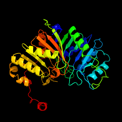
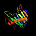
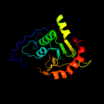
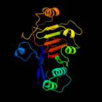
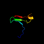
| 1 | c3hsiC_
|
|
 |
100.0 |
15 |
PDB header:transferase
Chain: C: PDB Molecule:phosphatidylserine synthase;
PDBTitle: crystal structure of phosphatidylserine synthase haemophilus2 influenzae rd kw20
|
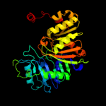
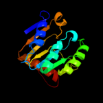
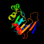
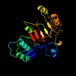
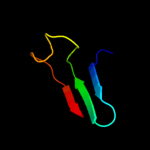
| 2 | c1v0sA_
|
|
 |
100.0 |
16 |
PDB header:hydrolase
Chain: A: PDB Molecule:phospholipase d;
PDBTitle: uninhibited form of phospholipase d from streptomyces sp.2 strain pmf
|
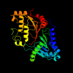
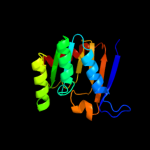
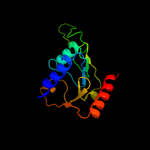
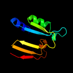
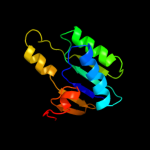
| 3 | c1xdoB_
|
|
 |
100.0 |
17 |
PDB header:transferase
Chain: B: PDB Molecule:polyphosphate kinase;
PDBTitle: crystal structure of escherichia coli polyphosphate kinase
|
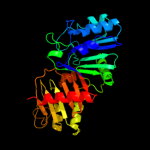
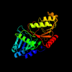
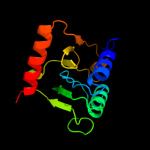
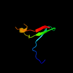
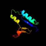
| 4 | d1v0wa1
|
|
 |
100.0 |
16 |
Fold:Phospholipase D/nuclease
Superfamily:Phospholipase D/nuclease
Family:Phospholipase D |
| 5 | d1xdpa3
|
|
 |
99.9 |
15 |
Fold:Phospholipase D/nuclease
Superfamily:Phospholipase D/nuclease
Family:Polyphosphate kinase C-terminal domain |
| 6 | d1byra_
|
|
 |
99.9 |
19 |
Fold:Phospholipase D/nuclease
Superfamily:Phospholipase D/nuclease
Family:Nuclease |
| 7 | c2o8rA_
|
|
 |
99.9 |
20 |
PDB header:transferase
Chain: A: PDB Molecule:polyphosphate kinase;
PDBTitle: crystal structure of polyphosphate kinase from2 porphyromonas gingivalis
|
| 8 | d1v0wa2
|
|
 |
99.9 |
16 |
Fold:Phospholipase D/nuclease
Superfamily:Phospholipase D/nuclease
Family:Phospholipase D |
| 9 | d2o8ra3
|
|
 |
99.0 |
12 |
Fold:Phospholipase D/nuclease
Superfamily:Phospholipase D/nuclease
Family:Polyphosphate kinase C-terminal domain |
| 10 | d1xdpa4
|
|
 |
99.0 |
15 |
Fold:Phospholipase D/nuclease
Superfamily:Phospholipase D/nuclease
Family:Polyphosphate kinase C-terminal domain |
| 11 | d2o8ra4
|
|
 |
98.7 |
18 |
Fold:Phospholipase D/nuclease
Superfamily:Phospholipase D/nuclease
Family:Polyphosphate kinase C-terminal domain |
| 12 | c1q32C_
|
|
 |
98.4 |
12 |
PDB header:replication,transcription,hydrolase
Chain: C: PDB Molecule:tyrosyl-dna phosphodiesterase;
PDBTitle: crystal structure analysis of the yeast tyrosyl-dna2 phosphodiesterase
|
| 13 | c3sq3C_
|
|
 |
98.1 |
13 |
PDB header:hydrolase
Chain: C: PDB Molecule:tyrosyl-dna phosphodiesterase 1;
PDBTitle: crystal structure analysis of the yeast tyrosyl-dna phosphodiesterase2 h182a mutant
|
| 14 | c2c1lA_
|
|
 |
97.7 |
25 |
PDB header:hydrolase
Chain: A: PDB Molecule:restriction endonuclease;
PDBTitle: structure of the bfii restriction endonuclease
|
| 15 | d1jy1a2
|
|
 |
96.8 |
23 |
Fold:Phospholipase D/nuclease
Superfamily:Phospholipase D/nuclease
Family:Tyrosyl-DNA phosphodiesterase TDP1 |
| 16 | c1nopB_
|
|
 |
96.7 |
28 |
PDB header:hydrolase/dna
Chain: B: PDB Molecule:tyrosyl-dna phosphodiesterase 1;
PDBTitle: crystal structure of human tyrosyl-dna phosphodiesterase2 (tdp1) in complex with vanadate, dna and a human3 topoisomerase i-derived peptide
|
| 17 | d1q32a2
|
|
 |
96.4 |
29 |
Fold:Phospholipase D/nuclease
Superfamily:Phospholipase D/nuclease
Family:Tyrosyl-DNA phosphodiesterase TDP1 |
| 18 | d1qzqa1
|
|
 |
90.6 |
12 |
Fold:Phospholipase D/nuclease
Superfamily:Phospholipase D/nuclease
Family:Tyrosyl-DNA phosphodiesterase TDP1 |
| 19 | d1jy1a1
|
|
 |
81.0 |
11 |
Fold:Phospholipase D/nuclease
Superfamily:Phospholipase D/nuclease
Family:Tyrosyl-DNA phosphodiesterase TDP1 |
| 20 | d2f5tx2
|
|
 |
78.8 |
15 |
Fold:Phospholipase D/nuclease
Superfamily:Phospholipase D/nuclease
Family:TrmB middle domain-like |
| 21 | c2f5tX_ |
|
not modelled |
71.8 |
15 |
PDB header:transcription
Chain: X: PDB Molecule:archaeal transcriptional regulator trmb;
PDBTitle: crystal structure of the sugar binding domain of the archaeal2 transcriptional regulator trmb
|
| 22 | d1q32a1 |
|
not modelled |
66.5 |
10 |
Fold:Phospholipase D/nuclease
Superfamily:Phospholipase D/nuclease
Family:Tyrosyl-DNA phosphodiesterase TDP1 |
| 23 | c2l82A_ |
|
not modelled |
42.6 |
19 |
PDB header:de novo protein
Chain: A: PDB Molecule:designed protein or32;
PDBTitle: solution nmr structure of de novo designed protein, p-loop ntpase2 fold, northeast structural genomics consortium target or32
|
| 24 | c2a5hC_ |
|
not modelled |
36.5 |
21 |
PDB header:isomerase
Chain: C: PDB Molecule:l-lysine 2,3-aminomutase;
PDBTitle: 2.1 angstrom x-ray crystal structure of lysine-2,3-aminomutase from2 clostridium subterminale sb4, with michaelis analog (l-alpha-lysine3 external aldimine form of pyridoxal-5'-phosphate).
|
| 25 | d1o98a1 |
|
not modelled |
34.2 |
26 |
Fold:2,3-Bisphosphoglycerate-independent phosphoglycerate mutase, substrate-binding domain
Superfamily:2,3-Bisphosphoglycerate-independent phosphoglycerate mutase, substrate-binding domain
Family:2,3-Bisphosphoglycerate-independent phosphoglycerate mutase, substrate-binding domain |
| 26 | c2o48X_ |
|
not modelled |
30.1 |
12 |
PDB header:oxidoreductase
Chain: X: PDB Molecule:dimeric dihydrodiol dehydrogenase;
PDBTitle: crystal structure of mammalian dimeric dihydrodiol dehydrogenase
|
| 27 | d1rvga_ |
|
not modelled |
28.0 |
25 |
Fold:TIM beta/alpha-barrel
Superfamily:Aldolase
Family:Class II FBP aldolase |
| 28 | d1gvfa_ |
|
not modelled |
26.2 |
15 |
Fold:TIM beta/alpha-barrel
Superfamily:Aldolase
Family:Class II FBP aldolase |
| 29 | d1gz0a2 |
|
not modelled |
20.0 |
24 |
Fold:Bacillus chorismate mutase-like
Superfamily:L30e-like
Family:RNA 2'-O ribose methyltransferase substrate binding domain |
| 30 | c2hqbA_ |
|
not modelled |
17.8 |
15 |
PDB header:transcription
Chain: A: PDB Molecule:transcriptional activator of comk gene;
PDBTitle: crystal structure of a transcriptional activator of comk2 gene from bacillus halodurans
|
| 31 | c1t3gB_ |
|
not modelled |
17.3 |
18 |
PDB header:membrane protein
Chain: B: PDB Molecule:x-linked interleukin-1 receptor accessory
PDBTitle: crystal structure of the toll/interleukin-1 receptor (tir)2 domain of human il-1rapl
|
| 32 | c3dy0B_ |
|
not modelled |
16.7 |
33 |
PDB header:blood clotting, hydrolase inhibitor
Chain: B: PDB Molecule:c-terminus plasma serine protease inhibitor;
PDBTitle: crystal structure of cleaved pci bound to heparin
|
| 33 | c3kgkA_ |
|
not modelled |
16.2 |
13 |
PDB header:chaperone
Chain: A: PDB Molecule:arsenical resistance operon trans-acting repressor arsd;
PDBTitle: crystal structure of arsd
|
| 34 | c3rfuC_ |
|
not modelled |
15.5 |
21 |
PDB header:hydrolase, membrane protein
Chain: C: PDB Molecule:copper efflux atpase;
PDBTitle: crystal structure of a copper-transporting pib-type atpase
|
| 35 | c3ecsD_ |
|
not modelled |
15.5 |
13 |
PDB header:translation
Chain: D: PDB Molecule:translation initiation factor eif-2b subunit
PDBTitle: crystal structure of human eif2b alpha
|
| 36 | c1lq8H_ |
|
not modelled |
15.4 |
33 |
PDB header:blood clotting
Chain: H: PDB Molecule:plasma serine protease inhibitor;
PDBTitle: crystal structure of cleaved protein c inhibitor
|
| 37 | c2rjoA_ |
|
not modelled |
15.1 |
16 |
PDB header:signaling protein
Chain: A: PDB Molecule:twin-arginine translocation pathway signal protein;
PDBTitle: crystal structure of twin-arginine translocation pathway signal2 protein from burkholderia phytofirmans
|
| 38 | c3gbcA_ |
|
not modelled |
15.1 |
19 |
PDB header:hydrolase
Chain: A: PDB Molecule:pyrazinamidase/nicotinamidas pnca;
PDBTitle: determination of the crystal structure of the pyrazinamidase from2 m.tuberculosis : a structure-function analysis for prediction3 resistance to pyrazinamide
|
| 39 | c2nytB_ |
|
not modelled |
14.4 |
12 |
PDB header:hydrolase
Chain: B: PDB Molecule:probable c->u-editing enzyme apobec-2;
PDBTitle: the apobec2 crystal structure and functional implications2 for aid
|
| 40 | d1gz0f2 |
|
not modelled |
14.2 |
24 |
Fold:Bacillus chorismate mutase-like
Superfamily:L30e-like
Family:RNA 2'-O ribose methyltransferase substrate binding domain |
| 41 | c3db2C_ |
|
not modelled |
13.7 |
10 |
PDB header:oxidoreductase
Chain: C: PDB Molecule:putative nadph-dependent oxidoreductase;
PDBTitle: crystal structure of a putative nadph-dependent oxidoreductase2 (dhaf_2064) from desulfitobacterium hafniense dcb-2 at 1.70 a3 resolution
|
| 42 | c3s99A_ |
|
not modelled |
13.2 |
19 |
PDB header:lipid binding protein
Chain: A: PDB Molecule:basic membrane lipoprotein;
PDBTitle: crystal structure of a basic membrane lipoprotein from brucella2 melitensis, iodide soak
|
| 43 | c3h75A_ |
|
not modelled |
12.4 |
10 |
PDB header:sugar binding protein
Chain: A: PDB Molecule:periplasmic sugar-binding domain protein;
PDBTitle: crystal structure of a periplasmic sugar-binding protein from the2 pseudomonas fluorescens
|
| 44 | c2kjwA_ |
|
not modelled |
11.9 |
12 |
PDB header:ribosomal protein
Chain: A: PDB Molecule:30s ribosomal protein s6;
PDBTitle: solution structure and backbone dynamics of the permutant2 p54-55
|
| 45 | d1bdga1 |
|
not modelled |
11.9 |
15 |
Fold:Ribonuclease H-like motif
Superfamily:Actin-like ATPase domain
Family:Hexokinase |
| 46 | c1o98A_ |
|
not modelled |
11.1 |
26 |
PDB header:isomerase
Chain: A: PDB Molecule:2,3-bisphosphoglycerate-independent
PDBTitle: 1.4a crystal structure of phosphoglycerate mutase from2 bacillus stearothermophilus complexed with3 2-phosphoglycerate
|
| 47 | d1acoa2 |
|
not modelled |
11.0 |
14 |
Fold:Aconitase iron-sulfur domain
Superfamily:Aconitase iron-sulfur domain
Family:Aconitase iron-sulfur domain |
| 48 | d2obba1 |
|
not modelled |
11.0 |
11 |
Fold:HAD-like
Superfamily:HAD-like
Family:BT0820-like |
| 49 | c2q4eB_ |
|
not modelled |
10.8 |
11 |
PDB header:oxidoreductase
Chain: B: PDB Molecule:probable oxidoreductase at4g09670;
PDBTitle: ensemble refinement of the protein crystal structure of gene product2 from arabidopsis thaliana at4g09670
|
| 50 | c2glxD_ |
|
not modelled |
9.9 |
13 |
PDB header:oxidoreductase
Chain: D: PDB Molecule:1,5-anhydro-d-fructose reductase;
PDBTitle: crystal structure analysis of bacterial 1,5-af reductase
|
| 51 | c2kl8A_ |
|
not modelled |
9.4 |
24 |
PDB header:de novo protein
Chain: A: PDB Molecule:or15;
PDBTitle: solution nmr structure of de novo designed ferredoxin-like2 fold protein, northeast structural genomics consortium3 target or15
|
| 52 | c3igzB_ |
|
not modelled |
9.3 |
26 |
PDB header:isomerase
Chain: B: PDB Molecule:cofactor-independent phosphoglycerate mutase;
PDBTitle: crystal structures of leishmania mexicana phosphoglycerate2 mutase at low cobalt concentration
|
| 53 | d1un8a4 |
|
not modelled |
9.2 |
16 |
Fold:DAK1/DegV-like
Superfamily:DAK1/DegV-like
Family:DAK1 |
| 54 | d1vb5a_ |
|
not modelled |
9.2 |
15 |
Fold:NagB/RpiA/CoA transferase-like
Superfamily:NagB/RpiA/CoA transferase-like
Family:IF2B-like |
| 55 | c2r60A_ |
|
not modelled |
8.8 |
28 |
PDB header:transferase
Chain: A: PDB Molecule:glycosyl transferase, group 1;
PDBTitle: structure of apo sucrose phosphate synthase (sps) of2 halothermothrix orenii
|
| 56 | c2ixaA_ |
|
not modelled |
8.4 |
13 |
PDB header:hydrolase
Chain: A: PDB Molecule:alpha-n-acetylgalactosaminidase;
PDBTitle: a-zyme, n-acetylgalactosaminidase
|
| 57 | c2b34C_ |
|
not modelled |
8.3 |
21 |
PDB header:hydrolase
Chain: C: PDB Molecule:mar1 ribonuclease;
PDBTitle: structure of mar1 ribonuclease from caenorhabditis elegans
|
| 58 | d1ydwa1 |
|
not modelled |
8.2 |
10 |
Fold:NAD(P)-binding Rossmann-fold domains
Superfamily:NAD(P)-binding Rossmann-fold domains
Family:Glyceraldehyde-3-phosphate dehydrogenase-like, N-terminal domain |
| 59 | d1x94a_ |
|
not modelled |
8.2 |
13 |
Fold:SIS domain
Superfamily:SIS domain
Family:mono-SIS domain |
| 60 | c3oziB_ |
|
not modelled |
8.0 |
20 |
PDB header:plant protein
Chain: B: PDB Molecule:l6tr;
PDBTitle: crystal structure of the tir domain from the flax disease resistance2 protein l6
|
| 61 | c2wltA_ |
|
not modelled |
7.9 |
18 |
PDB header:hydrolase
Chain: A: PDB Molecule:l-asparaginase;
PDBTitle: the crystal structure of helicobacter pylori l-asparaginase2 at 1.4 a resolution
|
| 62 | c2klnA_ |
|
not modelled |
7.7 |
11 |
PDB header:transport protein
Chain: A: PDB Molecule:probable sulphate-transport transmembrane protein, cog0659;
PDBTitle: solution structure of stas domain of rv1739c from m. tuberculosis
|
| 63 | c2x7mA_ |
|
not modelled |
7.7 |
8 |
PDB header:hydrolase
Chain: A: PDB Molecule:archaemetzincin;
PDBTitle: crystal structure of archaemetzincin (amza) from methanopyrus2 kandleri at 1.5 a resolution
|
| 64 | c3l2iB_ |
|
not modelled |
7.5 |
12 |
PDB header:lyase
Chain: B: PDB Molecule:3-dehydroquinate dehydratase;
PDBTitle: 1.85 angstrom crystal structure of the 3-dehydroquinate dehydratase2 (arod) from salmonella typhimurium lt2.
|
| 65 | c2krcA_ |
|
not modelled |
7.4 |
8 |
PDB header:transcription
Chain: A: PDB Molecule:dna-directed rna polymerase subunit delta;
PDBTitle: solution structure of the n-terminal domain of bacillus2 subtilis delta subunit of rna polymerase
|
| 66 | c2j37W_ |
|
not modelled |
7.1 |
11 |
PDB header:ribosome
Chain: W: PDB Molecule:signal recognition particle 54 kda protein
PDBTitle: model of mammalian srp bound to 80s rncs
|
| 67 | d1yaca_ |
|
not modelled |
7.1 |
21 |
Fold:Isochorismatase-like hydrolases
Superfamily:Isochorismatase-like hydrolases
Family:Isochorismatase-like hydrolases |
| 68 | c3a11D_ |
|
not modelled |
6.9 |
17 |
PDB header:isomerase
Chain: D: PDB Molecule:translation initiation factor eif-2b, delta
PDBTitle: crystal structure of ribose-1,5-bisphosphate isomerase from2 thermococcus kodakaraensis kod1
|
| 69 | c2y92A_ |
|
not modelled |
6.7 |
10 |
PDB header:immune system
Chain: A: PDB Molecule:toll/interleukin-1 receptor domain-containing adapter
PDBTitle: crystal structure of mal adaptor protein
|
| 70 | c3euwB_ |
|
not modelled |
6.6 |
12 |
PDB header:oxidoreductase
Chain: B: PDB Molecule:myo-inositol dehydrogenase;
PDBTitle: crystal structure of a myo-inositol dehydrogenase from corynebacterium2 glutamicum atcc 13032
|
| 71 | c1yd6A_ |
|
not modelled |
6.6 |
20 |
PDB header:dna binding protein
Chain: A: PDB Molecule:uvrc;
PDBTitle: crystal structure of the giy-yig n-terminal endonuclease2 domain of uvrc from bacillus caldotenax
|
| 72 | c2d6fA_ |
|
not modelled |
6.6 |
14 |
PDB header:ligase/rna
Chain: A: PDB Molecule:glutamyl-trna(gln) amidotransferase subunit d;
PDBTitle: crystal structure of glu-trna(gln) amidotransferase in the2 complex with trna(gln)
|
| 73 | d1v4sa1 |
|
not modelled |
6.5 |
20 |
Fold:Ribonuclease H-like motif
Superfamily:Actin-like ATPase domain
Family:Hexokinase |
| 74 | c5acnA_ |
|
not modelled |
6.4 |
16 |
PDB header:lyase(carbon-oxygen)
Chain: A: PDB Molecule:aconitase;
PDBTitle: structure of activated aconitase. formation of the (4fe-4s)2 cluster in the crystal
|
| 75 | c2iksA_ |
|
not modelled |
6.2 |
11 |
PDB header:transcription
Chain: A: PDB Molecule:dna-binding transcriptional dual regulator;
PDBTitle: crystal structure of n-terminal truncated dna-binding transcriptional2 dual regulator from escherichia coli k12
|
| 76 | d1nnsa_ |
|
not modelled |
6.2 |
19 |
Fold:Glutaminase/Asparaginase
Superfamily:Glutaminase/Asparaginase
Family:Glutaminase/Asparaginase |
| 77 | c3brsA_ |
|
not modelled |
6.2 |
17 |
PDB header:transport protein
Chain: A: PDB Molecule:periplasmic binding protein/laci transcriptional regulator;
PDBTitle: crystal structure of sugar transporter from clostridium2 phytofermentans
|
| 78 | c3hvbB_ |
|
not modelled |
6.1 |
14 |
PDB header:hydrolase
Chain: B: PDB Molecule:protein fimx;
PDBTitle: crystal structure of the dual-domain ggdef-eal module of2 fimx from pseudomonas aeruginosa
|
| 79 | c3pm6B_ |
|
not modelled |
5.8 |
16 |
PDB header:lyase
Chain: B: PDB Molecule:putative fructose-bisphosphate aldolase;
PDBTitle: crystal structure of a putative fructose-1,6-biphosphate aldolase from2 coccidioides immitis solved by combined sad mr
|
| 80 | c3i5mA_ |
|
not modelled |
5.8 |
18 |
PDB header:oxidoreductase
Chain: A: PDB Molecule:putative leucoanthocyanidin reductase 1;
PDBTitle: structure of the apo form of leucoanthocyanidin reductase from vitis2 vinifera
|
| 81 | c1l5jB_ |
|
not modelled |
5.7 |
16 |
PDB header:lyase
Chain: B: PDB Molecule:aconitate hydratase 2;
PDBTitle: crystal structure of e. coli aconitase b.
|
| 82 | c3q2kB_ |
|
not modelled |
5.7 |
16 |
PDB header:oxidoreductase
Chain: B: PDB Molecule:oxidoreductase;
PDBTitle: crystal structure of the wlba dehydrogenase from bordetella pertussis2 in complex with nadh and udp-glcnaca
|
| 83 | d2qalf1 |
|
not modelled |
5.7 |
12 |
Fold:Ferredoxin-like
Superfamily:Ribosomal protein S6
Family:Ribosomal protein S6 |
| 84 | c2qbbF_ |
|
not modelled |
5.7 |
12 |
PDB header:ribosome
Chain: F: PDB Molecule:30s ribosomal protein s6;
PDBTitle: crystal structure of the bacterial ribosome from2 escherichia coli in complex with gentamicin. this file3 contains the 30s subunit of the second 70s ribosome, with4 gentamicin bound. the entire crystal structure contains5 two 70s ribosomes and is described in remark 400.
|
| 85 | d1x9ga_ |
|
not modelled |
5.6 |
11 |
Fold:Isochorismatase-like hydrolases
Superfamily:Isochorismatase-like hydrolases
Family:Isochorismatase-like hydrolases |
| 86 | c3ezyB_ |
|
not modelled |
5.5 |
13 |
PDB header:structural genomics, unknown function
Chain: B: PDB Molecule:dehydrogenase;
PDBTitle: crystal structure of probable dehydrogenase tm_0414 from2 thermotoga maritima
|
| 87 | d2b5ic2 |
|
not modelled |
5.4 |
33 |
Fold:Immunoglobulin-like beta-sandwich
Superfamily:Fibronectin type III
Family:Fibronectin type III |
| 88 | c3brqA_ |
|
not modelled |
5.3 |
13 |
PDB header:transcription regulator
Chain: A: PDB Molecule:hth-type transcriptional regulator ascg;
PDBTitle: crystal structure of the escherichia coli transcriptional repressor2 ascg
|
| 89 | c3bleA_ |
|
not modelled |
5.3 |
10 |
PDB header:transferase
Chain: A: PDB Molecule:citramalate synthase from leptospira interrogans;
PDBTitle: crystal structure of the catalytic domain of licms in2 complexed with malonate
|
| 90 | c3ktbD_ |
|
not modelled |
5.2 |
21 |
PDB header:transcription regulator
Chain: D: PDB Molecule:arsenical resistance operon trans-acting repressor;
PDBTitle: crystal structure of arsenical resistance operon trans-acting2 repressor from bacteroides vulgatus atcc 8482
|
| 91 | c3c8fA_ |
|
not modelled |
5.1 |
12 |
PDB header:oxidoreductase
Chain: A: PDB Molecule:pyruvate formate-lyase 1-activating enzyme;
PDBTitle: 4fe-4s-pyruvate formate-lyase activating enzyme with2 partially disordered adomet
|