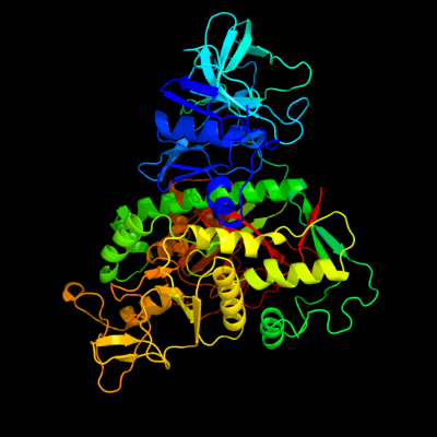
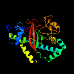
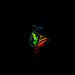
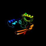
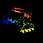
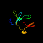
| 1 | c2ioaA_
|
|
 |
100.0 |
100 |
PDB header:ligase, hydrolase
Chain: A: PDB Molecule:bifunctional glutathionylspermidine
PDBTitle: e. coli bifunctional glutathionylspermidine2 synthetase/amidase incomplex with mg2+ and adp and3 phosphinate inhibitor
|
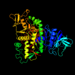
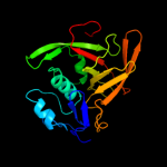
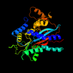
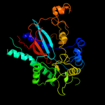
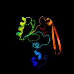
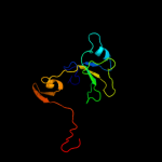
| 2 | c2vpmB_
|
|
 |
100.0 |
33 |
PDB header:ligase
Chain: B: PDB Molecule:trypanothione synthetase;
PDBTitle: trypanothione synthetase
|
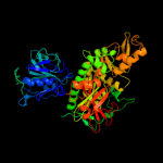
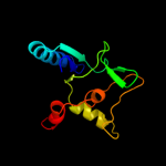
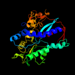
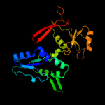
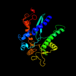
| 3 | d2io8a3
|
|
 |
100.0 |
98 |
Fold:ATP-grasp
Superfamily:Glutathione synthetase ATP-binding domain-like
Family:Glutathionylspermidine synthase ATP-binding domain-like |
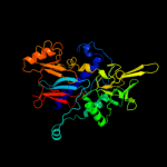
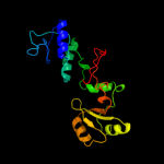
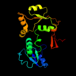
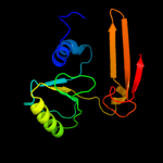
| 4 | d2io8a2
|
|
 |
100.0 |
98 |
Fold:Cysteine proteinases
Superfamily:Cysteine proteinases
Family:CHAP domain |
| 5 | d2io8a1
|
|
 |
100.0 |
74 |
Fold:PreATP-grasp domain
Superfamily:PreATP-grasp domain
Family:Glutathionylspermidine synthase substrate-binding domain-like |
| 6 | c3n6xA_
|
|
 |
99.7 |
16 |
PDB header:ligase
Chain: A: PDB Molecule:putative glutathionylspermidine synthase;
PDBTitle: crystal structure of a putative glutathionylspermidine synthase2 (mfla_0391) from methylobacillus flagellatus kt at 2.35 a resolution
|
| 7 | c2k3aA_
|
|
 |
98.6 |
18 |
PDB header:hydrolase
Chain: A: PDB Molecule:chap domain protein;
PDBTitle: nmr solution structure of staphylococcus saprophyticus chap2 (cysteine, histidine-dependent amidohydrolases/peptidases)3 domain protein. northeast structural genomics consortium4 target syr11
|
| 8 | c2hgsA_
|
|
 |
98.4 |
18 |
PDB header:amine/carboxylate ligase
Chain: A: PDB Molecule:protein (glutathione synthetase);
PDBTitle: human glutathione synthetase
|
| 9 | c3kalB_
|
|
 |
97.8 |
17 |
PDB header:ligase
Chain: B: PDB Molecule:homoglutathione synthetase;
PDBTitle: structure of homoglutathione synthetase from glycine max in2 closed conformation with homoglutathione, adp, a sulfate3 ion, and three magnesium ions bound
|
| 10 | c1gshA_
|
|
 |
97.4 |
14 |
PDB header:glutathione biosynthesis ligase
Chain: A: PDB Molecule:glutathione biosynthetic ligase;
PDBTitle: structure of escherichia coli glutathione synthetase at ph 7.5
|
| 11 | c1pk8D_
|
|
 |
97.1 |
14 |
PDB header:membrane protein
Chain: D: PDB Molecule:rat synapsin i;
PDBTitle: crystal structure of rat synapsin i c domain complexed to2 ca.atp
|
| 12 | c2wyoC_
|
|
 |
96.9 |
17 |
PDB header:ligase
Chain: C: PDB Molecule:glutathione synthetase;
PDBTitle: trypanosoma brucei glutathione synthetase
|
| 13 | c2qb5B_
|
|
 |
96.9 |
15 |
PDB header:transferase
Chain: B: PDB Molecule:inositol-tetrakisphosphate 1-kinase;
PDBTitle: crystal structure of human inositol 1,3,4-trisphosphate 5/6-kinase2 (itpk1) in complex with adp and mn2+
|
| 14 | c1uc8B_
|
|
 |
96.8 |
16 |
PDB header:biosynthetic protein
Chain: B: PDB Molecule:lysine biosynthesis enzyme;
PDBTitle: crystal structure of a lysine biosynthesis enzyme, lysx,2 from thermus thermophilus hb8
|
| 15 | c2p0aA_
|
|
 |
96.8 |
16 |
PDB header:neuropeptide
Chain: A: PDB Molecule:synapsin-3;
PDBTitle: the crystal structure of human synapsin iii (syn3) in complex with2 amppnp
|
| 16 | c1i7nA_
|
|
 |
96.7 |
14 |
PDB header:neuropeptide
Chain: A: PDB Molecule:synapsin ii;
PDBTitle: crystal structure analysis of the c domain of synapsin ii2 from rat brain
|
| 17 | c1m0tB_
|
|
 |
96.3 |
20 |
PDB header:ligase
Chain: B: PDB Molecule:glutathione synthetase;
PDBTitle: yeast glutathione synthase
|
| 18 | d1pk8a2
|
|
 |
96.1 |
15 |
Fold:ATP-grasp
Superfamily:Glutathione synthetase ATP-binding domain-like
Family:Synapsin C-terminal domain |
| 19 | c3t9aA_
|
|
 |
95.8 |
21 |
PDB header:transferase
Chain: A: PDB Molecule:inositol pyrophosphate kinase;
PDBTitle: crystal structure of the catalytic domain of human diphosphoinositol2 pentakisphosphate kinase 2 (ppip5k2) in complex with amppnp at ph 7.0
|
| 20 | c3ln6A_
|
|
 |
94.7 |
13 |
PDB header:ligase
Chain: A: PDB Molecule:glutathione biosynthesis bifunctional protein gshab;
PDBTitle: crystal structure of a bifunctional glutathione synthetase from2 streptococcus agalactiae
|
| 21 | d1i7na2 |
|
not modelled |
94.4 |
15 |
Fold:ATP-grasp
Superfamily:Glutathione synthetase ATP-binding domain-like
Family:Synapsin C-terminal domain |
| 22 | d1gsaa2 |
|
not modelled |
93.6 |
20 |
Fold:ATP-grasp
Superfamily:Glutathione synthetase ATP-binding domain-like
Family:ATP-binding domain of peptide synthetases |
| 23 | c3ln7A_ |
|
not modelled |
93.0 |
14 |
PDB header:ligase
Chain: A: PDB Molecule:glutathione biosynthesis bifunctional protein gshab;
PDBTitle: crystal structure of a bifunctional glutathione synthetase from2 pasteurella multocida
|
| 24 | c3bg5C_ |
|
not modelled |
90.5 |
18 |
PDB header:ligase
Chain: C: PDB Molecule:pyruvate carboxylase;
PDBTitle: crystal structure of staphylococcus aureus pyruvate2 carboxylase
|
| 25 | c3lp8A_ |
|
not modelled |
89.7 |
25 |
PDB header:ligase
Chain: A: PDB Molecule:phosphoribosylamine-glycine ligase;
PDBTitle: crystal structure of phosphoribosylamine-glycine ligase from2 ehrlichia chaffeensis
|
| 26 | c1kjjA_ |
|
not modelled |
89.4 |
12 |
PDB header:transferase
Chain: A: PDB Molecule:phosphoribosylglycinamide formyltransferase 2;
PDBTitle: crystal structure of glycniamide ribonucleotide2 transformylase in complex with mg-atp-gamma-s
|
| 27 | c2yyaB_ |
|
not modelled |
89.2 |
20 |
PDB header:ligase
Chain: B: PDB Molecule:phosphoribosylamine--glycine ligase;
PDBTitle: crystal structure of gar synthetase from aquifex aeolicus
|
| 28 | c1vkzA_ |
|
not modelled |
88.5 |
14 |
PDB header:ligase
Chain: A: PDB Molecule:phosphoribosylamine--glycine ligase;
PDBTitle: crystal structure of phosphoribosylamine--glycine ligase (tm1250) from2 thermotoga maritima at 2.30 a resolution
|
| 29 | c2hjwA_ |
|
not modelled |
88.0 |
17 |
PDB header:ligase
Chain: A: PDB Molecule:acetyl-coa carboxylase 2;
PDBTitle: crystal structure of the bc domain of acc2
|
| 30 | c2xd4A_ |
|
not modelled |
87.7 |
19 |
PDB header:ligase
Chain: A: PDB Molecule:phosphoribosylamine--glycine ligase;
PDBTitle: nucleotide-bound structures of bacillus subtilis glycinamide2 ribonucleotide synthetase
|
| 31 | d2if6a1 |
|
not modelled |
87.4 |
17 |
Fold:Cysteine proteinases
Superfamily:Cysteine proteinases
Family:YiiX-like |
| 32 | c1ulzA_ |
|
not modelled |
87.0 |
14 |
PDB header:ligase
Chain: A: PDB Molecule:pyruvate carboxylase n-terminal domain;
PDBTitle: crystal structure of the biotin carboxylase subunit of pyruvate2 carboxylase
|
| 33 | c2i80B_ |
|
not modelled |
86.9 |
18 |
PDB header:ligase
Chain: B: PDB Molecule:d-alanine-d-alanine ligase;
PDBTitle: allosteric inhibition of staphylococcus aureus d-alanine:d-alanine2 ligase revealed by crystallographic studies
|
| 34 | c2ip4A_ |
|
not modelled |
86.2 |
18 |
PDB header:ligase
Chain: A: PDB Molecule:phosphoribosylamine--glycine ligase;
PDBTitle: crystal structure of glycinamide ribonucleotide synthetase from2 thermus thermophilus hb8
|
| 35 | c3tinA_ |
|
not modelled |
86.1 |
12 |
PDB header:ligase
Chain: A: PDB Molecule:ttl protein;
PDBTitle: tubulin tyrosine ligase
|
| 36 | c1ehiB_ |
|
not modelled |
85.2 |
20 |
PDB header:ligase
Chain: B: PDB Molecule:d-alanine:d-lactate ligase;
PDBTitle: d-alanine:d-lactate ligase (lmddl2) of vancomycin-resistant2 leuconostoc mesenteroides
|
| 37 | c2dzdB_ |
|
not modelled |
84.3 |
12 |
PDB header:ligase
Chain: B: PDB Molecule:pyruvate carboxylase;
PDBTitle: crystal structure of the biotin carboxylase domain of2 pyruvate carboxylase
|
| 38 | c2vpqA_ |
|
not modelled |
84.0 |
12 |
PDB header:ligase
Chain: A: PDB Molecule:acetyl-coa carboxylase;
PDBTitle: crystal structure of biotin carboxylase from s. aureus2 complexed with amppnp
|
| 39 | c3se7A_ |
|
not modelled |
83.6 |
19 |
PDB header:ligase
Chain: A: PDB Molecule:vana;
PDBTitle: ancient vana
|
| 40 | c3gidB_ |
|
not modelled |
81.8 |
13 |
PDB header:ligase
Chain: B: PDB Molecule:acetyl-coa carboxylase 2;
PDBTitle: the biotin carboxylase (bc) domain of human acetyl-coa2 carboxylase 2 (acc2) in complex with soraphen a
|
| 41 | c2dlnA_ |
|
not modelled |
81.8 |
23 |
PDB header:ligase(peptidoglycan synthesis)
Chain: A: PDB Molecule:d-alanine--d-alanine ligase;
PDBTitle: vancomycin resistance: structure of d-alanine:d-alanine2 ligase at 2.3 angstroms resolution
|
| 42 | c2pvpB_ |
|
not modelled |
81.7 |
19 |
PDB header:ligase
Chain: B: PDB Molecule:d-alanine-d-alanine ligase;
PDBTitle: crystal structure of d-alanine-d-alanine ligase from helicobacter2 pylori
|
| 43 | c3tqtB_ |
|
not modelled |
80.7 |
16 |
PDB header:ligase
Chain: B: PDB Molecule:d-alanine--d-alanine ligase;
PDBTitle: structure of the d-alanine-d-alanine ligase from coxiella burnetii
|
| 44 | c3ouzA_ |
|
not modelled |
80.2 |
10 |
PDB header:ligase
Chain: A: PDB Molecule:biotin carboxylase;
PDBTitle: crystal structure of biotin carboxylase-adp complex from campylobacter2 jejuni
|
| 45 | c3g8cB_ |
|
not modelled |
79.7 |
14 |
PDB header:ligase
Chain: B: PDB Molecule:biotin carboxylase;
PDBTitle: crystal stucture of biotin carboxylase in complex with2 biotin, bicarbonate, adp and mg ion
|
| 46 | c3lwbA_ |
|
not modelled |
78.5 |
22 |
PDB header:ligase
Chain: A: PDB Molecule:d-alanine--d-alanine ligase;
PDBTitle: crystal structure of apo d-alanine:d-alanine ligase (ddl) from2 mycobacterium tuberculosis
|
| 47 | d1gsaa1 |
|
not modelled |
78.3 |
11 |
Fold:PreATP-grasp domain
Superfamily:PreATP-grasp domain
Family:Prokaryotic glutathione synthetase, N-terminal domain |
| 48 | d3etja3 |
|
not modelled |
76.6 |
16 |
Fold:ATP-grasp
Superfamily:Glutathione synthetase ATP-binding domain-like
Family:BC ATP-binding domain-like |
| 49 | d1e4ea2 |
|
not modelled |
75.3 |
13 |
Fold:ATP-grasp
Superfamily:Glutathione synthetase ATP-binding domain-like
Family:ATP-binding domain of peptide synthetases |
| 50 | c2zdqA_ |
|
not modelled |
75.3 |
10 |
PDB header:ligase
Chain: A: PDB Molecule:d-alanine--d-alanine ligase;
PDBTitle: crystal structure of d-alanine:d-alanine ligase with atp2 and d-alanine:d-alanine from thermus thermophius hb8
|
| 51 | c3r23B_ |
|
not modelled |
75.0 |
16 |
PDB header:ligase
Chain: B: PDB Molecule:d-alanine--d-alanine ligase;
PDBTitle: crystal structure of d-alanine--d-alanine ligase from bacillus2 anthracis
|
| 52 | d1uc8a2 |
|
not modelled |
74.8 |
27 |
Fold:ATP-grasp
Superfamily:Glutathione synthetase ATP-binding domain-like
Family:Lysine biosynthesis enzyme LysX ATP-binding domain |
| 53 | d1fs2b1 |
|
not modelled |
74.6 |
20 |
Fold:Skp1 dimerisation domain-like
Superfamily:Skp1 dimerisation domain-like
Family:Skp1 dimerisation domain-like |
| 54 | d1gsoa3 |
|
not modelled |
74.0 |
30 |
Fold:ATP-grasp
Superfamily:Glutathione synthetase ATP-binding domain-like
Family:BC ATP-binding domain-like |
| 55 | c1w96B_ |
|
not modelled |
72.8 |
20 |
PDB header:ligase
Chain: B: PDB Molecule:acetyl-coenzyme a carboxylase;
PDBTitle: crystal structure of biotin carboxylase domain of acetyl-2 coenzyme a carboxylase from saccharomyces cerevisiae in3 complex with soraphen a
|
| 56 | c2k1gA_ |
|
not modelled |
72.7 |
22 |
PDB header:lipoprotein
Chain: A: PDB Molecule:lipoprotein spr;
PDBTitle: solution nmr structure of lipoprotein spr from escherichia coli k12.2 northeast structural genomics target er541-37-162
|
| 57 | c3i12A_ |
|
not modelled |
72.5 |
11 |
PDB header:ligase
Chain: A: PDB Molecule:d-alanine-d-alanine ligase a;
PDBTitle: the crystal structure of the d-alanyl-alanine synthetase a from2 salmonella enterica subsp. enterica serovar typhimurium str. lt2
|
| 58 | d1iowa2 |
|
not modelled |
71.6 |
24 |
Fold:ATP-grasp
Superfamily:Glutathione synthetase ATP-binding domain-like
Family:ATP-binding domain of peptide synthetases |
| 59 | c3e5nA_ |
|
not modelled |
71.1 |
25 |
PDB header:ligase
Chain: A: PDB Molecule:d-alanine-d-alanine ligase a;
PDBTitle: crystal strucutre of d-alanine-d-alanine ligase from2 xanthomonas oryzae pv. oryzae kacc10331
|
| 60 | d1kjqa3 |
|
not modelled |
69.8 |
5 |
Fold:ATP-grasp
Superfamily:Glutathione synthetase ATP-binding domain-like
Family:BC ATP-binding domain-like |
| 61 | d1ehia2 |
|
not modelled |
69.6 |
24 |
Fold:ATP-grasp
Superfamily:Glutathione synthetase ATP-binding domain-like
Family:ATP-binding domain of peptide synthetases |
| 62 | d2j9ga3 |
|
not modelled |
68.0 |
20 |
Fold:ATP-grasp
Superfamily:Glutathione synthetase ATP-binding domain-like
Family:BC ATP-binding domain-like |
| 63 | c3kw0D_ |
|
not modelled |
67.0 |
33 |
PDB header:hydrolase
Chain: D: PDB Molecule:cysteine peptidase;
PDBTitle: crystal structure of cysteine peptidase (np_982244.1) from bacillus2 cereus atcc 10987 at 2.50 a resolution
|
| 64 | c3df7A_ |
|
not modelled |
64.5 |
14 |
PDB header:structural genomics, unknown function
Chain: A: PDB Molecule:putative atp-grasp superfamily protein;
PDBTitle: crystal structure of a putative atp-grasp superfamily2 protein from archaeoglobus fulgidus
|
| 65 | c1e4eB_ |
|
not modelled |
64.5 |
13 |
PDB header:ligase
Chain: B: PDB Molecule:vancomycin/teicoplanin a-type resistance protein vana;
PDBTitle: d-alanyl-d-lacate ligase
|
| 66 | d1vkza3 |
|
not modelled |
63.6 |
17 |
Fold:ATP-grasp
Superfamily:Glutathione synthetase ATP-binding domain-like
Family:BC ATP-binding domain-like |
| 67 | d1w96a3 |
|
not modelled |
62.8 |
13 |
Fold:ATP-grasp
Superfamily:Glutathione synthetase ATP-binding domain-like
Family:BC ATP-binding domain-like |
| 68 | c2cqyA_ |
|
not modelled |
60.6 |
16 |
PDB header:ligase
Chain: A: PDB Molecule:propionyl-coa carboxylase alpha chain,
PDBTitle: solution structure of b domain from human propionyl-coa2 carboxylase alpha subunit
|
| 69 | c2gpwC_ |
|
not modelled |
59.8 |
13 |
PDB header:ligase
Chain: C: PDB Molecule:biotin carboxylase;
PDBTitle: crystal structure of the biotin carboxylase subunit, f363a2 mutant, of acetyl-coa carboxylase from escherichia coli.
|
| 70 | c3pbiA_ |
|
not modelled |
59.3 |
27 |
PDB header:hydrolase
Chain: A: PDB Molecule:invasion protein;
PDBTitle: structure of the peptidoglycan hydrolase ripb (rv1478) from2 mycobacterium tuberculosis at 1.6 resolution
|
| 71 | c3k3pA_ |
|
not modelled |
58.6 |
18 |
PDB header:ligase
Chain: A: PDB Molecule:d-alanine--d-alanine ligase;
PDBTitle: crystal structure of the apo form of d-alanine:d-alanine ligase (ddl)2 from streptococcus mutans
|
| 72 | d2evra2 |
|
not modelled |
58.4 |
27 |
Fold:Cysteine proteinases
Superfamily:Cysteine proteinases
Family:NlpC/P60 |
| 73 | c3orqA_ |
|
not modelled |
55.0 |
8 |
PDB header:ligase,biosynthetic protein
Chain: A: PDB Molecule:n5-carboxyaminoimidazole ribonucleotide synthetase;
PDBTitle: crystal structure of n5-carboxyaminoimidazole synthetase from2 staphylococcus aureus complexed with adp
|
| 74 | d1nexa1 |
|
not modelled |
53.3 |
19 |
Fold:Skp1 dimerisation domain-like
Superfamily:Skp1 dimerisation domain-like
Family:Skp1 dimerisation domain-like |
| 75 | d1a9xa6 |
|
not modelled |
51.3 |
8 |
Fold:ATP-grasp
Superfamily:Glutathione synthetase ATP-binding domain-like
Family:BC ATP-binding domain-like |
| 76 | d1vbva1 |
|
not modelled |
49.3 |
23 |
Fold:SH3-like barrel
Superfamily:YccV-like
Family:YccV-like |
| 77 | d2ovra1 |
|
not modelled |
49.0 |
21 |
Fold:Skp1 dimerisation domain-like
Superfamily:Skp1 dimerisation domain-like
Family:Skp1 dimerisation domain-like |
| 78 | c2ys6A_ |
|
not modelled |
47.1 |
35 |
PDB header:ligase
Chain: A: PDB Molecule:phosphoribosylglycinamide synthetase;
PDBTitle: crystal structure of gar synthetase from geobacillus kaustophilus
|
| 79 | c2fg0B_ |
|
not modelled |
46.6 |
29 |
PDB header:hydrolase
Chain: B: PDB Molecule:cog0791: cell wall-associated hydrolases (invasion-
PDBTitle: crystal structure of a putative gamma-d-glutamyl-l-diamino acid2 endopeptidase (npun_r0659) from nostoc punctiforme pcc 73102 at 1.793 a resolution
|
| 80 | c2p1nD_ |
|
not modelled |
45.5 |
24 |
PDB header:signaling protein
Chain: D: PDB Molecule:skp1-like protein 1a;
PDBTitle: mechanism of auxin perception by the tir1 ubiqutin ligase
|
| 81 | c1z2pX_ |
|
not modelled |
45.0 |
19 |
PDB header:transferase
Chain: X: PDB Molecule:inositol 1,3,4-trisphosphate 5/6-kinase;
PDBTitle: inositol 1,3,4-trisphosphate 5/6-kinase in complex with mg2+/amp-2 pcp/ins(1,3,4)p3
|
| 82 | d1a9xa5 |
|
not modelled |
42.0 |
8 |
Fold:ATP-grasp
Superfamily:Glutathione synthetase ATP-binding domain-like
Family:BC ATP-binding domain-like |
| 83 | c1nexC_ |
|
not modelled |
42.0 |
18 |
PDB header:ligase, cell cycle
Chain: C: PDB Molecule:centromere dna-binding protein complex cbf3
PDBTitle: crystal structure of scskp1-sccdc4-cpd peptide complex
|
| 84 | c3s4wB_ |
|
not modelled |
40.9 |
32 |
PDB header:dna binding protein
Chain: B: PDB Molecule:fanconi anemia group d2 protein homolog;
PDBTitle: structure of the fanci-fancd2 complex
|
| 85 | c2dwcB_ |
|
not modelled |
40.7 |
4 |
PDB header:transferase
Chain: B: PDB Molecule:433aa long hypothetical phosphoribosylglycinamide formyl
PDBTitle: crystal structure of probable phosphoribosylglycinamide formyl2 transferase from pyrococcus horikoshii ot3 complexed with adp
|
| 86 | d2hgsa4 |
|
not modelled |
40.3 |
19 |
Fold:ATP-grasp
Superfamily:Glutathione synthetase ATP-binding domain-like
Family:Eukaryotic glutathione synthetase ATP-binding domain |
| 87 | c3n6rK_ |
|
not modelled |
39.6 |
25 |
PDB header:ligase
Chain: K: PDB Molecule:propionyl-coa carboxylase, alpha subunit;
PDBTitle: crystal structure of the holoenzyme of propionyl-coa carboxylase (pcc)
|
| 88 | c3i86A_ |
|
not modelled |
39.2 |
17 |
PDB header:hydrolase
Chain: A: PDB Molecule:putative uncharacterized protein;
PDBTitle: crystal structure of the p60 domain from m. avium subspecies2 paratuberculosis antigen map1204
|
| 89 | d2auaa1 |
|
not modelled |
38.0 |
40 |
Fold:ADP-ribosylation
Superfamily:ADP-ribosylation
Family:BC2332-like |
| 90 | d1irza_ |
|
not modelled |
37.8 |
27 |
Fold:DNA/RNA-binding 3-helical bundle
Superfamily:Homeodomain-like
Family:GARP response regulators |
| 91 | c2xivA_ |
|
not modelled |
36.8 |
18 |
PDB header:structural protein
Chain: A: PDB Molecule:hypothetical invasion protein;
PDBTitle: structure of rv1477, hypothetical invasion protein of2 mycobacterium tuberculosis
|
| 92 | d2r85a2 |
|
not modelled |
35.5 |
13 |
Fold:ATP-grasp
Superfamily:Glutathione synthetase ATP-binding domain-like
Family:PurP ATP-binding domain-like |
| 93 | c3gt2A_ |
|
not modelled |
34.6 |
19 |
PDB header:unknown function
Chain: A: PDB Molecule:putative uncharacterized protein;
PDBTitle: crystal structure of the p60 domain from m. avium2 paratuberculosis antigen map1272c
|
| 94 | c1gsoA_ |
|
not modelled |
32.4 |
33 |
PDB header:ligase
Chain: A: PDB Molecule:protein (glycinamide ribonucleotide synthetase);
PDBTitle: glycinamide ribonucleotide synthetase (gar-syn) from e.2 coli.
|
| 95 | c3al6A_ |
|
not modelled |
32.1 |
10 |
PDB header:unknown function
Chain: A: PDB Molecule:jmjc domain-containing protein c2orf60;
PDBTitle: crystal structure of human tyw5
|
| 96 | d2r7ka2 |
|
not modelled |
32.0 |
21 |
Fold:ATP-grasp
Superfamily:Glutathione synthetase ATP-binding domain-like
Family:PurP ATP-binding domain-like |
| 97 | d2oo3a1 |
|
not modelled |
31.8 |
19 |
Fold:S-adenosyl-L-methionine-dependent methyltransferases
Superfamily:S-adenosyl-L-methionine-dependent methyltransferases
Family:LPG1296-like |
| 98 | c3kopB_ |
|
not modelled |
30.9 |
17 |
PDB header:structural genomics, unknown function
Chain: B: PDB Molecule:uncharacterized protein;
PDBTitle: crystal structure of protein with a cyclophilin-like fold2 (yp_831253.1) from arthrobacter sp. fb24 at 1.90 a resolution
|
| 99 | c2qk4A_ |
|
not modelled |
30.7 |
26 |
PDB header:ligase
Chain: A: PDB Molecule:trifunctional purine biosynthetic protein adenosine-3;
PDBTitle: human glycinamide ribonucleotide synthetase
|
| 100 | c3h41A_ |
|
not modelled |
28.8 |
17 |
PDB header:hydrolase
Chain: A: PDB Molecule:nlp/p60 family protein;
PDBTitle: crystal structure of a nlpc/p60 family protein (bce_2878) from2 bacillus cereus atcc 10987 at 1.79 a resolution
|
| 101 | d1saza1 |
|
not modelled |
27.0 |
18 |
Fold:Ribonuclease H-like motif
Superfamily:Actin-like ATPase domain
Family:Acetokinase-like |
| 102 | c3etjB_ |
|
not modelled |
26.2 |
16 |
PDB header:lyase
Chain: B: PDB Molecule:phosphoribosylaminoimidazole carboxylase atpase
PDBTitle: crystal structure e. coli purk in complex with mg, adp, and2 pi
|
| 103 | d1vlya1 |
|
not modelled |
25.2 |
27 |
Fold:Elongation factor/aminomethyltransferase common domain
Superfamily:Aminomethyltransferase beta-barrel domain
Family:Aminomethyltransferase beta-barrel domain |
| 104 | c3heiI_ |
|
not modelled |
25.0 |
18 |
PDB header:transferase/signaling protein
Chain: I: PDB Molecule:ephrin type-a receptor 2;
PDBTitle: ligand recognition by a-class eph receptors: crystal structures of the2 epha2 ligand-binding domain and the epha2/ephrin-a1 complex
|
| 105 | d1ulza3 |
|
not modelled |
24.0 |
21 |
Fold:ATP-grasp
Superfamily:Glutathione synthetase ATP-binding domain-like
Family:BC ATP-binding domain-like |
| 106 | c3q2oB_ |
|
not modelled |
24.0 |
5 |
PDB header:lyase
Chain: B: PDB Molecule:phosphoribosylaminoimidazole carboxylase, atpase subunit;
PDBTitle: crystal structure of purk: n5-carboxyaminoimidazole ribonucleotide2 synthetase
|
| 107 | c2kytA_ |
|
not modelled |
23.8 |
35 |
PDB header:hydrolase
Chain: A: PDB Molecule:group xvi phospholipase a2;
PDBTitle: solution struture of the h-rev107 n-terminal domain
|
| 108 | d1t3da_ |
|
not modelled |
23.6 |
15 |
Fold:Single-stranded left-handed beta-helix
Superfamily:Trimeric LpxA-like enzymes
Family:Serine acetyltransferase |
| 109 | c2z04A_ |
|
not modelled |
23.6 |
10 |
PDB header:lyase
Chain: A: PDB Molecule:phosphoribosylaminoimidazole carboxylase atpase
PDBTitle: crystal structure of phosphoribosylaminoimidazole2 carboxylase atpase subunit from aquifex aeolicus
|
| 110 | c3kvpB_ |
|
not modelled |
23.6 |
57 |
PDB header:structural genomics, unknown function
Chain: B: PDB Molecule:uncharacterized protein ymzc;
PDBTitle: crystal structure of uncharacterized protein ymzc precursor2 from bacillus subtilis, northeast structural genomics3 consortium target sr378a
|
| 111 | c2pn1A_ |
|
not modelled |
22.8 |
9 |
PDB header:ligase
Chain: A: PDB Molecule:carbamoylphosphate synthase large subunit;
PDBTitle: crystal structure of carbamoylphosphate synthase large subunit (split2 gene in mj) (zp_00538348.1) from exiguobacterium sp. 255-15 at 2.00 a3 resolution
|
| 112 | c2ovqA_ |
|
not modelled |
21.5 |
21 |
PDB header:transcription/cell cycle
Chain: A: PDB Molecule:s-phase kinase-associated protein 1a;
PDBTitle: structure of the skp1-fbw7-cyclinedegc complex
|
| 113 | c2p1gA_ |
|
not modelled |
20.6 |
14 |
PDB header:structural genomics, unknown function
Chain: A: PDB Molecule:putative xylanase;
PDBTitle: crystal structure of a putative xylanase from bacteroides fragilis
|