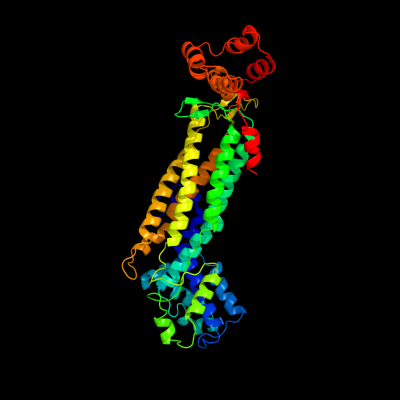
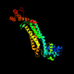
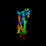
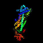
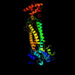
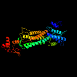
| 1 | c2ptsA_
|
|
 |
100.0 |
95 |
PDB header:lyase
Chain: A: PDB Molecule:adenylosuccinate lyase;
PDBTitle: crystal structure of wild type escherichia coli adenylosuccinate lyase
|
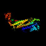
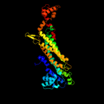
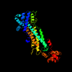
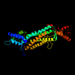
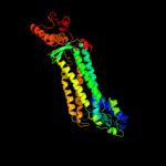
| 2 | c3bhgA_
|
|
 |
100.0 |
55 |
PDB header:lyase
Chain: A: PDB Molecule:adenylosuccinate lyase;
PDBTitle: crystal structure of adenylosuccinate lyase from legionella2 pneumophila
|
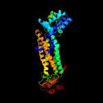
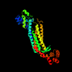
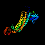
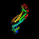
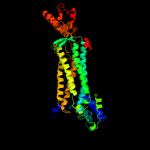
| 3 | c2qgaC_
|
|
 |
100.0 |
46 |
PDB header:lyase
Chain: C: PDB Molecule:adenylosuccinate lyase;
PDBTitle: plasmodium vivax adenylosuccinate lyase pv003765 with amp bound
|
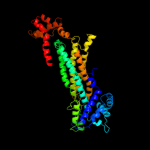
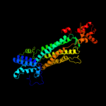
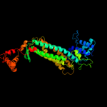
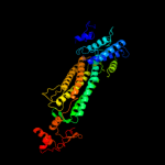
| 4 | c2vd6B_
|
|
 |
100.0 |
18 |
PDB header:lyase
Chain: B: PDB Molecule:adenylosuccinate lyase;
PDBTitle: human adenylosuccinate lyase in complex with its substrate2 n6-(1,2-dicarboxyethyl)-amp, and its products amp and3 fumarate.
|
| 5 | c2pfmA_
|
|
 |
100.0 |
22 |
PDB header:lyase
Chain: A: PDB Molecule:adenylosuccinate lyase;
PDBTitle: crystal structure of adenylosuccinate lyase (purb) from bacillus2 anthracis
|
| 6 | d1c3ca_
|
|
 |
100.0 |
21 |
Fold:L-aspartase-like
Superfamily:L-aspartase-like
Family:L-aspartase/fumarase |
| 7 | c1yisA_
|
|
 |
100.0 |
17 |
PDB header:lyase
Chain: A: PDB Molecule:adenylosuccinate lyase;
PDBTitle: structural genomics of caenorhabditis elegans: adenylosuccinate lyase
|
| 8 | d1re5a_
|
|
 |
100.0 |
21 |
Fold:L-aspartase-like
Superfamily:L-aspartase-like
Family:L-aspartase/fumarase |
| 9 | d1i0aa_
|
|
 |
100.0 |
18 |
Fold:L-aspartase-like
Superfamily:L-aspartase-like
Family:L-aspartase/fumarase |
| 10 | d1tj7a_
|
|
 |
100.0 |
17 |
Fold:L-aspartase-like
Superfamily:L-aspartase-like
Family:L-aspartase/fumarase |
| 11 | d1q5na_
|
|
 |
100.0 |
18 |
Fold:L-aspartase-like
Superfamily:L-aspartase-like
Family:L-aspartase/fumarase |
| 12 | d1hy0a_
|
|
 |
100.0 |
17 |
Fold:L-aspartase-like
Superfamily:L-aspartase-like
Family:L-aspartase/fumarase |
| 13 | d1dofa_
|
|
 |
100.0 |
24 |
Fold:L-aspartase-like
Superfamily:L-aspartase-like
Family:L-aspartase/fumarase |
| 14 | d1k62a_
|
|
 |
100.0 |
18 |
Fold:L-aspartase-like
Superfamily:L-aspartase-like
Family:L-aspartase/fumarase |
| 15 | d1j3ua_
|
|
 |
100.0 |
15 |
Fold:L-aspartase-like
Superfamily:L-aspartase-like
Family:L-aspartase/fumarase |
| 16 | c2e9fC_
|
|
 |
100.0 |
18 |
PDB header:lyase
Chain: C: PDB Molecule:argininosuccinate lyase;
PDBTitle: crystal structure of t.th.hb8 argininosuccinate lyase complexed with2 l-arginine
|
| 17 | d1tjva_
|
|
 |
100.0 |
17 |
Fold:L-aspartase-like
Superfamily:L-aspartase-like
Family:L-aspartase/fumarase |
| 18 | c3c8tA_
|
|
 |
100.0 |
20 |
PDB header:lyase
Chain: A: PDB Molecule:fumarate lyase;
PDBTitle: crystal structure of fumarate lyase from mesorhizobium sp. bnc1
|
| 19 | c1yfmA_
|
|
 |
100.0 |
16 |
PDB header:lyase
Chain: A: PDB Molecule:fumarase;
PDBTitle: recombinant yeast fumarase
|
| 20 | d1yfma_
|
|
 |
100.0 |
16 |
Fold:L-aspartase-like
Superfamily:L-aspartase-like
Family:L-aspartase/fumarase |
| 21 | d1fuoa_ |
|
not modelled |
100.0 |
17 |
Fold:L-aspartase-like
Superfamily:L-aspartase-like
Family:L-aspartase/fumarase |
| 22 | d1vdka_ |
|
not modelled |
100.0 |
18 |
Fold:L-aspartase-like
Superfamily:L-aspartase-like
Family:L-aspartase/fumarase |
| 23 | d1jswa_ |
|
not modelled |
100.0 |
15 |
Fold:L-aspartase-like
Superfamily:L-aspartase-like
Family:L-aspartase/fumarase |
| 24 | c3no9C_ |
|
not modelled |
100.0 |
15 |
PDB header:lyase
Chain: C: PDB Molecule:fumarate hydratase class ii;
PDBTitle: crystal structure of apo fumarate hydratase from mycobacterium2 tuberculosis
|
| 25 | c3ocfB_ |
|
not modelled |
100.0 |
16 |
PDB header:lyase
Chain: B: PDB Molecule:fumarate lyase:delta crystallin;
PDBTitle: crystal structure of fumarate lyase:delta crystallin from brucella2 melitensis in native form
|
| 26 | d1jswc_ |
|
not modelled |
100.0 |
14 |
Fold:L-aspartase-like
Superfamily:L-aspartase-like
Family:L-aspartase/fumarase |
| 27 | c3e04C_ |
|
not modelled |
100.0 |
15 |
PDB header:lyase
Chain: C: PDB Molecule:fumarate hydratase;
PDBTitle: crystal structure of human fumarate hydratase
|
| 28 | c2fenA_ |
|
not modelled |
100.0 |
21 |
PDB header:isomerase
Chain: A: PDB Molecule:3-carboxy-cis,cis-muconate lactonizing enzyme;
PDBTitle: 3-carboxy-cis,cis-muconate lactonizing enzyme from agrobacterium2 radiobacter s2
|
| 29 | c3gtdB_ |
|
not modelled |
100.0 |
15 |
PDB header:lyase
Chain: B: PDB Molecule:fumarate hydratase class ii;
PDBTitle: 2.4 angstrom crystal structure of fumarate hydratase from rickettsia2 prowazekii
|
| 30 | d1f1oa_ |
|
not modelled |
100.0 |
24 |
Fold:L-aspartase-like
Superfamily:L-aspartase-like
Family:L-aspartase/fumarase |
| 31 | c3r6yG_ |
|
not modelled |
100.0 |
14 |
PDB header:lyase
Chain: G: PDB Molecule:aspartase;
PDBTitle: crystal structure of chymotrypsin-treated aspartase from bacillus sp.2 ym55-1
|
| 32 | c2ctoA_ |
|
not modelled |
28.3 |
50 |
PDB header:structural genomics, unknown function
Chain: A: PDB Molecule:novel protein;
PDBTitle: solution structure of the hmg box like domain from human2 hypothetical protein flj14904
|
| 33 | d1rp4a_ |
|
not modelled |
18.4 |
11 |
Fold:ERO1-like
Superfamily:ERO1-like
Family:ERO1-like |
| 34 | c3ahrA_ |
|
not modelled |
15.6 |
17 |
PDB header:oxidoreductase
Chain: A: PDB Molecule:ero1-like protein alpha;
PDBTitle: inactive human ero1
|
| 35 | c2xgvA_ |
|
not modelled |
15.0 |
23 |
PDB header:viral protein
Chain: A: PDB Molecule:psiv capsid n-terminal domain;
PDBTitle: structure of the n-terminal domain of capsid protein from2 rabbit endogenous lentivirus (relik)
|
| 36 | c1wjvA_ |
|
not modelled |
14.2 |
56 |
PDB header:dna binding protein
Chain: A: PDB Molecule:cell growth regulating nucleolar protein lyar;
PDBTitle: solution structure of the n-terminal zinc finger domain of2 mouse cell growth regulating nucleolar protein lyar
|
| 37 | c2ev9B_ |
|
not modelled |
13.1 |
43 |
PDB header:oxidoreductase
Chain: B: PDB Molecule:shikimate 5-dehydrogenase;
PDBTitle: crystal structure of shikimate 5-dehydrogenase (aroe) from thermus2 thermophilus hb8 in complex with nadp(h) and shikimate
|
| 38 | c3fbtB_ |
|
not modelled |
12.7 |
33 |
PDB header:oxidoreductase, lyase
Chain: B: PDB Molecule:chorismate mutase and shikimate 5-dehydrogenase
PDBTitle: crystal structure of a chorismate mutase/shikimate 5-2 dehydrogenase fusion protein from clostridium3 acetobutylicum
|
| 39 | c3bbnC_ |
|
not modelled |
12.7 |
57 |
PDB header:ribosome
Chain: C: PDB Molecule:ribosomal protein s3;
PDBTitle: homology model for the spinach chloroplast 30s subunit2 fitted to 9.4a cryo-em map of the 70s chlororibosome.
|
| 40 | d1nvta2 |
|
not modelled |
12.2 |
29 |
Fold:Aminoacid dehydrogenase-like, N-terminal domain
Superfamily:Aminoacid dehydrogenase-like, N-terminal domain
Family:Shikimate dehydrogenase-like |
| 41 | d2cb2a1 |
|
not modelled |
11.3 |
33 |
Fold:Ferredoxin-like
Superfamily:Dimeric alpha+beta barrel
Family:SOR-like |
| 42 | d1v5pa_ |
|
not modelled |
10.7 |
50 |
Fold:PH domain-like barrel
Superfamily:PH domain-like
Family:Pleckstrin-homology domain (PH domain) |
| 43 | d1vi2a2 |
|
not modelled |
10.4 |
14 |
Fold:Aminoacid dehydrogenase-like, N-terminal domain
Superfamily:Aminoacid dehydrogenase-like, N-terminal domain
Family:Shikimate dehydrogenase-like |
| 44 | d1g3pa2 |
|
not modelled |
9.6 |
33 |
Fold:N-terminal domains of the minor coat protein g3p
Superfamily:N-terminal domains of the minor coat protein g3p
Family:N-terminal domains of the minor coat protein g3p |
| 45 | d1npya2 |
|
not modelled |
9.3 |
23 |
Fold:Aminoacid dehydrogenase-like, N-terminal domain
Superfamily:Aminoacid dehydrogenase-like, N-terminal domain
Family:Shikimate dehydrogenase-like |
| 46 | c3egqB_ |
|
not modelled |
9.3 |
8 |
PDB header:transcription
Chain: B: PDB Molecule:tetr family transcriptional regulator;
PDBTitle: crystal structure of a tetr-family transcriptional regulator (af_1817)2 from archaeoglobus fulgidus at 2.55 a resolution
|
| 47 | d1p77a2 |
|
not modelled |
9.1 |
20 |
Fold:Aminoacid dehydrogenase-like, N-terminal domain
Superfamily:Aminoacid dehydrogenase-like, N-terminal domain
Family:Shikimate dehydrogenase-like |
| 48 | c3dikA_ |
|
not modelled |
8.9 |
22 |
PDB header:viral protein
Chain: A: PDB Molecule:capsid protein p24;
PDBTitle: pseudo-atomic model of the hiv-1 ca hexameric lattice
|
| 49 | c1nvtA_ |
|
not modelled |
8.8 |
27 |
PDB header:oxidoreductase
Chain: A: PDB Molecule:shikimate 5'-dehydrogenase;
PDBTitle: crystal structure of shikimate dehydrogenase (aroe or2 mj1084) in complex with nadp+
|
| 50 | c3pwzA_ |
|
not modelled |
8.7 |
27 |
PDB header:oxidoreductase
Chain: A: PDB Molecule:shikimate dehydrogenase 3;
PDBTitle: crystal structure of an ael1 enzyme from pseudomonas putida
|
| 51 | c1p74B_ |
|
not modelled |
8.3 |
20 |
PDB header:oxidoreductase
Chain: B: PDB Molecule:shikimate 5-dehydrogenase;
PDBTitle: crystal structure of shikimate dehydrogenase (aroe) from2 haemophilus influenzae
|
| 52 | c1dipA_ |
|
not modelled |
7.9 |
14 |
PDB header:acetylation
Chain: A: PDB Molecule:delta-sleep-inducing peptide immunoreactive
PDBTitle: the solution structure of porcine delta-sleep-inducing2 peptide immunoreactive peptide, nmr, 10 structures
|
| 53 | d2qalc1 |
|
not modelled |
7.7 |
38 |
Fold:Alpha-lytic protease prodomain-like
Superfamily:Prokaryotic type KH domain (KH-domain type II)
Family:Prokaryotic type KH domain (KH-domain type II) |
| 54 | c3u62A_ |
|
not modelled |
7.6 |
57 |
PDB header:oxidoreductase
Chain: A: PDB Molecule:shikimate dehydrogenase;
PDBTitle: crystal structure of shikimate dehydrogenase from thermotoga maritima
|
| 55 | d1lbua1 |
|
not modelled |
7.6 |
23 |
Fold:PGBD-like
Superfamily:PGBD-like
Family:Peptidoglycan binding domain, PGBD |
| 56 | c3pgjB_ |
|
not modelled |
7.5 |
27 |
PDB header:oxidoreductase
Chain: B: PDB Molecule:shikimate dehydrogenase;
PDBTitle: 2.49 angstrom resolution crystal structure of shikimate 5-2 dehydrogenase (aroe) from vibrio cholerae o1 biovar eltor str. n169613 in complex with shikimate
|
| 57 | c3tozA_ |
|
not modelled |
7.4 |
27 |
PDB header:oxidoreductase
Chain: A: PDB Molecule:shikimate dehydrogenase;
PDBTitle: 2.2 angstrom crystal structure of shikimate 5-dehydrogenase from2 listeria monocytogenes in complex with nad.
|
| 58 | c2hk8B_ |
|
not modelled |
7.4 |
20 |
PDB header:oxidoreductase
Chain: B: PDB Molecule:shikimate dehydrogenase;
PDBTitle: crystal structure of shikimate dehydrogenase from aquifex2 aeolicus at 2.35 angstrom resolution
|
| 59 | d1wjva1 |
|
not modelled |
7.4 |
63 |
Fold:beta-beta-alpha zinc fingers
Superfamily:beta-beta-alpha zinc fingers
Family:C2HC finger |
| 60 | d1nyta2 |
|
not modelled |
7.4 |
29 |
Fold:Aminoacid dehydrogenase-like, N-terminal domain
Superfamily:Aminoacid dehydrogenase-like, N-terminal domain
Family:Shikimate dehydrogenase-like |
| 61 | c1ekuA_ |
|
not modelled |
7.3 |
31 |
PDB header:immune system
Chain: A: PDB Molecule:interferon gamma;
PDBTitle: crystal structure of a biologically active single chain2 mutant of human ifn-gamma
|
| 62 | c2eggA_ |
|
not modelled |
7.3 |
57 |
PDB header:oxidoreductase
Chain: A: PDB Molecule:shikimate 5-dehydrogenase;
PDBTitle: crystal structure of shikimate 5-dehydrogenase (aroe) from2 geobacillus kaustophilus
|
| 63 | d1prtb1 |
|
not modelled |
7.1 |
42 |
Fold:OB-fold
Superfamily:Bacterial enterotoxins
Family:Bacterial AB5 toxins, B-subunits |
| 64 | c2choA_ |
|
not modelled |
6.5 |
83 |
PDB header:hydrolase
Chain: A: PDB Molecule:glucosaminidase;
PDBTitle: bacteroides thetaiotaomicron hexosaminidase with o-2 glcnacase activity
|
| 65 | d1u5ta1 |
|
not modelled |
6.5 |
3 |
Fold:DNA/RNA-binding 3-helical bundle
Superfamily:"Winged helix" DNA-binding domain
Family:Vacuolar sorting protein domain |
| 66 | c3o8qB_ |
|
not modelled |
6.5 |
27 |
PDB header:oxidoreductase
Chain: B: PDB Molecule:shikimate 5-dehydrogenase i alpha;
PDBTitle: 1.45 angstrom resolution crystal structure of shikimate 5-2 dehydrogenase (aroe) from vibrio cholerae
|
| 67 | c2zmeA_ |
|
not modelled |
6.4 |
4 |
PDB header:protein transport
Chain: A: PDB Molecule:vacuolar-sorting protein snf8;
PDBTitle: integrated structural and functional model of the human escrt-ii2 complex
|
| 68 | c3oakC_ |
|
not modelled |
6.4 |
21 |
PDB header:transcription
Chain: C: PDB Molecule:transcription elongation factor spt6;
PDBTitle: crystal structure of a spn1 (iws1)-spt6 complex
|
| 69 | c1npyA_ |
|
not modelled |
6.4 |
43 |
PDB header:structural genomics, unknown function
Chain: A: PDB Molecule:hypothetical shikimate 5-dehydrogenase-like
PDBTitle: structure of shikimate 5-dehydrogenase-like protein hi0607
|
| 70 | c1jmtB_ |
|
not modelled |
6.4 |
29 |
PDB header:rna binding protein
Chain: B: PDB Molecule:splicing factor u2af 65 kda subunit;
PDBTitle: x-ray structure of a core u2af65/u2af35 heterodimer
|
| 71 | c2dinA_ |
|
not modelled |
6.2 |
32 |
PDB header:dna binding protein
Chain: A: PDB Molecule:cell division cycle 5-like protein;
PDBTitle: solution structure of the myb_dna-binding domain of human2 cell division cycle 5-like protein
|
| 72 | d2ibge1 |
|
not modelled |
6.2 |
11 |
Fold:Hedgehog/DD-peptidase
Superfamily:Hedgehog/DD-peptidase
Family:Hedgehog (development protein), N-terminal signaling domain |
| 73 | d2uubc1 |
|
not modelled |
6.1 |
50 |
Fold:Alpha-lytic protease prodomain-like
Superfamily:Prokaryotic type KH domain (KH-domain type II)
Family:Prokaryotic type KH domain (KH-domain type II) |
| 74 | c3no7A_ |
|
not modelled |
6.0 |
23 |
PDB header:dna binding protein
Chain: A: PDB Molecule:putative plasmid related protein;
PDBTitle: crystal structure of the centromere-binding protein parb from plasmid2 pcxc100
|
| 75 | c1nytC_ |
|
not modelled |
6.0 |
27 |
PDB header:oxidoreductase
Chain: C: PDB Molecule:shikimate 5-dehydrogenase;
PDBTitle: shikimate dehydrogenase aroe complexed with nadp+
|
| 76 | c3cuqA_ |
|
not modelled |
5.9 |
4 |
PDB header:protein transport
Chain: A: PDB Molecule:vacuolar-sorting protein snf8;
PDBTitle: integrated structural and functional model of the human escrt-ii2 complex
|
| 77 | c2nloA_ |
|
not modelled |
5.8 |
20 |
PDB header:oxidoreductase
Chain: A: PDB Molecule:shikimate dehydrogenase;
PDBTitle: crystal structure of the quinate dehydrogenase from corynebacterium2 glutamicum
|
| 78 | d1jfla2 |
|
not modelled |
5.7 |
12 |
Fold:ATC-like
Superfamily:Aspartate/glutamate racemase
Family:Aspartate/glutamate racemase |
| 79 | c2dchX_ |
|
not modelled |
5.7 |
36 |
PDB header:hydrolase
Chain: X: PDB Molecule:putative homing endonuclease;
PDBTitle: crystal structure of archaeal intron-encoded homing endonuclease i-2 tsp061i
|
| 80 | c1vi2B_ |
|
not modelled |
5.6 |
15 |
PDB header:oxidoreductase
Chain: B: PDB Molecule:shikimate 5-dehydrogenase 2;
PDBTitle: crystal structure of shikimate-5-dehydrogenase with nad
|
| 81 | d1cg2a2 |
|
not modelled |
5.6 |
22 |
Fold:Ferredoxin-like
Superfamily:Bacterial exopeptidase dimerisation domain
Family:Bacterial exopeptidase dimerisation domain |
| 82 | c2qv5A_ |
|
not modelled |
5.6 |
7 |
PDB header:structural genomics, unknown function
Chain: A: PDB Molecule:uncharacterized protein atu2773;
PDBTitle: crystal structure of uncharacterized protein atu2773 from2 agrobacterium tumefaciens c58
|
| 83 | c1iijA_ |
|
not modelled |
5.5 |
50 |
PDB header:signaling protein
Chain: A: PDB Molecule:erbb-2 receptor protein-tyrosine kinase;
PDBTitle: solution structure of the neu/erbb-2 membrane spanning2 segment
|
| 84 | c3hsbB_ |
|
not modelled |
5.5 |
43 |
PDB header:rna binding protein/rna
Chain: B: PDB Molecule:protein hfq;
PDBTitle: crystal structure of ymah (hfq) from bacillus subtilis in complex with2 an rna aptamer
|
| 85 | d2i9xa1 |
|
not modelled |
5.2 |
43 |
Fold:SpoVG-like
Superfamily:SpoVG-like
Family:SpoVG-like |
| 86 | c2i9zB_ |
|
not modelled |
5.2 |
43 |
PDB header:structural genomics, unknown function
Chain: B: PDB Molecule:putative septation protein spovg;
PDBTitle: structural genomics, the crystal structure of full-length spovg from2 staphylococcus epidermidis atcc 12228
|
| 87 | c1s1iC_ |
|
not modelled |
5.2 |
15 |
PDB header:ribosome
Chain: C: PDB Molecule:60s ribosomal protein l3;
PDBTitle: structure of the ribosomal 80s-eef2-sordarin complex from2 yeast obtained by docking atomic models for rna and protein3 components into a 11.7 a cryo-em map. this file, 1s1i,4 contains 60s subunit. the 40s ribosomal subunit is in file5 1s1h.
|
| 88 | c2jq1A_ |
|
not modelled |
5.1 |
33 |
PDB header:antimicrobial protein
Chain: A: PDB Molecule:phylloseptin-3;
PDBTitle: phylloseptin-3
|