1 c3rfzB_
100.0
31
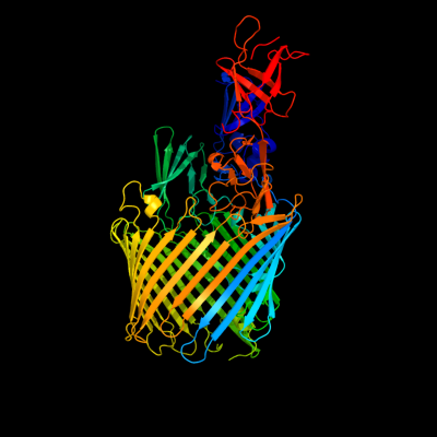
PDB header: cell adhesion/transport/chaperoneChain: B: PDB Molecule: outer membrane usher protein, type 1 fimbrial synthesis;PDBTitle: crystal structure of the fimd usher bound to its cognate fimc:fimh2 substrate
2 c3ohnA_
100.0
33
PDB header: membrane proteinChain: A: PDB Molecule: outer membrane usher protein fimd;PDBTitle: crystal structure of the fimd translocation domain
3 c2vqiA_
100.0
27
PDB header: transportChain: A: PDB Molecule: outer membrane usher protein papc;PDBTitle: structure of the p pilus usher (papc) translocation pore
4 d1zdva1
99.9
31
Fold: FimD N-terminal domain-likeSuperfamily: FimD N-terminal domain-likeFamily: Usher N-domain5 d3bwud1
99.9
23
Fold: FimD N-terminal domain-likeSuperfamily: FimD N-terminal domain-likeFamily: Usher N-domain6 c3fcgB_
99.9
39
PDB header: membrane protein, protein transportChain: B: PDB Molecule: f1 capsule-anchoring protein;PDBTitle: crystal structure analysis of the middle domain of the2 caf1a usher
7 d1zdxa1
99.8
26
Fold: FimD N-terminal domain-likeSuperfamily: FimD N-terminal domain-likeFamily: Usher N-domain8 c3l48B_
99.7
18
PDB header: transport proteinChain: B: PDB Molecule: outer membrane usher protein papc;PDBTitle: crystal structure of the c-terminal domain of the papc usher
9 c2xetB_
99.7
18
PDB header: transport proteinChain: B: PDB Molecule: f1 capsule-anchoring protein;PDBTitle: conserved hydrophobic clusters on the surface of the caf1a usher2 c-terminal domain are important for f1 antigen assembly
10 c3pe9B_
95.0
16
PDB header: unknown functionChain: B: PDB Molecule: fibronectin(iii)-like module;PDBTitle: structures of clostridium thermocellum cbha fibronectin(iii)-like2 modules
11 c3pdgA_
94.8
14
PDB header: unknown functionChain: A: PDB Molecule: fibronectin(iii)-like module;PDBTitle: structures of clostridium thermocellum cbha fibronectin(iii)-like2 modules
12 c3pe9D_
90.5
16
PDB header: unknown functionChain: D: PDB Molecule: fibronectin(iii)-like module;PDBTitle: structures of clostridium thermocellum cbha fibronectin(iii)-like2 modules
13 c1h8lA_
90.4
11
PDB header: carboxypeptidaseChain: A: PDB Molecule: carboxypeptidase gp180 residues 503-882;PDBTitle: duck carboxypeptidase d domain ii in complex with gemsa
14 d1h8la1
90.2
10
Fold: Prealbumin-likeSuperfamily: Carboxypeptidase regulatory domain-likeFamily: Carboxypeptidase regulatory domain15 c2nsmA_
89.2
14
PDB header: hydrolaseChain: A: PDB Molecule: carboxypeptidase n catalytic chain;PDBTitle: crystal structure of the human carboxypeptidase n (kininase i)2 catalytic domain
16 c3pe9C_
86.9
16
PDB header: unknown functionChain: C: PDB Molecule: fibronectin(iii)-like module;PDBTitle: structures of clostridium thermocellum cbha fibronectin(iii)-like2 modules
17 c3pe9A_
86.9
16
PDB header: unknown functionChain: A: PDB Molecule: fibronectin(iii)-like module;PDBTitle: structures of clostridium thermocellum cbha fibronectin(iii)-like2 modules
18 d1w0na_
85.5
21
Fold: Galactose-binding domain-likeSuperfamily: Galactose-binding domain-likeFamily: Family 36 carbohydrate binding module, CBM3619 d1uwya1
85.4
7
Fold: Prealbumin-likeSuperfamily: Carboxypeptidase regulatory domain-likeFamily: Carboxypeptidase regulatory domain20 c2x5pA_
84.1
22
PDB header: protein bindingChain: A: PDB Molecule: fibronectin binding protein;PDBTitle: crystal structure of the streptococcus pyogenes fibronectin binding2 protein fbab-b
21 c1d2pA_
not modelled
82.5
20
PDB header: structural proteinChain: A: PDB Molecule: collagen adhesin;PDBTitle: crystal structure of two b repeat units (b1b2) of the2 collagen binding protein (cna) of staphylococcus aureus
22 c3mn8A_
not modelled
82.1
11
PDB header: hydrolaseChain: A: PDB Molecule: lp15968p;PDBTitle: structure of drosophila melanogaster carboxypeptidase d isoform 1b2 short
23 c1uwyA_
not modelled
81.8
8
PDB header: hydrolaseChain: A: PDB Molecule: carboxypeptidase m;PDBTitle: crystal structure of human carboxypeptidase m
24 c2vnvC_
not modelled
79.4
11
PDB header: sugar-binding proteinChain: C: PDB Molecule: bcla;PDBTitle: crystal structure of bcla lectin from burkholderia2 cenocepacia in complex with alpha-methyl-mannoside at 1.73 angstrom resolution
25 d3pccm_
not modelled
64.9
17
Fold: Prealbumin-likeSuperfamily: Aromatic compound dioxygenaseFamily: Aromatic compound dioxygenase26 d2burb1
not modelled
63.6
20
Fold: Prealbumin-likeSuperfamily: Aromatic compound dioxygenaseFamily: Aromatic compound dioxygenase27 c2boiA_
not modelled
63.2
13
PDB header: lectinChain: A: PDB Molecule: cv-iil lectin;PDBTitle: 1.1a structure of chromobacterium violaceum lectin cv2l in2 complex with alpha-methyl-fucoside
28 c2xr4A_
not modelled
63.1
16
PDB header: sugar binding proteinChain: A: PDB Molecule: lectin;PDBTitle: c-terminal domain of bc2l-c lectin from burkholderia cenocepacia
29 d1uzva_
not modelled
54.0
14
Fold: Calcium-mediated lectinSuperfamily: Calcium-mediated lectinFamily: Calcium-mediated lectin30 c1u00A_
not modelled
52.6
18
PDB header: chaperoneChain: A: PDB Molecule: chaperone protein hsca;PDBTitle: hsca substrate binding domain complexed with the iscu2 recognition peptide elppvkihc
31 c3e8vA_
not modelled
50.7
14
PDB header: structural genomics, unknown functionChain: A: PDB Molecule: possible transglutaminase-family protein;PDBTitle: crystal structure of a possible transglutaminase-family2 protein proteolytic fragment from bacteroides fragilis
32 c3bryB_
not modelled
50.0
19
PDB header: transport proteinChain: B: PDB Molecule: tbux;PDBTitle: crystal structure of the ralstonia pickettii toluene2 transporter tbux
33 c3dpqE_
not modelled
49.8
21
PDB header: chaperone, peptide binding proteinChain: E: PDB Molecule: chaperone protein dnak;PDBTitle: crystal structure of the substrate binding domain of e.2 coli dnak in complex with a long pyrrhocoricin-derived3 inhibitor peptide (form b)
34 c1bprA_
not modelled
49.5
21
PDB header: molecular chaperoneChain: A: PDB Molecule: dnak;PDBTitle: nmr structure of the substrate binding domain of dnak,2 minimized average structure
35 c3b9eA_
not modelled
49.3
13
PDB header: hydrolaseChain: A: PDB Molecule: chitinase a;PDBTitle: crystal structure of inactive mutant e315m chitinase a from2 vibrio harveyi
36 c2bpbA_
not modelled
49.0
19
PDB header: oxidoreductaseChain: A: PDB Molecule: sulfite\:cytochrome c oxidoreductase subunit a;PDBTitle: sulfite dehydrogenase from starkeya novella
37 d1nkga1
not modelled
48.5
7
Fold: Prealbumin-likeSuperfamily: Starch-binding domain-likeFamily: Rhamnogalacturonase B, RhgB, middle domain38 c1ug9A_
not modelled
48.2
25
PDB header: hydrolaseChain: A: PDB Molecule: glucodextranase;PDBTitle: crystal structure of glucodextranase from arthrobacter globiformis i42
39 c3c12A_
not modelled
47.1
22
PDB header: biosynthetic proteinChain: A: PDB Molecule: flagellar protein;PDBTitle: crystal structure of flgd from xanthomonas campestris:2 insights into the hook capping essential for flagellar3 assembly
40 c2op6A_
not modelled
46.4
18
PDB header: peptide binding proteinChain: A: PDB Molecule: heat shock 70 kda protein d;PDBTitle: peptide-binding domain of heat shock 70 kda protein d precursor from2 c.elegans
41 c3n8eA_
not modelled
45.3
18
PDB header: chaperoneChain: A: PDB Molecule: stress-70 protein, mitochondrial;PDBTitle: substrate binding domain of the human heat shock 70kda protein 92 (mortalin)
42 c3fn9B_
not modelled
42.9
15
PDB header: hydrolaseChain: B: PDB Molecule: putative beta-galactosidase;PDBTitle: crystal structure of putative beta-galactosidase from bacteroides2 fragilis
43 d1v8ha1
not modelled
41.7
21
Fold: Immunoglobulin-like beta-sandwichSuperfamily: E set domainsFamily: SoxZ-like44 c3cmgA_
not modelled
40.9
15
PDB header: hydrolaseChain: A: PDB Molecule: putative beta-galactosidase;PDBTitle: crystal structure of putative beta-galactosidase from bacteroides2 fragilis
45 d1edqa1
not modelled
39.5
17
Fold: Immunoglobulin-like beta-sandwichSuperfamily: E set domainsFamily: E-set domains of sugar-utilizing enzymes46 c1nkgA_
not modelled
37.8
17
PDB header: lyaseChain: A: PDB Molecule: rhamnogalacturonase b;PDBTitle: rhamnogalacturonan lyase from aspergillus aculeatus
47 d1u00a2
not modelled
36.8
23
Fold: Heat shock protein 70kD (HSP70), peptide-binding domainSuperfamily: Heat shock protein 70kD (HSP70), peptide-binding domainFamily: Heat shock protein 70kD (HSP70), peptide-binding domain48 d2chha1
not modelled
34.3
13
Fold: Calcium-mediated lectinSuperfamily: Calcium-mediated lectinFamily: Calcium-mediated lectin49 d1dkza2
not modelled
33.0
21
Fold: Heat shock protein 70kD (HSP70), peptide-binding domainSuperfamily: Heat shock protein 70kD (HSP70), peptide-binding domainFamily: Heat shock protein 70kD (HSP70), peptide-binding domain50 c2a9dB_
not modelled
32.2
20
PDB header: oxidoreductaseChain: B: PDB Molecule: sulfite oxidase;PDBTitle: crystal structure of recombinant chicken sulfite oxidase with arg at2 residue 161
51 c3dqgC_
not modelled
32.2
19
PDB header: chaperoneChain: C: PDB Molecule: heat shock 70 kda protein f;PDBTitle: peptide-binding domain of heat shock 70 kda protein f, mitochondrial2 precursor, from caenorhabditis elegans.
52 d2vzsa4
not modelled
31.3
19
Fold: Galactose-binding domain-likeSuperfamily: Galactose-binding domain-likeFamily: beta-Galactosidase/glucuronidase, N-terminal domain53 d1yuwa1
not modelled
31.2
20
Fold: Heat shock protein 70kD (HSP70), peptide-binding domainSuperfamily: Heat shock protein 70kD (HSP70), peptide-binding domainFamily: Heat shock protein 70kD (HSP70), peptide-binding domain54 d1ci3m2
not modelled
31.1
10
Fold: Barrel-sandwich hybridSuperfamily: Rudiment single hybrid motifFamily: Cytochrome f, small domain55 d1aoza2
not modelled
28.7
18
Fold: Cupredoxin-likeSuperfamily: CupredoxinsFamily: Multidomain cupredoxins56 c2r32A_
not modelled
28.6
29
PDB header: immune systemChain: A: PDB Molecule: gcn4-pii/tumor necrosis factor ligandPDBTitle: crystal structure of human gitrl variant
57 d1e2wa2
not modelled
27.3
11
Fold: Barrel-sandwich hybridSuperfamily: Rudiment single hybrid motifFamily: Cytochrome f, small domain58 c2oxgE_
not modelled
26.7
25
PDB header: transport proteinChain: E: PDB Molecule: soxz protein;PDBTitle: the soxyz complex of paracoccus pantotrophus
59 d2a9da1
not modelled
26.2
25
Fold: Immunoglobulin-like beta-sandwichSuperfamily: E set domainsFamily: Molybdenum-containing oxidoreductases-like dimerisation domain60 c3pddA_
not modelled
25.1
16
PDB header: unknown functionChain: A: PDB Molecule: glycoside hydrolase, family 9;PDBTitle: structures of clostridium thermocellum cbha fibronectin(iii)-like2 modules
61 c2ww8A_
not modelled
24.7
6
PDB header: cell adhesionChain: A: PDB Molecule: cell wall surface anchor family protein;PDBTitle: structure of the pilus adhesin (rrga) from streptococcus2 pneumoniae
62 c3k1dA_
not modelled
23.0
15
PDB header: transferaseChain: A: PDB Molecule: 1,4-alpha-glucan-branching enzyme;PDBTitle: crystal structure of glycogen branching enzyme synonym: 1,4-alpha-d-2 glucan:1,4-alpha-d-glucan 6-glucosyl-transferase from mycobacterium3 tuberculosis h37rv
63 d2dj4a1
not modelled
22.0
13
Fold: Immunoglobulin-like beta-sandwichSuperfamily: E set domainsFamily: Filamin repeat (rod domain)64 d3pcca_
not modelled
21.9
11
Fold: Prealbumin-likeSuperfamily: Aromatic compound dioxygenaseFamily: Aromatic compound dioxygenase65 d1o75a2
not modelled
21.2
27
Fold: Immunoglobulin-like beta-sandwichSuperfamily: Tp47 lipoprotein, middle and C-terminal domainsFamily: Tp47 lipoprotein, middle and C-terminal domains66 c3rghA_
not modelled
21.2
33
PDB header: cell adhesionChain: A: PDB Molecule: filamin-a;PDBTitle: structure of filamin a immunoglobulin-like repeat 10 from homo sapiens
67 c2lgeA_
not modelled
21.0
23
PDB header: metal binding proteinChain: A: PDB Molecule: uncharacterized protein;PDBTitle: nmr structure of the calcium-bound form of the protein yp_001302112.12 from parabacteroides distasonis
68 d2je8a4
not modelled
21.0
13
Fold: Galactose-binding domain-likeSuperfamily: Galactose-binding domain-likeFamily: beta-Galactosidase/glucuronidase, N-terminal domain69 d1wlha1
not modelled
20.2
20
Fold: Immunoglobulin-like beta-sandwichSuperfamily: E set domainsFamily: Filamin repeat (rod domain)70 d2bp3a1
not modelled
20.2
40
Fold: Immunoglobulin-like beta-sandwichSuperfamily: E set domainsFamily: Filamin repeat (rod domain)71 c3hj8A_
not modelled
20.2
12
PDB header: oxidoreductaseChain: A: PDB Molecule: catechol 1,2-dioxygenase;PDBTitle: crystal structure determination of catechol 1,2-dioxygenase from2 rhodococcus opacus 1cp in complex with 4-chlorocatechol
72 c2yujA_
not modelled
19.5
10
PDB header: protein bindingChain: A: PDB Molecule: ubiquitin fusion degradation 1-like;PDBTitle: solution structure of human ubiquitin fusion degradation2 protein 1 homolog ufd1
73 c2l3bA_
not modelled
19.1
13
PDB header: structural genomics, unknown functionChain: A: PDB Molecule: conserved protein found in conjugate transposon;PDBTitle: solution nmr structure of the bt_0084 lipoprotein from bacteroides2 thetaiotaomicron, northeast structural genomics consortium target3 btr376
74 d1ogpa1
not modelled
18.7
20
Fold: Immunoglobulin-like beta-sandwichSuperfamily: E set domainsFamily: Molybdenum-containing oxidoreductases-like dimerisation domain75 c2pdtD_
not modelled
17.5
20
PDB header: circadian clock proteinChain: D: PDB Molecule: vivid pas protein vvd;PDBTitle: 2.3 angstrom structure of phosphodiesterase treated vivid
76 d2d7oa1
not modelled
16.8
27
Fold: Immunoglobulin-like beta-sandwichSuperfamily: E set domainsFamily: Filamin repeat (rod domain)77 d2e9ia1
not modelled
16.7
13
Fold: Immunoglobulin-like beta-sandwichSuperfamily: E set domainsFamily: Filamin repeat (rod domain)78 d2dmca1
not modelled
16.5
14
Fold: Immunoglobulin-like beta-sandwichSuperfamily: E set domainsFamily: Filamin repeat (rod domain)79 c1zc1A_
not modelled
16.4
13
PDB header: protein turnoverChain: A: PDB Molecule: ubiquitin fusion degradation protein 1;PDBTitle: ufd1 exhibits the aaa-atpase fold with two distinct2 ubiquitin interaction sites
80 c3d33B_
not modelled
16.3
14
PDB header: unknown functionChain: B: PDB Molecule: domain of unknown function with an immunoglobulin-likePDBTitle: crystal structure of a duf3244 family protein with an immunoglobulin-2 like beta-sandwich fold (bvu_0276) from bacteroides vulgatus atcc3 8482 at 1.70 a resolution
81 c2jxmB_
not modelled
16.1
12
PDB header: electron transportChain: B: PDB Molecule: cytochrome f;PDBTitle: ensemble of twenty structures of the prochlorothrix2 hollandica plastocyanin- cytochrome f complex
82 d1ulva2
not modelled
16.1
33
Fold: Immunoglobulin-like beta-sandwichSuperfamily: E set domainsFamily: E-set domains of sugar-utilizing enzymes83 c2crvA_
not modelled
15.5
15
PDB header: translationChain: A: PDB Molecule: translation initiation factor if-2;PDBTitle: solution structure of c-terminal domain of mitochondrial2 translational initiationfactor 2
84 d1qfha1
not modelled
15.5
22
Fold: Immunoglobulin-like beta-sandwichSuperfamily: E set domainsFamily: Filamin repeat (rod domain)85 d1yq2a3
not modelled
15.4
18
Fold: Galactose-binding domain-likeSuperfamily: Galactose-binding domain-likeFamily: beta-Galactosidase/glucuronidase, N-terminal domain86 d2slia1
not modelled
15.4
16
Fold: Concanavalin A-like lectins/glucanasesSuperfamily: Concanavalin A-like lectins/glucanasesFamily: Leech intramolecular trans-sialidase, N-terminal domain87 d1uura3
not modelled
15.2
26
Fold: SH2-likeSuperfamily: SH2 domainFamily: SH2 domain88 c3girA_
not modelled
14.6
21
PDB header: transferaseChain: A: PDB Molecule: aminomethyltransferase;PDBTitle: crystal structure of glycine cleavage system2 aminomethyltransferase t from bartonella henselae
89 d2dica1
not modelled
14.3
33
Fold: Immunoglobulin-like beta-sandwichSuperfamily: E set domainsFamily: Filamin repeat (rod domain)90 c2brqB_
not modelled
14.1
27
PDB header: structural proteinChain: B: PDB Molecule: filamin a;PDBTitle: crystal structure of the filamin a repeat 21 complexed with2 the integrin beta7 cytoplasmic tail peptide
91 c2ds4A_
not modelled
13.8
27
PDB header: protein bindingChain: A: PDB Molecule: tripartite motif protein 45;PDBTitle: solution structure of the filamin domain from human2 tripartite motif protein 45
92 d2j3sa2
not modelled
13.6
13
Fold: Immunoglobulin-like beta-sandwichSuperfamily: E set domainsFamily: Filamin repeat (rod domain)93 d2di8a1
not modelled
13.5
20
Fold: Immunoglobulin-like beta-sandwichSuperfamily: E set domainsFamily: Filamin repeat (rod domain)94 d2w0pa1
not modelled
13.4
27
Fold: Immunoglobulin-like beta-sandwichSuperfamily: E set domainsFamily: Filamin repeat (rod domain)95 d2nn6i1
not modelled
13.4
16
Fold: OB-foldSuperfamily: Nucleic acid-binding proteinsFamily: Cold shock DNA-binding domain-like96 d2diaa1
not modelled
13.3
13
Fold: Immunoglobulin-like beta-sandwichSuperfamily: E set domainsFamily: Filamin repeat (rod domain)97 d2d7pa1
not modelled
13.3
27
Fold: Immunoglobulin-like beta-sandwichSuperfamily: E set domainsFamily: Filamin repeat (rod domain)98 d2dmba1
not modelled
13.2
27
Fold: Immunoglobulin-like beta-sandwichSuperfamily: E set domainsFamily: Filamin repeat (rod domain)99 c1w2tE_
not modelled
13.2
16
PDB header: hydrolaseChain: E: PDB Molecule: beta fructosidase;PDBTitle: beta-fructosidase from thermotoga maritima in complex with2 raffinose