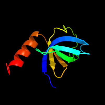
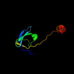
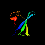
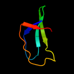
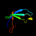
1 c3d3rA_
100.0
32
PDB header: chaperoneChain: A: PDB Molecule: hydrogenase assembly chaperone hypc/hupf;PDBTitle: crystal structure of the hydrogenase assembly chaperone hypc/hupf2 family protein from shewanella oneidensis mr-1
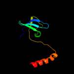
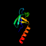
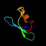
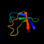
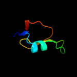
2 d2ot2a1
99.9
57
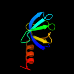
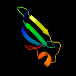
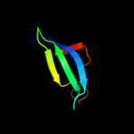
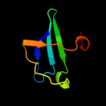
Fold: OB-foldSuperfamily: HupF/HypC-likeFamily: HupF/HypC-like3 d3d3ra1
99.9
32
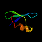
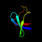
Fold: OB-foldSuperfamily: HupF/HypC-likeFamily: HupF/HypC-like4 d2z1ca1
99.9
38
Fold: OB-foldSuperfamily: HupF/HypC-likeFamily: HupF/HypC-like5 d1rl2a2
80.6
28
Fold: OB-foldSuperfamily: Nucleic acid-binding proteinsFamily: Cold shock DNA-binding domain-like6 c2rcnA_
67.5
26
PDB header: hydrolaseChain: A: PDB Molecule: probable gtpase engc;PDBTitle: crystal structure of the ribosomal interacting gtpase yjeq from the2 enterobacterial species salmonella typhimurium.
7 d1vqoa2
66.7
24
Fold: OB-foldSuperfamily: Nucleic acid-binding proteinsFamily: Cold shock DNA-binding domain-like8 c4a2iV_
65.4
26
PDB header: ribosome/hydrolaseChain: V: PDB Molecule: putative ribosome biogenesis gtpase rsga;PDBTitle: cryo-electron microscopy structure of the 30s subunit in complex with2 the yjeq biogenesis factor
9 d2zjra2
56.1
34
Fold: OB-foldSuperfamily: Nucleic acid-binding proteinsFamily: Cold shock DNA-binding domain-like10 c1u0lB_
55.1
18
PDB header: hydrolaseChain: B: PDB Molecule: probable gtpase engc;PDBTitle: crystal structure of yjeq from thermotoga maritima
11 c2dgyA_
54.3
16
PDB header: translationChain: A: PDB Molecule: mgc11102 protein;PDBTitle: solution structure of the eukaryotic initiation factor 1a2 in mgc11102 protein
12 c2yv5A_
54.1
17
PDB header: hydrolaseChain: A: PDB Molecule: yjeq protein;PDBTitle: crystal structure of yjeq from aquifex aeolicus
13 c3mxnB_
53.7
18
PDB header: replicationChain: B: PDB Molecule: recq-mediated genome instability protein 2;PDBTitle: crystal structure of the rmi core complex
14 c1g0dA_
43.4
33
PDB header: transferaseChain: A: PDB Molecule: protein-glutamine gamma-glutamyltransferase;PDBTitle: crystal structure of red sea bream transglutaminase
15 c1kv3F_
42.5
44
PDB header: transferaseChain: F: PDB Molecule: protein-glutamine gamma-glutamyltransferase;PDBTitle: human tissue transglutaminase in gdp bound form
16 c1l9mB_
42.3
33
PDB header: transferaseChain: B: PDB Molecule: protein-glutamine glutamyltransferase e3;PDBTitle: three-dimensional structure of the human transglutaminase 32 enzyme: binding of calcium ions change structure for3 activation
17 c1f13A_
40.0
33
PDB header: coagulation factorChain: A: PDB Molecule: cellular coagulation factor xiii zymogen;PDBTitle: recombinant human cellular coagulation factor xiii
18 c2oqkA_
39.7
34
PDB header: translationChain: A: PDB Molecule: putative translation initiation factor eif-1a;PDBTitle: crystal structure of putative cryptosporidium parvum translation2 initiation factor eif-1a
19 d2j01d2
37.2
27
Fold: OB-foldSuperfamily: Nucleic acid-binding proteinsFamily: Cold shock DNA-binding domain-like20 d1ny4a_
35.6
17
Fold: OB-foldSuperfamily: Nucleic acid-binding proteinsFamily: Cold shock DNA-binding domain-like21 d2qamc2
not modelled
35.4
22
Fold: OB-foldSuperfamily: Nucleic acid-binding proteinsFamily: Cold shock DNA-binding domain-like22 d1jt8a_
not modelled
34.9
16
Fold: OB-foldSuperfamily: Nucleic acid-binding proteinsFamily: Cold shock DNA-binding domain-like23 d1g0da4
not modelled
33.7
30
Fold: Cysteine proteinasesSuperfamily: Cysteine proteinasesFamily: Transglutaminase core24 d1d7qa_
not modelled
33.3
19
Fold: OB-foldSuperfamily: Nucleic acid-binding proteinsFamily: Cold shock DNA-binding domain-like25 d1njjb1
not modelled
33.1
33
Fold: Domain of alpha and beta subunits of F1 ATP synthase-likeSuperfamily: Alanine racemase C-terminal domain-likeFamily: Eukaryotic ODC-like26 d2q3za4
not modelled
32.8
44
Fold: Cysteine proteinasesSuperfamily: Cysteine proteinasesFamily: Transglutaminase core27 d1ex0a4
not modelled
31.3
30
Fold: Cysteine proteinasesSuperfamily: Cysteine proteinasesFamily: Transglutaminase core28 c1t9hA_
not modelled
31.2
9
PDB header: hydrolaseChain: A: PDB Molecule: probable gtpase engc;PDBTitle: the crystal structure of yloq, a circularly permuted gtpase.
29 d1vjja4
not modelled
29.6
30
Fold: Cysteine proteinasesSuperfamily: Cysteine proteinasesFamily: Transglutaminase core30 c2xzm1_
not modelled
29.1
19
PDB header: ribosomeChain: 1: PDB Molecule: ribosomal protein s28e containing protein;PDBTitle: crystal structure of the eukaryotic 40s ribosomal2 subunit in complex with initiation factor 1. this file3 contains the 40s subunit and initiation factor for4 molecule 1
31 d2toda1
not modelled
28.4
33
Fold: Domain of alpha and beta subunits of F1 ATP synthase-likeSuperfamily: Alanine racemase C-terminal domain-likeFamily: Eukaryotic ODC-like32 c1rl2A_
not modelled
28.3
17
PDB header: ribosomal proteinChain: A: PDB Molecule: protein (ribosomal protein l2);PDBTitle: ribosomal protein l2 rna-binding domain from bacillus2 stearothermophilus
33 d1d7ka1
not modelled
27.1
33
Fold: Domain of alpha and beta subunits of F1 ATP synthase-likeSuperfamily: Alanine racemase C-terminal domain-likeFamily: Eukaryotic ODC-like34 d1ne3a_
not modelled
26.7
18
Fold: OB-foldSuperfamily: Nucleic acid-binding proteinsFamily: Cold shock DNA-binding domain-like35 c1njjC_
not modelled
23.3
33
PDB header: lyaseChain: C: PDB Molecule: ornithine decarboxylase;PDBTitle: crystal structure determination of t. brucei ornithine2 decarboxylase bound to d-ornithine and to g418
36 c3i4oA_
not modelled
22.8
21
PDB header: translationChain: A: PDB Molecule: translation initiation factor if-1;PDBTitle: crystal structure of translation initiation factor 1 from2 mycobacterium tuberculosis
37 c3fp9E_
not modelled
22.8
24
PDB header: hydrolaseChain: E: PDB Molecule: proteasome-associated atpase;PDBTitle: crystal structure of intern domain of proteasome-associated2 atpase, mycobacterium tuberculosis
38 d1f3ta1
not modelled
22.3
33
Fold: Domain of alpha and beta subunits of F1 ATP synthase-likeSuperfamily: Alanine racemase C-terminal domain-likeFamily: Eukaryotic ODC-like39 c2hc8A_
not modelled
22.2
24
PDB header: transport proteinChain: A: PDB Molecule: cation-transporting atpase, p-type;PDBTitle: structure of the a. fulgidus copa a-domain
40 c1oy5B_
not modelled
20.2
22
PDB header: transferaseChain: B: PDB Molecule: trna (guanine-n(1)-)-methyltransferase;PDBTitle: crystal structure of trna (m1g37) methyltransferase from aquifex2 aeolicus
41 d1u0la1
not modelled
19.7
18
Fold: OB-foldSuperfamily: Nucleic acid-binding proteinsFamily: Cold shock DNA-binding domain-like42 d1knwa1
not modelled
19.5
25
Fold: Domain of alpha and beta subunits of F1 ATP synthase-likeSuperfamily: Alanine racemase C-terminal domain-likeFamily: Eukaryotic ODC-like43 c2qghA_
not modelled
19.5
17
PDB header: lyaseChain: A: PDB Molecule: diaminopimelate decarboxylase;PDBTitle: crystal structure of diaminopimelate decarboxylase from helicobacter2 pylori complexed with l-lysine
44 d1t9ha1
not modelled
19.1
14
Fold: OB-foldSuperfamily: Nucleic acid-binding proteinsFamily: Cold shock DNA-binding domain-like45 d7odca1
not modelled
18.6
33
Fold: Domain of alpha and beta subunits of F1 ATP synthase-likeSuperfamily: Alanine racemase C-terminal domain-likeFamily: Eukaryotic ODC-like46 d1oy5a_
not modelled
18.3
22
Fold: alpha/beta knotSuperfamily: alpha/beta knotFamily: tRNA(m1G37)-methyltransferase TrmD47 c1xniI_
not modelled
17.8
19
PDB header: cell cycleChain: I: PDB Molecule: tumor suppressor p53-binding protein 1;PDBTitle: tandem tudor domain of 53bp1
48 d1twia1
not modelled
17.8
8
Fold: Domain of alpha and beta subunits of F1 ATP synthase-likeSuperfamily: Alanine racemase C-terminal domain-likeFamily: Eukaryotic ODC-like49 c2p3eA_
not modelled
16.9
17
PDB header: lyaseChain: A: PDB Molecule: diaminopimelate decarboxylase;PDBTitle: crystal structure of aq1208 from aquifex aeolicus
50 c1kqsA_
not modelled
16.5
24
PDB header: ribosomeChain: A: PDB Molecule: ribosomal protein l2;PDBTitle: the haloarcula marismortui 50s complexed with a2 pretranslocational intermediate in protein synthesis
51 c2nvaH_
not modelled
15.5
33
PDB header: lyaseChain: H: PDB Molecule: arginine decarboxylase, a207r protein;PDBTitle: the x-ray crystal structure of the paramecium bursaria2 chlorella virus arginine decarboxylase bound to agmatine
52 c1knwA_
not modelled
14.9
25
PDB header: lyaseChain: A: PDB Molecule: diaminopimelate decarboxylase;PDBTitle: crystal structure of diaminopimelate decarboxylase
53 d3pnpa_
not modelled
14.4
22
Fold: Phosphorylase/hydrolase-likeSuperfamily: Purine and uridine phosphorylasesFamily: Purine and uridine phosphorylases54 c2yxxA_
not modelled
14.3
25
PDB header: lyaseChain: A: PDB Molecule: diaminopimelate decarboxylase;PDBTitle: crystal structure analysis of diaminopimelate decarboxylate (lysa)
55 c1yr3A_
not modelled
13.7
15
PDB header: transferaseChain: A: PDB Molecule: xanthosine phosphorylase;PDBTitle: escherichia coli purine nucleoside phosphorylase ii, the2 product of the xapa gene
56 d1p3ha_
not modelled
13.6
28
Fold: GroES-likeSuperfamily: GroES-likeFamily: GroES57 d1p3qq_
not modelled
13.5
33
Fold: RuvA C-terminal domain-likeSuperfamily: UBA-likeFamily: CUE domain58 c1w4sA_
not modelled
13.4
38
PDB header: nuclear proteinChain: A: PDB Molecule: polybromo 1 protein;PDBTitle: crystal structure of the proximal bah domain of polybromo
59 d1aono_
not modelled
13.3
24
Fold: GroES-likeSuperfamily: GroES-likeFamily: GroES60 d1cb0a_
not modelled
13.2
11
Fold: Phosphorylase/hydrolase-likeSuperfamily: Purine and uridine phosphorylasesFamily: Purine and uridine phosphorylases61 d1fr3a_
not modelled
12.9
11
Fold: OB-foldSuperfamily: MOP-likeFamily: Molybdate/tungstate binding protein MOP62 d2id1a1
not modelled
12.8
27
Fold: NucleotidyltransferaseSuperfamily: NucleotidyltransferaseFamily: Iojap/YbeB-like63 d1uala_
not modelled
12.7
38
Fold: alpha/beta knotSuperfamily: alpha/beta knotFamily: tRNA(m1G37)-methyltransferase TrmD64 d2a7ya1
not modelled
12.6
22
Fold: SH3-like barrelSuperfamily: Cell growth inhibitor/plasmid maintenance toxic componentFamily: Rv2302-like65 c2a7yA_
not modelled
12.6
22
PDB header: structural genomics, unknown functionChain: A: PDB Molecule: hypothetical protein rv2302/mt2359;PDBTitle: solution structure of the conserved hypothetical protein2 rv2302 from the bacterium mycobacterium tuberculosis
66 d1vmka_
not modelled
12.5
30
Fold: Phosphorylase/hydrolase-likeSuperfamily: Purine and uridine phosphorylasesFamily: Purine and uridine phosphorylases67 c2akwB_
not modelled
12.0
15
PDB header: ligaseChain: B: PDB Molecule: phenylalanyl-trna synthetase beta chain;PDBTitle: crystal structure of t.thermophilus phenylalanyl-trna synthetase2 complexed with p-cl-phenylalanine
68 c2pljA_
not modelled
11.7
25
PDB header: lyaseChain: A: PDB Molecule: lysine/ornithine decarboxylase;PDBTitle: crystal structure of lysine/ornithine decarboxylase complexed with2 putrescine from vibrio vulnificus
69 c2k29A_
not modelled
11.5
25
PDB header: transcriptionChain: A: PDB Molecule: antitoxin relb;PDBTitle: structure of the dbd domain of e. coli antitoxin relb
70 d1ntga_
not modelled
11.4
17
Fold: OB-foldSuperfamily: Nucleic acid-binding proteinsFamily: Myf domain71 d1pqwa_
not modelled
11.3
31
Fold: NAD(P)-binding Rossmann-fold domainsSuperfamily: NAD(P)-binding Rossmann-fold domainsFamily: Alcohol dehydrogenase-like, C-terminal domain72 c3n29A_
not modelled
11.2
8
PDB header: lyaseChain: A: PDB Molecule: carboxynorspermidine decarboxylase;PDBTitle: crystal structure of carboxynorspermidine decarboxylase complexed with2 norspermidine from campylobacter jejuni
73 c3knuD_
not modelled
11.2
33
PDB header: transferaseChain: D: PDB Molecule: trna (guanine-n(1)-)-methyltransferase;PDBTitle: crystal structure of trna (guanine-n1)-methyltransferase from2 anaplasma phagocytophilum
74 d1g2oa_
not modelled
11.1
22
Fold: Phosphorylase/hydrolase-likeSuperfamily: Purine and uridine phosphorylasesFamily: Purine and uridine phosphorylases75 c3upsA_
not modelled
11.0
19
PDB header: structural genomics, unknown functionChain: A: PDB Molecule: iojap-like protein;PDBTitle: crystal structure of iojap-like protein from zymomonas mobilis
76 c3quvB_
not modelled
10.9
33
PDB header: transferaseChain: B: PDB Molecule: trna (guanine-n(1)-)-methyltransferase;PDBTitle: crystal structure of a trna-guanine-n1-methyltransferase from2 mycobacterium abscessus
77 c3khsB_
not modelled
10.8
26
PDB header: hydrolaseChain: B: PDB Molecule: purine nucleoside phosphorylase;PDBTitle: crystal structure of grouper iridovirus purine nucleoside2 phosphorylase
78 d1v3va2
not modelled
10.8
31
Fold: NAD(P)-binding Rossmann-fold domainsSuperfamily: NAD(P)-binding Rossmann-fold domainsFamily: Alcohol dehydrogenase-like, C-terminal domain79 c2j66A_
not modelled
10.7
45
PDB header: lyaseChain: A: PDB Molecule: btrk;PDBTitle: structural characterisation of btrk decarboxylase from2 butirosin biosynthesis
80 c3ky7A_
not modelled
10.7
21
PDB header: transferaseChain: A: PDB Molecule: trna (guanine-n(1)-)-methyltransferase;PDBTitle: 2.35 angstrom resolution crystal structure of a putative trna2 (guanine-7-)-methyltransferase (trmd) from staphylococcus aureus3 subsp. aureus mrsa252
81 c3e6eC_
not modelled
10.6
15
PDB header: isomeraseChain: C: PDB Molecule: alanine racemase;PDBTitle: crystal structure of alanine racemase from e.faecalis2 complex with cycloserine
82 d2fzwa2
not modelled
10.6
15
Fold: NAD(P)-binding Rossmann-fold domainsSuperfamily: NAD(P)-binding Rossmann-fold domainsFamily: Alcohol dehydrogenase-like, C-terminal domain83 d1p9pa_
not modelled
10.5
33
Fold: alpha/beta knotSuperfamily: alpha/beta knotFamily: tRNA(m1G37)-methyltransferase TrmD84 c1ltlE_
not modelled
10.4
22
PDB header: replicationChain: E: PDB Molecule: dna replication initiator (cdc21/cdc54);PDBTitle: the dodecamer structure of mcm from archaeal m.2 thermoautotrophicum
85 c2equA_
not modelled
10.4
17
PDB header: protein bindingChain: A: PDB Molecule: phd finger protein 20-like 1;PDBTitle: solution structure of the tudor domain of phd finger2 protein 20-like 1
86 c1tufA_
not modelled
10.4
8
PDB header: lyaseChain: A: PDB Molecule: diaminopimelate decarboxylase;PDBTitle: crystal structure of diaminopimelate decarboxylase from m.2 jannaschi
87 c3iefA_
not modelled
10.3
22
PDB header: transferase, rna binding proteinChain: A: PDB Molecule: trna (guanine-n(1)-)-methyltransferase;PDBTitle: crystal structure of trna guanine-n1-methyltransferase from2 bartonella henselae using mpcs.
88 c3ggsA_
not modelled
10.2
22
PDB header: transferaseChain: A: PDB Molecule: purine nucleoside phosphorylase;PDBTitle: human purine nucleoside phosphorylase double mutant e201q,n243d2 complexed with 2-fluoro-2'-deoxyadenosine
89 c3mt1B_
not modelled
10.2
8
PDB header: lyaseChain: B: PDB Molecule: putative carboxynorspermidine decarboxylase protein;PDBTitle: crystal structure of putative carboxynorspermidine decarboxylase2 protein from sinorhizobium meliloti
90 c2p4sA_
not modelled
10.1
19
PDB header: transferaseChain: A: PDB Molecule: purine nucleoside phosphorylase;PDBTitle: structure of purine nucleoside phosphorylase from anopheles gambiae in2 complex with dadme-immh
91 d2fsqa1
not modelled
10.0
6
Fold: LigT-likeSuperfamily: LigT-likeFamily: Atu0111-like92 d2hd3a1
not modelled
10.0
33
Fold: OB-foldSuperfamily: EutN/CcmL-likeFamily: EutN/CcmL-like93 d2qw7a1
not modelled
9.9
24
Fold: OB-foldSuperfamily: EutN/CcmL-likeFamily: EutN/CcmL-like94 c3oo2A_
not modelled
9.8
16
PDB header: isomeraseChain: A: PDB Molecule: alanine racemase 1;PDBTitle: 2.37 angstrom resolution crystal structure of an alanine racemase2 (alr) from staphylococcus aureus subsp. aureus col
95 d2soba_
not modelled
9.7
9
Fold: OB-foldSuperfamily: Staphylococcal nucleaseFamily: Staphylococcal nuclease96 d1ltla_
not modelled
9.6
21
Fold: OB-foldSuperfamily: Nucleic acid-binding proteinsFamily: DNA replication initiator (cdc21/cdc54) N-terminal domain97 d2nn6h1
not modelled
9.5
16
Fold: OB-foldSuperfamily: Nucleic acid-binding proteinsFamily: Cold shock DNA-binding domain-like98 c2opkC_
not modelled
9.4
27
PDB header: isomeraseChain: C: PDB Molecule: hypothetical protein;PDBTitle: crystal structure of a putative mannose-6-phosphate isomerase2 (reut_a1446) from ralstonia eutropha jmp134 at 2.10 a resolution
99 c2zkra_
not modelled
9.4
25
PDB header: ribosomal protein/rnaChain: A: PDB Molecule: rna expansion segment es3;PDBTitle: structure of a mammalian ribosomal 60s subunit within an2 80s complex obtained by docking homology models of the rna3 and proteins into an 8.7 a cryo-em map