| 1 | c3pdkB_
|
|
 |
100.0 |
46 |
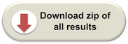
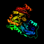
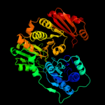
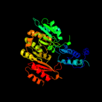
PDB header:isomerase
Chain: B: PDB Molecule:phosphoglucosamine mutase;
PDBTitle: crystal structure of phosphoglucosamine mutase from b. anthracis
|
| 2 | c1wqaB_
|
|
 |
100.0 |
33 |
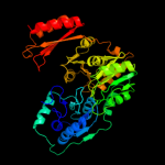
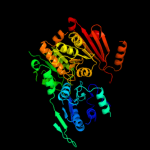
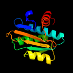
PDB header:isomerase
Chain: B: PDB Molecule:phospho-sugar mutase;
PDBTitle: crystal structure of pyrococcus horikoshii2 phosphomannomutase/phosphoglucomutase complexed with mg2+
|
| 3 | c2f7lA_
|
|
 |
100.0 |
28 |
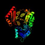
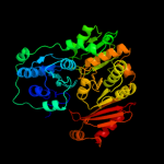
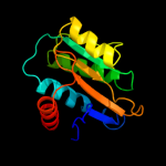
PDB header:isomerase
Chain: A: PDB Molecule:455aa long hypothetical phospho-sugar mutase;
PDBTitle: crystal structure of sulfolobus tokodaii2 phosphomannomutase/phosphoglucomutase
|
| 4 | c3i3wB_
|
|
 |
100.0 |
43 |
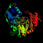
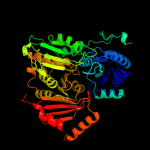
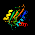
PDB header:isomerase
Chain: B: PDB Molecule:phosphoglucosamine mutase;
PDBTitle: structure of a phosphoglucosamine mutase from francisella tularensis
|
| 5 | c3c04A_
|
|
 |
100.0 |
26 |
PDB header:isomerase
Chain: A: PDB Molecule:phosphomannomutase/phosphoglucomutase;
PDBTitle: structure of the p368g mutant of pmm/pgm from p. aeruginosa
|
| 6 | c1tuoA_
|
|
 |
100.0 |
27 |
PDB header:biosynthetic protein
Chain: A: PDB Molecule:putative phosphomannomutase;
PDBTitle: crystal structure of putative phosphomannomutase from2 thermus thermophilus hb8
|
| 7 | c2fuvB_
|
|
 |
100.0 |
21 |
PDB header:isomerase
Chain: B: PDB Molecule:phosphoglucomutase;
PDBTitle: phosphoglucomutase from salmonella typhimurium.
|
| 8 | c1c4gB_
|
|
 |
100.0 |
23 |
PDB header:transferase
Chain: B: PDB Molecule:protein (alpha-d-glucose 1-phosphate
PDBTitle: phosphoglucomutase vanadate based transition state analog2 complex
|
| 9 | c2z0fA_
|
|
 |
100.0 |
22 |
PDB header:isomerase
Chain: A: PDB Molecule:putative phosphoglucomutase;
PDBTitle: crystal structure of putative phosphoglucomutase from thermus2 thermophilus hb8
|
| 10 | c1kfiA_
|
|
 |
100.0 |
20 |
PDB header:isomerase
Chain: A: PDB Molecule:phosphoglucomutase 1;
PDBTitle: crystal structure of the exocytosis-sensitive2 phosphoprotein, pp63/parafusin (phosphoglucomutase) from3 paramecium
|
| 11 | c2dkdA_
|
|
 |
100.0 |
24 |
PDB header:isomerase
Chain: A: PDB Molecule:phosphoacetylglucosamine mutase;
PDBTitle: crystal structure of n-acetylglucosamine-phosphate mutase,2 a member of the alpha-d-phosphohexomutase superfamily, in3 the product complex
|
| 12 | d1p5dx1
|
|
 |
100.0 |
26 |
Fold:Phosphoglucomutase, first 3 domains
Superfamily:Phosphoglucomutase, first 3 domains
Family:Phosphoglucomutase, first 3 domains |
| 13 | d1kfia1
|
|
 |
100.0 |
26 |
Fold:Phosphoglucomutase, first 3 domains
Superfamily:Phosphoglucomutase, first 3 domains
Family:Phosphoglucomutase, first 3 domains |
| 14 | d3pmga1
|
|
 |
100.0 |
27 |
Fold:Phosphoglucomutase, first 3 domains
Superfamily:Phosphoglucomutase, first 3 domains
Family:Phosphoglucomutase, first 3 domains |
| 15 | d1p5dx3
|
|
 |
99.9 |
19 |
Fold:Phosphoglucomutase, first 3 domains
Superfamily:Phosphoglucomutase, first 3 domains
Family:Phosphoglucomutase, first 3 domains |
| 16 | d3pmga3
|
|
 |
99.9 |
17 |
Fold:Phosphoglucomutase, first 3 domains
Superfamily:Phosphoglucomutase, first 3 domains
Family:Phosphoglucomutase, first 3 domains |
| 17 | d1p5dx2
|
|
 |
99.9 |
40 |
Fold:Phosphoglucomutase, first 3 domains
Superfamily:Phosphoglucomutase, first 3 domains
Family:Phosphoglucomutase, first 3 domains |
| 18 | d1kfia3
|
|
 |
99.9 |
18 |
Fold:Phosphoglucomutase, first 3 domains
Superfamily:Phosphoglucomutase, first 3 domains
Family:Phosphoglucomutase, first 3 domains |
| 19 | d3pmga2
|
|
 |
99.9 |
29 |
Fold:Phosphoglucomutase, first 3 domains
Superfamily:Phosphoglucomutase, first 3 domains
Family:Phosphoglucomutase, first 3 domains |
| 20 | d1kfia2
|
|
 |
99.8 |
20 |
Fold:Phosphoglucomutase, first 3 domains
Superfamily:Phosphoglucomutase, first 3 domains
Family:Phosphoglucomutase, first 3 domains |
| 21 | d1p5dx4 |
|
not modelled |
99.6 |
19 |
Fold:TBP-like
Superfamily:Phosphoglucomutase, C-terminal domain
Family:Phosphoglucomutase, C-terminal domain |
| 22 | d1wjwa_ |
|
not modelled |
99.4 |
18 |
Fold:TBP-like
Superfamily:Phosphoglucomutase, C-terminal domain
Family:Phosphoglucomutase, C-terminal domain |
| 23 | d1kfia4 |
|
not modelled |
95.2 |
18 |
Fold:TBP-like
Superfamily:Phosphoglucomutase, C-terminal domain
Family:Phosphoglucomutase, C-terminal domain |
| 24 | d3pmga4 |
|
not modelled |
94.9 |
18 |
Fold:TBP-like
Superfamily:Phosphoglucomutase, C-terminal domain
Family:Phosphoglucomutase, C-terminal domain |
| 25 | c3he8A_ |
|
not modelled |
90.8 |
30 |
PDB header:isomerase
Chain: A: PDB Molecule:ribose-5-phosphate isomerase;
PDBTitle: structural study of clostridium thermocellum ribose-5-phosphate2 isomerase b
|
| 26 | d1nn4a_ |
|
not modelled |
89.2 |
28 |
Fold:Ribose/Galactose isomerase RpiB/AlsB
Superfamily:Ribose/Galactose isomerase RpiB/AlsB
Family:Ribose/Galactose isomerase RpiB/AlsB |
| 27 | c3s5pA_ |
|
not modelled |
89.2 |
19 |
PDB header:isomerase
Chain: A: PDB Molecule:ribose 5-phosphate isomerase;
PDBTitle: crystal structure of ribose-5-phosphate isomerase b rpib from giardia2 lamblia
|
| 28 | c3k7pA_ |
|
not modelled |
88.8 |
24 |
PDB header:isomerase
Chain: A: PDB Molecule:ribose 5-phosphate isomerase;
PDBTitle: structure of mutant of ribose 5-phosphate isomerase type b from2 trypanosoma cruzi.
|
| 29 | c3m1pA_ |
|
not modelled |
87.5 |
24 |
PDB header:isomerase
Chain: A: PDB Molecule:ribose 5-phosphate isomerase;
PDBTitle: structure of ribose 5-phosphate isomerase type b from trypanosoma2 cruzi, soaked with allose-6-phosphate
|
| 30 | d3bula2 |
|
not modelled |
86.6 |
14 |
Fold:Flavodoxin-like
Superfamily:Cobalamin (vitamin B12)-binding domain
Family:Cobalamin (vitamin B12)-binding domain |
| 31 | d2vvpa1 |
|
not modelled |
84.2 |
28 |
Fold:Ribose/Galactose isomerase RpiB/AlsB
Superfamily:Ribose/Galactose isomerase RpiB/AlsB
Family:Ribose/Galactose isomerase RpiB/AlsB |
| 32 | c3qayC_ |
|
not modelled |
77.5 |
18 |
PDB header:lyase
Chain: C: PDB Molecule:endolysin;
PDBTitle: catalytic domain of cd27l endolysin targeting clostridia difficile
|
| 33 | c1k98A_ |
|
not modelled |
75.9 |
15 |
PDB header:transferase
Chain: A: PDB Molecule:methionine synthase;
PDBTitle: adomet complex of meth c-terminal fragment
|
| 34 | c2g04B_ |
|
not modelled |
72.4 |
30 |
PDB header:isomerase
Chain: B: PDB Molecule:probable fatty-acid-coa racemase far;
PDBTitle: crystal structure of fatty acid-coa racemase from mycobacterium2 tuberculosis h37rv
|
| 35 | c2yxbA_ |
|
not modelled |
71.4 |
16 |
PDB header:isomerase
Chain: A: PDB Molecule:coenzyme b12-dependent mutase;
PDBTitle: crystal structure of the methylmalonyl-coa mutase alpha-subunit from2 aeropyrum pernix
|
| 36 | d1ycga1 |
|
not modelled |
71.1 |
27 |
Fold:Flavodoxin-like
Superfamily:Flavoproteins
Family:Flavodoxin-related |
| 37 | d1o1xa_ |
|
not modelled |
71.0 |
25 |
Fold:Ribose/Galactose isomerase RpiB/AlsB
Superfamily:Ribose/Galactose isomerase RpiB/AlsB
Family:Ribose/Galactose isomerase RpiB/AlsB |
| 38 | d1ccwa_ |
|
not modelled |
69.7 |
18 |
Fold:Flavodoxin-like
Superfamily:Cobalamin (vitamin B12)-binding domain
Family:Cobalamin (vitamin B12)-binding domain |
| 39 | d1x74a1 |
|
not modelled |
68.3 |
39 |
Fold:CoA-transferase family III (CaiB/BaiF)
Superfamily:CoA-transferase family III (CaiB/BaiF)
Family:CoA-transferase family III (CaiB/BaiF) |
| 40 | c1bmtB_ |
|
not modelled |
67.4 |
13 |
PDB header:methyltransferase
Chain: B: PDB Molecule:methionine synthase;
PDBTitle: how a protein binds b12: a 3.o angstrom x-ray structure of2 the b12-binding domains of methionine synthase
|
| 41 | c3sz8D_ |
|
not modelled |
66.6 |
9 |
PDB header:transferase
Chain: D: PDB Molecule:2-dehydro-3-deoxyphosphooctonate aldolase 2;
PDBTitle: crystal structure of 2-dehydro-3-deoxyphosphooctonate aldolase from2 burkholderia pseudomallei
|
| 42 | d1xk7a1 |
|
not modelled |
66.3 |
33 |
Fold:CoA-transferase family III (CaiB/BaiF)
Superfamily:CoA-transferase family III (CaiB/BaiF)
Family:CoA-transferase family III (CaiB/BaiF) |
| 43 | c2i2xD_ |
|
not modelled |
64.0 |
13 |
PDB header:transferase
Chain: D: PDB Molecule:methyltransferase 1;
PDBTitle: crystal structure of methanol:cobalamin methyltransferase complex2 mtabc from methanosarcina barkeri
|
| 44 | c3hlyA_ |
|
not modelled |
63.3 |
14 |
PDB header:flavoprotein
Chain: A: PDB Molecule:flavodoxin-like domain;
PDBTitle: crystal structure of the flavodoxin-like domain from2 synechococcus sp q5mzp6_synp6 protein. northeast structural3 genomics consortium target snr135d.
|
| 45 | c2ppwA_ |
|
not modelled |
63.2 |
12 |
PDB header:isomerase
Chain: A: PDB Molecule:conserved domain protein;
PDBTitle: the crystal structure of uncharacterized ribose 5-phosphate isomerase2 rpib from streptococcus pneumoniae
|
| 46 | c2ohiB_ |
|
not modelled |
63.0 |
12 |
PDB header:oxidoreductase
Chain: B: PDB Molecule:type a flavoprotein fpra;
PDBTitle: crystal structure of coenzyme f420h2 oxidase (fpra), a diiron2 flavoprotein, reduced state
|
| 47 | c1xa3B_ |
|
not modelled |
62.8 |
33 |
PDB header:transferase
Chain: B: PDB Molecule:crotonobetainyl-coa:carnitine coa-transferase;
PDBTitle: crystal structure of caib, a type iii coa transferase in2 carnitine metabolism
|
| 48 | c3qd5B_ |
|
not modelled |
62.7 |
26 |
PDB header:isomerase
Chain: B: PDB Molecule:putative ribose-5-phosphate isomerase;
PDBTitle: crystal structure of a putative ribose-5-phosphate isomerase from2 coccidioides immitis solved by combined iodide ion sad and mr
|
| 49 | d2vjma1 |
|
not modelled |
60.5 |
30 |
Fold:CoA-transferase family III (CaiB/BaiF)
Superfamily:CoA-transferase family III (CaiB/BaiF)
Family:CoA-transferase family III (CaiB/BaiF) |
| 50 | d1e5da1 |
|
not modelled |
58.8 |
20 |
Fold:Flavodoxin-like
Superfamily:Flavoproteins
Family:Flavodoxin-related |
| 51 | c1y80A_ |
|
not modelled |
58.3 |
13 |
PDB header:structural genomics, unknown function
Chain: A: PDB Molecule:predicted cobalamin binding protein;
PDBTitle: structure of a corrinoid (factor iiim)-binding protein from2 moorella thermoacetica
|
| 52 | d1q7ea_ |
|
not modelled |
57.9 |
30 |
Fold:CoA-transferase family III (CaiB/BaiF)
Superfamily:CoA-transferase family III (CaiB/BaiF)
Family:CoA-transferase family III (CaiB/BaiF) |
| 53 | c3c5yD_ |
|
not modelled |
56.5 |
17 |
PDB header:isomerase
Chain: D: PDB Molecule:ribose/galactose isomerase;
PDBTitle: crystal structure of a putative ribose 5-phosphate isomerase2 (saro_3514) from novosphingobium aromaticivorans dsm at 1.81 a3 resolution
|
| 54 | c3ne8A_ |
|
not modelled |
54.0 |
17 |
PDB header:hydrolase
Chain: A: PDB Molecule:n-acetylmuramoyl-l-alanine amidase;
PDBTitle: the crystal structure of a domain from n-acetylmuramoyl-l-alanine2 amidase of bartonella henselae str. houston-1
|
| 55 | c2q9uB_ |
|
not modelled |
53.4 |
15 |
PDB header:oxidoreductase
Chain: B: PDB Molecule:a-type flavoprotein;
PDBTitle: crystal structure of the flavodiiron protein from giardia2 intestinalis
|
| 56 | c3nvtA_ |
|
not modelled |
49.1 |
12 |
PDB header:transferase/isomerase
Chain: A: PDB Molecule:3-deoxy-d-arabino-heptulosonate 7-phosphate synthase;
PDBTitle: 1.95 angstrom crystal structure of a bifunctional 3-deoxy-7-2 phosphoheptulonate synthase/chorismate mutase (aroa) from listeria3 monocytogenes egd-e
|
| 57 | d1b1ca_ |
|
not modelled |
48.1 |
10 |
Fold:Flavodoxin-like
Superfamily:Flavoproteins
Family:Cytochrome p450 reductase N-terminal domain-like |
| 58 | c2yh5A_ |
|
not modelled |
46.0 |
13 |
PDB header:lipid binding protein
Chain: A: PDB Molecule:dapx protein;
PDBTitle: structure of the c-terminal domain of bamc
|
| 59 | d1dxha2 |
|
not modelled |
43.8 |
15 |
Fold:ATC-like
Superfamily:Aspartate/ornithine carbamoyltransferase
Family:Aspartate/ornithine carbamoyltransferase |
| 60 | c1e5dA_ |
|
not modelled |
42.9 |
15 |
PDB header:oxidoreductase
Chain: A: PDB Molecule:rubredoxin\:oxygen oxidoreductase;
PDBTitle: rubredoxin oxygen:oxidoreductase (roo) from anaerobe2 desulfovibrio gigas
|
| 61 | d1d9ea_ |
|
not modelled |
41.6 |
9 |
Fold:TIM beta/alpha-barrel
Superfamily:Aldolase
Family:Class I DAHP synthetase |
| 62 | c3ezxA_ |
|
not modelled |
40.5 |
12 |
PDB header:transferase
Chain: A: PDB Molecule:monomethylamine corrinoid protein 1;
PDBTitle: structure of methanosarcina barkeri monomethylamine2 corrinoid protein
|
| 63 | c3fniA_ |
|
not modelled |
38.7 |
15 |
PDB header:oxidoreductase
Chain: A: PDB Molecule:putative diflavin flavoprotein a 3;
PDBTitle: crystal structure of a diflavin flavoprotein a3 (all3895) from nostoc2 sp., northeast structural genomics consortium target nsr431a
|
| 64 | d3eeqa2 |
|
not modelled |
38.2 |
10 |
Fold:CbiG N-terminal domain-like
Superfamily:CbiG N-terminal domain-like
Family:CbiG N-terminal domain-like |
| 65 | d1xrsb1 |
|
not modelled |
37.5 |
13 |
Fold:Flavodoxin-like
Superfamily:Cobalamin (vitamin B12)-binding domain
Family:Cobalamin (vitamin B12)-binding domain |
| 66 | d1vmea1 |
|
not modelled |
35.2 |
9 |
Fold:Flavodoxin-like
Superfamily:Flavoproteins
Family:Flavodoxin-related |
| 67 | c2pfsA_ |
|
not modelled |
34.3 |
10 |
PDB header:structural genomics, unknown function
Chain: A: PDB Molecule:universal stress protein;
PDBTitle: crystal structure of universal stress protein from nitrosomonas2 europaea
|
| 68 | c1xrsB_ |
|
not modelled |
33.6 |
13 |
PDB header:isomerase
Chain: B: PDB Molecule:d-lysine 5,6-aminomutase beta subunit;
PDBTitle: crystal structure of lysine 5,6-aminomutase in complex with plp,2 cobalamin, and 5'-deoxyadenosine
|
| 69 | c3onoA_ |
|
not modelled |
32.4 |
8 |
PDB header:isomerase
Chain: A: PDB Molecule:ribose/galactose isomerase;
PDBTitle: crystal structure of ribose-5-phosphate isomerase lacab_rpib from2 vibrio parahaemolyticus
|
| 70 | c3lopA_ |
|
not modelled |
32.1 |
16 |
PDB header:substrate binding protein
Chain: A: PDB Molecule:substrate binding periplasmic protein;
PDBTitle: crystal structure of substrate-binding periplasmic protein2 (pbp) from ralstonia solanacearum
|
| 71 | c2rejA_ |
|
not modelled |
31.6 |
6 |
PDB header:choline-binding protein
Chain: A: PDB Molecule:putative glycine betaine abc transporter protein;
PDBTitle: abc-transporter choline binding protein in unliganded semi-2 closed conformation
|
| 72 | d7reqa2 |
|
not modelled |
31.0 |
10 |
Fold:Flavodoxin-like
Superfamily:Cobalamin (vitamin B12)-binding domain
Family:Cobalamin (vitamin B12)-binding domain |
| 73 | c3n0wA_ |
|
not modelled |
30.6 |
13 |
PDB header:transport protein
Chain: A: PDB Molecule:abc branched chain amino acid family transporter,
PDBTitle: crystal structure of a branched chain amino acid abc transporter2 periplasmic ligand-binding protein (bxe_c0949) from burkholderia3 xenovorans lb400 at 1.88 a resolution
|
| 74 | c2zkiH_ |
|
not modelled |
30.2 |
16 |
PDB header:transcription
Chain: H: PDB Molecule:199aa long hypothetical trp repressor binding
PDBTitle: crystal structure of hypothetical trp repressor binding2 protein from sul folobus tokodaii (st0872)
|
| 75 | d2cc0a1 |
|
not modelled |
29.0 |
20 |
Fold:7-stranded beta/alpha barrel
Superfamily:Glycoside hydrolase/deacetylase
Family:NodB-like polysaccharide deacetylase |
| 76 | d1fmfa_ |
|
not modelled |
28.0 |
22 |
Fold:Flavodoxin-like
Superfamily:Cobalamin (vitamin B12)-binding domain
Family:Cobalamin (vitamin B12)-binding domain |
| 77 | d1ws6a1 |
|
not modelled |
27.1 |
18 |
Fold:S-adenosyl-L-methionine-dependent methyltransferases
Superfamily:S-adenosyl-L-methionine-dependent methyltransferases
Family:YhhF-like |
| 78 | d1tubb1 |
|
not modelled |
27.0 |
4 |
Fold:Tubulin nucleotide-binding domain-like
Superfamily:Tubulin nucleotide-binding domain-like
Family:Tubulin, GTPase domain |
| 79 | c2zf8A_ |
|
not modelled |
27.0 |
9 |
PDB header:structural protein
Chain: A: PDB Molecule:component of sodium-driven polar flagellar motor;
PDBTitle: crystal structure of moty
|
| 80 | d2p1ra1 |
|
not modelled |
24.7 |
17 |
Fold:NAD kinase/diacylglycerol kinase-like
Superfamily:NAD kinase/diacylglycerol kinase-like
Family:Diacylglycerol kinase-like |
| 81 | c3k6qB_ |
|
not modelled |
24.5 |
13 |
PDB header:ligand binding protein
Chain: B: PDB Molecule:putative ligand binding protein;
PDBTitle: crystal structure of an antitoxin part of a putative toxin/antitoxin2 system (swol_0700) from syntrophomonas wolfei subsp. wolfei at 1.80 a3 resolution
|
| 82 | c3qqzA_ |
|
not modelled |
24.3 |
0 |
PDB header:metal binding protein
Chain: A: PDB Molecule:putative uncharacterized protein yjik;
PDBTitle: crystal structure of the c-terminal domain of the yjik protein from2 escherichia coli cft073
|
| 83 | d1duvg2 |
|
not modelled |
23.9 |
14 |
Fold:ATC-like
Superfamily:Aspartate/ornithine carbamoyltransferase
Family:Aspartate/ornithine carbamoyltransferase |
| 84 | d2ebfx2 |
|
not modelled |
22.5 |
10 |
Fold:EreA/ChaN-like
Superfamily:EreA/ChaN-like
Family:PMT domain-like |
| 85 | d2btoa1 |
|
not modelled |
22.5 |
10 |
Fold:Tubulin nucleotide-binding domain-like
Superfamily:Tubulin nucleotide-binding domain-like
Family:Tubulin, GTPase domain |
| 86 | d1ml4a2 |
|
not modelled |
22.2 |
17 |
Fold:ATC-like
Superfamily:Aspartate/ornithine carbamoyltransferase
Family:Aspartate/ornithine carbamoyltransferase |
| 87 | d1qo0a_ |
|
not modelled |
22.2 |
14 |
Fold:Periplasmic binding protein-like I
Superfamily:Periplasmic binding protein-like I
Family:L-arabinose binding protein-like |
| 88 | d1q77a_ |
|
not modelled |
21.8 |
4 |
Fold:Adenine nucleotide alpha hydrolase-like
Superfamily:Adenine nucleotide alpha hydrolases-like
Family:Universal stress protein-like |
| 89 | c3ff1B_ |
|
not modelled |
21.4 |
13 |
PDB header:isomerase
Chain: B: PDB Molecule:glucose-6-phosphate isomerase;
PDBTitle: structure of glucose 6-phosphate isomerase from staphylococcus aureus
|
| 90 | d1a9xa4 |
|
not modelled |
21.4 |
25 |
Fold:PreATP-grasp domain
Superfamily:PreATP-grasp domain
Family:BC N-terminal domain-like |
| 91 | c3hjtB_ |
|
not modelled |
21.3 |
15 |
PDB header:cell adhesion, transport protein
Chain: B: PDB Molecule:lmb;
PDBTitle: structure of laminin binding protein (lmb) of streptococcus2 agalactiae a bifunctional protein with adhesin and metal3 transporting activity
|
| 92 | d2a5la1 |
|
not modelled |
21.1 |
16 |
Fold:Flavodoxin-like
Superfamily:Flavoproteins
Family:WrbA-like |
| 93 | c3s3tD_ |
|
not modelled |
20.6 |
5 |
PDB header:chaperone
Chain: D: PDB Molecule:nucleotide-binding protein, universal stress protein uspa
PDBTitle: universal stress protein uspa from lactobacillus plantarum
|
| 94 | d2z3va1 |
|
not modelled |
20.3 |
13 |
Fold:Adenine nucleotide alpha hydrolase-like
Superfamily:Adenine nucleotide alpha hydrolases-like
Family:Universal stress protein-like |
| 95 | d1yt8a4 |
|
not modelled |
19.9 |
13 |
Fold:Rhodanese/Cell cycle control phosphatase
Superfamily:Rhodanese/Cell cycle control phosphatase
Family:Multidomain sulfurtransferase (rhodanese) |
| 96 | d1vp8a_ |
|
not modelled |
19.8 |
20 |
Fold:Pyruvate kinase C-terminal domain-like
Superfamily:PK C-terminal domain-like
Family:MTH1675-like |
| 97 | c2x41A_ |
|
not modelled |
19.7 |
17 |
PDB header:hydrolase
Chain: A: PDB Molecule:beta-glucosidase;
PDBTitle: structure of beta-glucosidase 3b from thermotoga neapolitana2 in complex with glucose
|
| 98 | d1of8a_ |
|
not modelled |
19.7 |
17 |
Fold:TIM beta/alpha-barrel
Superfamily:Aldolase
Family:Class I DAHP synthetase |
| 99 | d1ak2a1 |
|
not modelled |
19.5 |
13 |
Fold:P-loop containing nucleoside triphosphate hydrolases
Superfamily:P-loop containing nucleoside triphosphate hydrolases
Family:Nucleotide and nucleoside kinases |