1 c2qzsA_
100.0
100
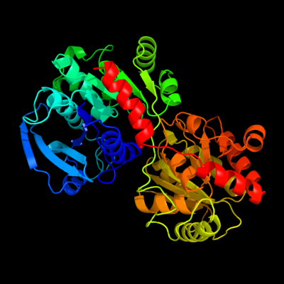
PDB header: transferaseChain: A: PDB Molecule: glycogen synthase;PDBTitle: crystal structure of wild-type e.coli gs in complex with adp2 and glucose(wtgsb)
2 d1rzua_
100.0
43
Fold: UDP-Glycosyltransferase/glycogen phosphorylaseSuperfamily: UDP-Glycosyltransferase/glycogen phosphorylaseFamily: Glycosyl transferases group 13 d2bisa1
100.0
24
Fold: UDP-Glycosyltransferase/glycogen phosphorylaseSuperfamily: UDP-Glycosyltransferase/glycogen phosphorylaseFamily: Glycosyl transferases group 14 c3o3cD_
100.0
18
PDB header: transferaseChain: D: PDB Molecule: glycogen [starch] synthase isoform 2;PDBTitle: glycogen synthase basal state udp complex
5 c3nb0A_
100.0
18
PDB header: transferaseChain: A: PDB Molecule: glycogen [starch] synthase isoform 2;PDBTitle: glucose-6-phosphate activated form of yeast glycogen synthase
6 c3s29C_
100.0
15
PDB header: transferaseChain: C: PDB Molecule: sucrose synthase 1;PDBTitle: the crystal structure of sucrose synthase-1 from arabidopsis thaliana2 and its functional implications.
7 c3c4vB_
100.0
19
PDB header: transferaseChain: B: PDB Molecule: predicted glycosyltransferases;PDBTitle: structure of the retaining glycosyltransferase msha:the2 first step in mycothiol biosynthesis. organism:3 corynebacterium glutamicum : complex with udp and 1l-ins-1-4 p.
8 c2r60A_
100.0
17
PDB header: transferaseChain: A: PDB Molecule: glycosyl transferase, group 1;PDBTitle: structure of apo sucrose phosphate synthase (sps) of2 halothermothrix orenii
9 c3okaA_
100.0
20
PDB header: transferaseChain: A: PDB Molecule: gdp-mannose-dependent alpha-(1-6)-phosphatidylinositolPDBTitle: crystal structure of corynebacterium glutamicum pimb' in complex with2 gdp-man (triclinic crystal form)
10 c2jjmH_
100.0
20
PDB header: transferaseChain: H: PDB Molecule: glycosyl transferase, group 1 family protein;PDBTitle: crystal structure of a family gt4 glycosyltransferase from2 bacillus anthracis orf ba1558.
11 c2gejA_
100.0
19
PDB header: transferaseChain: A: PDB Molecule: phosphatidylinositol mannosyltransferase (pima);PDBTitle: crystal structure of phosphatidylinositol mannosyltransferase (pima)2 from mycobacterium smegmatis in complex with gdp-man
12 c2xmpB_
100.0
19
PDB header: sugar binding proteinChain: B: PDB Molecule: trehalose-synthase tret;PDBTitle: crystal structure of trehalose synthase tret mutant e326a2 from p.horishiki in complex with udp
13 c3oy2A_
100.0
17
PDB header: viral protein,transferaseChain: A: PDB Molecule: glycosyltransferase b736l;PDBTitle: crystal structure of a putative glycosyltransferase from paramecium2 bursaria chlorella virus ny2a
14 c2x6rA_
100.0
18
PDB header: isomeraseChain: A: PDB Molecule: trehalose-synthase tret;PDBTitle: crystal structure of trehalose synthase tret from p.2 horikoshi produced by soaking in trehalose
15 c1uquB_
100.0
15
PDB header: synthaseChain: B: PDB Molecule: alpha, alpha-trehalose-phosphate synthase;PDBTitle: trehalose-6-phosphate from e. coli bound with udp-glucose.
16 d1uqta_
100.0
14
Fold: UDP-Glycosyltransferase/glycogen phosphorylaseSuperfamily: UDP-Glycosyltransferase/glycogen phosphorylaseFamily: Trehalose-6-phosphate synthase, OtsA17 d2iw1a1
100.0
17
Fold: UDP-Glycosyltransferase/glycogen phosphorylaseSuperfamily: UDP-Glycosyltransferase/glycogen phosphorylaseFamily: Glycosyl transferases group 118 c2iv3B_
100.0
21
PDB header: transferaseChain: B: PDB Molecule: glycosyltransferase;PDBTitle: crystal structure of avigt4, a glycosyltransferase involved2 in avilamycin a biosynthesis
19 c2x0dA_
100.0
10
PDB header: transferaseChain: A: PDB Molecule: wsaf;PDBTitle: apo structure of wsaf
20 c2q6vA_
100.0
15
PDB header: transferaseChain: A: PDB Molecule: glucuronosyltransferase gumk;PDBTitle: crystal structure of gumk in complex with udp
21 c3rhzB_
not modelled
100.0
13
PDB header: transferaseChain: B: PDB Molecule: nucleotide sugar synthetase-like protein;PDBTitle: structure and functional analysis of a new subfamily of2 glycosyltransferases required for glycosylation of serine-rich3 streptococcal adhesions
22 d1f6da_
not modelled
100.0
16
Fold: UDP-Glycosyltransferase/glycogen phosphorylaseSuperfamily: UDP-Glycosyltransferase/glycogen phosphorylaseFamily: UDP-N-acetylglucosamine 2-epimerase23 c3ot5D_
not modelled
100.0
14
PDB header: isomeraseChain: D: PDB Molecule: udp-n-acetylglucosamine 2-epimerase;PDBTitle: 2.2 angstrom resolution crystal structure of putative udp-n-2 acetylglucosamine 2-epimerase from listeria monocytogenes
24 c3dzcA_
not modelled
100.0
16
PDB header: isomeraseChain: A: PDB Molecule: udp-n-acetylglucosamine 2-epimerase;PDBTitle: 2.35 angstrom resolution structure of wecb (vc0917), a udp-n-2 acetylglucosamine 2-epimerase from vibrio cholerae.
25 d1v4va_
not modelled
99.9
14
Fold: UDP-Glycosyltransferase/glycogen phosphorylaseSuperfamily: UDP-Glycosyltransferase/glycogen phosphorylaseFamily: UDP-N-acetylglucosamine 2-epimerase26 d1o6ca_
not modelled
99.9
14
Fold: UDP-Glycosyltransferase/glycogen phosphorylaseSuperfamily: UDP-Glycosyltransferase/glycogen phosphorylaseFamily: UDP-N-acetylglucosamine 2-epimerase27 c3ia7A_
not modelled
99.9
12
PDB header: transferaseChain: A: PDB Molecule: calg4;PDBTitle: crystal structure of calg4, the calicheamicin glycosyltransferase
28 c3iaaB_
not modelled
99.9
13
PDB header: transferaseChain: B: PDB Molecule: calg2;PDBTitle: crystal structure of calg2, calicheamicin glycosyltransferase, tdp2 bound form
29 d1f0ka_
not modelled
99.9
17
Fold: UDP-Glycosyltransferase/glycogen phosphorylaseSuperfamily: UDP-Glycosyltransferase/glycogen phosphorylaseFamily: Peptidoglycan biosynthesis glycosyltransferase MurG30 c2xcuC_
not modelled
99.9
13
PDB header: transferaseChain: C: PDB Molecule: 3-deoxy-d-manno-2-octulosonic acid transferase;PDBTitle: membrane-embedded monofunctional glycosyltransferase waaa of aquifex2 aeolicus, comlex with cmp
31 c3othB_
not modelled
99.9
16
PDB header: transferase/antibioticChain: B: PDB Molecule: calg1;PDBTitle: crystal structure of calg1, calicheamicin glycostyltransferase, tdp2 and calicheamicin alpha3i bound form
32 c2iyaB_
not modelled
99.9
11
PDB header: transferaseChain: B: PDB Molecule: oleandomycin glycosyltransferase;PDBTitle: the crystal structure of macrolide glycosyltransferases: a2 blueprint for antibiotic engineering
33 c2p6pB_
not modelled
99.9
13
PDB header: transferaseChain: B: PDB Molecule: glycosyl transferase;PDBTitle: x-ray crystal structure of c-c bond-forming dtdp-d-olivose-transferase2 urdgt2
34 c2iyfA_
not modelled
99.8
16
PDB header: transferaseChain: A: PDB Molecule: oleandomycin glycosyltransferase;PDBTitle: the crystal structure of macrolide glycosyltransferases: a2 blueprint for antibiotic engineering
35 d2f9fa1
not modelled
99.8
15
Fold: UDP-Glycosyltransferase/glycogen phosphorylaseSuperfamily: UDP-Glycosyltransferase/glycogen phosphorylaseFamily: Glycosyl transferases group 136 c2vsnB_
not modelled
99.8
13
PDB header: transferaseChain: B: PDB Molecule: xcogt;PDBTitle: structure and topological arrangement of an o-glcnac2 transferase homolog: insight into molecular control of3 intracellular glycosylation
37 d2bfwa1
not modelled
99.8
27
Fold: UDP-Glycosyltransferase/glycogen phosphorylaseSuperfamily: UDP-Glycosyltransferase/glycogen phosphorylaseFamily: Glycosyl transferases group 138 d1iira_
not modelled
99.7
14
Fold: UDP-Glycosyltransferase/glycogen phosphorylaseSuperfamily: UDP-Glycosyltransferase/glycogen phosphorylaseFamily: Gtf glycosyltransferase39 c3d0qB_
not modelled
99.7
13
PDB header: transferaseChain: B: PDB Molecule: protein calg3;PDBTitle: crystal structure of calg3 from micromonospora echinospora determined2 in space group i222
40 d1rrva_
not modelled
99.7
14
Fold: UDP-Glycosyltransferase/glycogen phosphorylaseSuperfamily: UDP-Glycosyltransferase/glycogen phosphorylaseFamily: Gtf glycosyltransferase41 c3pe3D_
not modelled
99.7
16
PDB header: transferaseChain: D: PDB Molecule: udp-n-acetylglucosamine--peptide n-PDBTitle: structure of human o-glcnac transferase and its complex with a peptide2 substrate
42 d1pn3a_
not modelled
99.6
15
Fold: UDP-Glycosyltransferase/glycogen phosphorylaseSuperfamily: UDP-Glycosyltransferase/glycogen phosphorylaseFamily: Gtf glycosyltransferase43 c3qhpB_
not modelled
99.6
16
PDB header: transferaseChain: B: PDB Molecule: type 1 capsular polysaccharide biosynthesis protein jPDBTitle: crystal structure of the catalytic domain of cholesterol-alpha-2 glucosyltransferase from helicobacter pylori
44 d2acva1
not modelled
99.3
9
Fold: UDP-Glycosyltransferase/glycogen phosphorylaseSuperfamily: UDP-Glycosyltransferase/glycogen phosphorylaseFamily: UDPGT-like45 c2c4mA_
not modelled
99.3
16
PDB header: transferaseChain: A: PDB Molecule: glycogen phosphorylase;PDBTitle: starch phosphorylase: structural studies explain oxyanion-2 dependent kinetic stability and regulatory control.
46 c3ddsB_
not modelled
99.2
17
PDB header: transferaseChain: B: PDB Molecule: glycogen phosphorylase, liver form;PDBTitle: crystal structure of glycogen phosphorylase complexed with an2 anthranilimide based inhibitor gsk261
47 c3hbjA_
not modelled
99.2
10
PDB header: transferaseChain: A: PDB Molecule: flavonoid 3-o-glucosyltransferase;PDBTitle: structure of ugt78g1 complexed with udp
48 d2atia1
not modelled
99.2
17
Fold: UDP-Glycosyltransferase/glycogen phosphorylaseSuperfamily: UDP-Glycosyltransferase/glycogen phosphorylaseFamily: Oligosaccharide phosphorylase49 d1l5wa_
not modelled
99.1
16
Fold: UDP-Glycosyltransferase/glycogen phosphorylaseSuperfamily: UDP-Glycosyltransferase/glycogen phosphorylaseFamily: Oligosaccharide phosphorylase50 d1ygpa_
not modelled
99.0
17
Fold: UDP-Glycosyltransferase/glycogen phosphorylaseSuperfamily: UDP-Glycosyltransferase/glycogen phosphorylaseFamily: Oligosaccharide phosphorylase51 d2c1xa1
not modelled
98.9
11
Fold: UDP-Glycosyltransferase/glycogen phosphorylaseSuperfamily: UDP-Glycosyltransferase/glycogen phosphorylaseFamily: UDPGT-like52 d2gj4a1
not modelled
98.9
17
Fold: UDP-Glycosyltransferase/glycogen phosphorylaseSuperfamily: UDP-Glycosyltransferase/glycogen phosphorylaseFamily: Oligosaccharide phosphorylase53 d2vcha1
not modelled
98.7
9
Fold: UDP-Glycosyltransferase/glycogen phosphorylaseSuperfamily: UDP-Glycosyltransferase/glycogen phosphorylaseFamily: UDPGT-like54 d2pq6a1
not modelled
98.6
7
Fold: UDP-Glycosyltransferase/glycogen phosphorylaseSuperfamily: UDP-Glycosyltransferase/glycogen phosphorylaseFamily: UDPGT-like55 c3hbmA_
not modelled
98.3
10
PDB header: hydrolaseChain: A: PDB Molecule: udp-sugar hydrolase;PDBTitle: crystal structure of pseg from campylobacter jejuni
56 c3q3hA_
not modelled
98.3
11
PDB header: transferaseChain: A: PDB Molecule: hmw1c-like glycosyltransferase;PDBTitle: crystal structure of the actinobacillus pleuropneumoniae hmw1c2 glycosyltransferase in complex with udp-glc
57 c3l7mC_
not modelled
97.4
10
PDB header: structural proteinChain: C: PDB Molecule: teichoic acid biosynthesis protein f;PDBTitle: structure of the wall teichoic acid polymerase tagf, h548a
58 c2o6lA_
not modelled
97.1
7
PDB header: transferaseChain: A: PDB Molecule: udp-glucuronosyltransferase 2b7;PDBTitle: crystal structure of the udp-glucuronic acid binding domain2 of the human drug metabolizing udp-glucuronosyltransferase3 2b7
59 c2h1fB_
not modelled
96.4
15
PDB header: transferaseChain: B: PDB Molecule: lipopolysaccharide heptosyltransferase-1;PDBTitle: e. coli heptosyltransferase waac with adp
60 d1pswa_
not modelled
95.3
13
Fold: UDP-Glycosyltransferase/glycogen phosphorylaseSuperfamily: UDP-Glycosyltransferase/glycogen phosphorylaseFamily: ADP-heptose LPS heptosyltransferase II61 d1jaya_
not modelled
95.1
27
Fold: NAD(P)-binding Rossmann-fold domainsSuperfamily: NAD(P)-binding Rossmann-fold domainsFamily: 6-phosphogluconate dehydrogenase-like, N-terminal domain62 d1udca_
not modelled
94.8
28
Fold: NAD(P)-binding Rossmann-fold domainsSuperfamily: NAD(P)-binding Rossmann-fold domainsFamily: Tyrosine-dependent oxidoreductases63 c2pzlB_
not modelled
94.7
20
PDB header: sugar binding proteinChain: B: PDB Molecule: putative nucleotide sugar epimerase/ dehydratase;PDBTitle: crystal structure of the bordetella bronchiseptica enzyme2 wbmg in complex with nad and udp
64 c2p5uC_
not modelled
94.7
30
PDB header: isomeraseChain: C: PDB Molecule: udp-glucose 4-epimerase;PDBTitle: crystal structure of thermus thermophilus hb8 udp-glucose 4-2 epimerase complex with nad
65 c3icpA_
not modelled
94.6
23
PDB header: isomeraseChain: A: PDB Molecule: nad-dependent epimerase/dehydratase;PDBTitle: crystal structure of udp-galactose 4-epimerase
66 c3m2pD_
not modelled
94.6
20
PDB header: isomeraseChain: D: PDB Molecule: udp-n-acetylglucosamine 4-epimerase;PDBTitle: the crystal structure of udp-n-acetylglucosamine 4-epimerase2 from bacillus cereus
67 c2x4gA_
not modelled
94.0
23
PDB header: isomeraseChain: A: PDB Molecule: nucleoside-diphosphate-sugar epimerase;PDBTitle: crystal structure of pa4631, a nucleoside-diphosphate-sugar2 epimerase from pseudomonas aeruginosa
68 c2pk3B_
not modelled
94.0
18
PDB header: oxidoreductaseChain: B: PDB Molecule: gdp-6-deoxy-d-lyxo-4-hexulose reductase;PDBTitle: crystal structure of a gdp-4-keto-6-deoxy-d-mannose reductase
69 d2c5aa1
not modelled
93.9
8
Fold: NAD(P)-binding Rossmann-fold domainsSuperfamily: NAD(P)-binding Rossmann-fold domainsFamily: Tyrosine-dependent oxidoreductases70 d2f1ka2
not modelled
93.7
19
Fold: NAD(P)-binding Rossmann-fold domainsSuperfamily: NAD(P)-binding Rossmann-fold domainsFamily: 6-phosphogluconate dehydrogenase-like, N-terminal domain71 c2ofpB_
not modelled
93.6
20
PDB header: oxidoreductaseChain: B: PDB Molecule: ketopantoate reductase;PDBTitle: crystal structure of escherichia coli ketopantoate2 reductase in a ternary complex with nadp+ and pantoate
72 c1gshA_
not modelled
93.4
13
PDB header: glutathione biosynthesis ligaseChain: A: PDB Molecule: glutathione biosynthetic ligase;PDBTitle: structure of escherichia coli glutathione synthetase at ph 7.5
73 c3l4bG_
not modelled
93.4
13
PDB header: transport proteinChain: G: PDB Molecule: trka k+ channel protien tm1088b;PDBTitle: crystal structure of an octomeric two-subunit trka k+ channel ring2 gating assembly, tm1088a:tm1088b, from thermotoga maritima
74 c3io3A_
not modelled
93.2
9
PDB header: chaperoneChain: A: PDB Molecule: deha2d07832p;PDBTitle: get3 with adp from d. hansenii in closed form
75 d1txga2
not modelled
93.2
31
Fold: NAD(P)-binding Rossmann-fold domainsSuperfamily: NAD(P)-binding Rossmann-fold domainsFamily: 6-phosphogluconate dehydrogenase-like, N-terminal domain76 d1fjha_
not modelled
93.1
11
Fold: NAD(P)-binding Rossmann-fold domainsSuperfamily: NAD(P)-binding Rossmann-fold domainsFamily: Tyrosine-dependent oxidoreductases77 c3oh8A_
not modelled
92.9
24
PDB header: isomeraseChain: A: PDB Molecule: nucleoside-diphosphate sugar epimerase (sula family);PDBTitle: crystal structure of the nucleoside-diphosphate sugar epimerase from2 corynebacterium glutamicum. northeast structural genomics consortium3 target cgr91
78 d1vl0a_
not modelled
92.8
15
Fold: NAD(P)-binding Rossmann-fold domainsSuperfamily: NAD(P)-binding Rossmann-fold domainsFamily: Tyrosine-dependent oxidoreductases79 d1ks9a2
not modelled
92.8
20
Fold: NAD(P)-binding Rossmann-fold domainsSuperfamily: NAD(P)-binding Rossmann-fold domainsFamily: 6-phosphogluconate dehydrogenase-like, N-terminal domain80 d1ydga_
not modelled
92.7
23
Fold: Flavodoxin-likeSuperfamily: FlavoproteinsFamily: WrbA-like81 d1gsaa1
not modelled
92.7
13
Fold: PreATP-grasp domainSuperfamily: PreATP-grasp domainFamily: Prokaryotic glutathione synthetase, N-terminal domain82 c1ks9A_
not modelled
92.6
20
PDB header: oxidoreductaseChain: A: PDB Molecule: 2-dehydropantoate 2-reductase;PDBTitle: ketopantoate reductase from escherichia coli
83 c2ggsB_
not modelled
92.6
17
PDB header: oxidoreductaseChain: B: PDB Molecule: 273aa long hypothetical dtdp-4-dehydrorhamnosePDBTitle: crystal structure of hypothetical dtdp-4-dehydrorhamnose2 reductase from sulfolobus tokodaii
84 d2blla1
not modelled
92.6
17
Fold: NAD(P)-binding Rossmann-fold domainsSuperfamily: NAD(P)-binding Rossmann-fold domainsFamily: Tyrosine-dependent oxidoreductases85 c3fmfA_
not modelled
92.6
37
PDB header: ligaseChain: A: PDB Molecule: dethiobiotin synthetase;PDBTitle: crystal structure of mycobacterium tuberculosis dethiobiotin2 synthetase complexed with 7,8 diaminopelargonic acid carbamate
86 c3ibgF_
not modelled
92.3
15
PDB header: hydrolaseChain: F: PDB Molecule: atpase, subunit of the get complex;PDBTitle: crystal structure of aspergillus fumigatus get3 with bound2 adp
87 c3tovB_
not modelled
92.3
10
PDB header: transferaseChain: B: PDB Molecule: glycosyl transferase family 9;PDBTitle: the crystal structure of the glycosyl transferase family 9 from2 veillonella parvula dsm 2008
88 c2wooC_
not modelled
92.3
14
PDB header: hydrolaseChain: C: PDB Molecule: atpase get3;PDBTitle: nucleotide-free form of s. pombe get3
89 d1kewa_
not modelled
92.2
29
Fold: NAD(P)-binding Rossmann-fold domainsSuperfamily: NAD(P)-binding Rossmann-fold domainsFamily: Tyrosine-dependent oxidoreductases90 c2f1kD_
not modelled
92.1
19
PDB header: oxidoreductaseChain: D: PDB Molecule: prephenate dehydrogenase;PDBTitle: crystal structure of synechocystis arogenate dehydrogenase
91 c3l77A_
not modelled
92.0
21
PDB header: oxidoreductaseChain: A: PDB Molecule: short-chain alcohol dehydrogenase;PDBTitle: x-ray structure alcohol dehydrogenase from archaeon thermococcus2 sibiricus complexed with 5-hydroxy-nadp
92 d1mv8a2
not modelled
92.0
22
Fold: NAD(P)-binding Rossmann-fold domainsSuperfamily: NAD(P)-binding Rossmann-fold domainsFamily: 6-phosphogluconate dehydrogenase-like, N-terminal domain93 c2hunB_
not modelled
92.0
18
PDB header: lyaseChain: B: PDB Molecule: 336aa long hypothetical dtdp-glucose 4,6-dehydratase;PDBTitle: crystal structure of hypothetical protein ph0414 from pyrococcus2 horikoshii ot3
94 d2d1pa1
not modelled
91.9
24
Fold: DsrEFH-likeSuperfamily: DsrEFH-likeFamily: DsrEF-like95 c2q1wC_
not modelled
91.9
12
PDB header: sugar binding proteinChain: C: PDB Molecule: putative nucleotide sugar epimerase/ dehydratase;PDBTitle: crystal structure of the bordetella bronchiseptica enzyme wbmh in2 complex with nad+
96 c2ixdB_
not modelled
91.8
9
PDB header: hydrolaseChain: B: PDB Molecule: lmbe-related protein;PDBTitle: crystal structure of the putative deacetylase bc1534 from2 bacilus cereus
97 c3dojA_
not modelled
91.7
28
PDB header: oxidoreductaseChain: A: PDB Molecule: dehydrogenase-like protein;PDBTitle: structure of glyoxylate reductase 1 from arabidopsis2 (atglyr1)
98 d1bxka_
not modelled
91.6
28
Fold: NAD(P)-binding Rossmann-fold domainsSuperfamily: NAD(P)-binding Rossmann-fold domainsFamily: Tyrosine-dependent oxidoreductases99 c1hyqA_
not modelled
91.3
26
PDB header: cell cycleChain: A: PDB Molecule: cell division inhibitor (mind-1);PDBTitle: mind bacterial cell division regulator from a. fulgidus
100 d1hyqa_
not modelled
91.3
26
Fold: P-loop containing nucleoside triphosphate hydrolasesSuperfamily: P-loop containing nucleoside triphosphate hydrolasesFamily: Nitrogenase iron protein-like101 d1j9ja_
not modelled
91.3
18
Fold: SurE-likeSuperfamily: SurE-likeFamily: SurE-like102 d2hy5a1
not modelled
91.1
22
Fold: DsrEFH-likeSuperfamily: DsrEFH-likeFamily: DsrEF-like103 c3g0oA_
not modelled
91.0
16
PDB header: oxidoreductaseChain: A: PDB Molecule: 3-hydroxyisobutyrate dehydrogenase;PDBTitle: crystal structure of 3-hydroxyisobutyrate dehydrogenase2 (ygbj) from salmonella typhimurium
104 c3g17H_
not modelled
91.0
9
PDB header: structural genomics, unknown functionChain: H: PDB Molecule: similar to 2-dehydropantoate 2-reductase;PDBTitle: structure of putative 2-dehydropantoate 2-reductase from2 staphylococcus aureus
105 c2gf2B_
not modelled
90.7
16
PDB header: oxidoreductaseChain: B: PDB Molecule: 3-hydroxyisobutyrate dehydrogenase;PDBTitle: crystal structure of human hydroxyisobutyrate dehydrogenase
106 d1pgja2
not modelled
90.5
25
Fold: NAD(P)-binding Rossmann-fold domainsSuperfamily: NAD(P)-binding Rossmann-fold domainsFamily: 6-phosphogluconate dehydrogenase-like, N-terminal domain107 d1n2sa_
not modelled
90.3
23
Fold: NAD(P)-binding Rossmann-fold domainsSuperfamily: NAD(P)-binding Rossmann-fold domainsFamily: Tyrosine-dependent oxidoreductases108 d2afhe1
not modelled
90.2
27
Fold: P-loop containing nucleoside triphosphate hydrolasesSuperfamily: P-loop containing nucleoside triphosphate hydrolasesFamily: Nitrogenase iron protein-like109 c3kjgB_
not modelled
90.1
24
PDB header: hydrolase, metal binding proteinChain: B: PDB Molecule: co dehydrogenase/acetyl-coa synthase complex, accessoryPDBTitle: adp-bound state of cooc1
110 d1lssa_
not modelled
90.0
19
Fold: NAD(P)-binding Rossmann-fold domainsSuperfamily: NAD(P)-binding Rossmann-fold domainsFamily: Potassium channel NAD-binding domain111 d1oc2a_
not modelled
89.9
13
Fold: NAD(P)-binding Rossmann-fold domainsSuperfamily: NAD(P)-binding Rossmann-fold domainsFamily: Tyrosine-dependent oxidoreductases112 d1uana_
not modelled
89.9
19
Fold: LmbE-likeSuperfamily: LmbE-likeFamily: LmbE-like113 c1txgA_
not modelled
89.7
28
PDB header: oxidoreductaseChain: A: PDB Molecule: glycerol-3-phosphate dehydrogenase [nad(p)+];PDBTitle: structure of glycerol-3-phosphate dehydrogenase from archaeoglobus2 fulgidus
114 c3ckyA_
not modelled
89.7
13
PDB header: oxidoreductaseChain: A: PDB Molecule: 2-hydroxymethyl glutarate dehydrogenase;PDBTitle: structural and kinetic properties of a beta-hydroxyacid dehydrogenase2 involved in nicotinate fermentation
115 c3ghyA_
not modelled
89.5
30
PDB header: oxidoreductaseChain: A: PDB Molecule: ketopantoate reductase protein;PDBTitle: crystal structure of a putative ketopantoate reductase from ralstonia2 solanacearum molk2
116 c3sc6F_
not modelled
89.4
18
PDB header: oxidoreductaseChain: F: PDB Molecule: dtdp-4-dehydrorhamnose reductase;PDBTitle: 2.65 angstrom resolution crystal structure of dtdp-4-dehydrorhamnose2 reductase (rfbd) from bacillus anthracis str. ames in complex with3 nadp
117 c3i4fD_
not modelled
89.2
18
PDB header: oxidoreductaseChain: D: PDB Molecule: 3-oxoacyl-[acyl-carrier protein] reductase;PDBTitle: structure of putative 3-oxoacyl-reductase from bacillus thuringiensis
118 c1i36A_
not modelled
89.1
21
PDB header: structural genomics, unknown functionChain: A: PDB Molecule: conserved hypothetical protein mth1747;PDBTitle: structure of conserved protein mth1747 of unknown function2 reveals structural similarity with 3-hydroxyacid3 dehydrogenases
119 d1i36a2
not modelled
88.6
21
Fold: NAD(P)-binding Rossmann-fold domainsSuperfamily: NAD(P)-binding Rossmann-fold domainsFamily: 6-phosphogluconate dehydrogenase-like, N-terminal domain120 c2v4oB_
not modelled
88.6
26
PDB header: hydrolaseChain: B: PDB Molecule: multifunctional protein sur e;PDBTitle: crystal structure of salmonella typhimurium sure at 2.752 angstrom resolution in monoclinic form