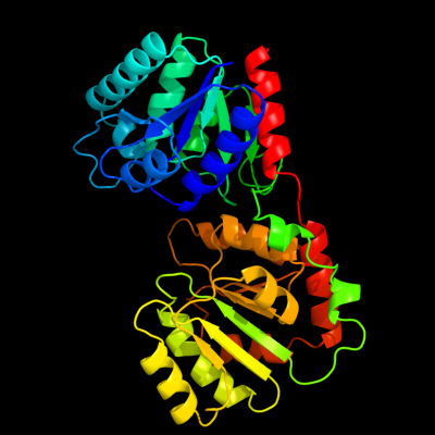
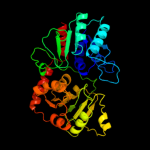
| 1 | d1f0ka_
|
|
 |
100.0 |
100 |
Fold:UDP-Glycosyltransferase/glycogen phosphorylase
Superfamily:UDP-Glycosyltransferase/glycogen phosphorylase
Family:Peptidoglycan biosynthesis glycosyltransferase MurG |
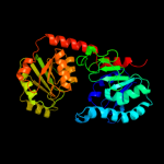
| 2 | c3ia7A_
|
|
 |
100.0 |
15 |
PDB header:transferase
Chain: A: PDB Molecule:calg4;
PDBTitle: crystal structure of calg4, the calicheamicin glycosyltransferase
|
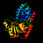
| 3 | c3iaaB_
|
|
 |
100.0 |
15 |
PDB header:transferase
Chain: B: PDB Molecule:calg2;
PDBTitle: crystal structure of calg2, calicheamicin glycosyltransferase, tdp2 bound form
|
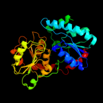
| 4 | c2iyfA_
|
|
 |
100.0 |
20 |
PDB header:transferase
Chain: A: PDB Molecule:oleandomycin glycosyltransferase;
PDBTitle: the crystal structure of macrolide glycosyltransferases: a2 blueprint for antibiotic engineering
|
| 5 | c2iyaB_
|
|
 |
100.0 |
12 |
PDB header:transferase
Chain: B: PDB Molecule:oleandomycin glycosyltransferase;
PDBTitle: the crystal structure of macrolide glycosyltransferases: a2 blueprint for antibiotic engineering
|
| 6 | c3othB_
|
|
 |
100.0 |
17 |
PDB header:transferase/antibiotic
Chain: B: PDB Molecule:calg1;
PDBTitle: crystal structure of calg1, calicheamicin glycostyltransferase, tdp2 and calicheamicin alpha3i bound form
|
| 7 | c2p6pB_
|
|
 |
100.0 |
15 |
PDB header:transferase
Chain: B: PDB Molecule:glycosyl transferase;
PDBTitle: x-ray crystal structure of c-c bond-forming dtdp-d-olivose-transferase2 urdgt2
|
| 8 | d1iira_
|
|
 |
100.0 |
15 |
Fold:UDP-Glycosyltransferase/glycogen phosphorylase
Superfamily:UDP-Glycosyltransferase/glycogen phosphorylase
Family:Gtf glycosyltransferase |
| 9 | d1rrva_
|
|
 |
100.0 |
16 |
Fold:UDP-Glycosyltransferase/glycogen phosphorylase
Superfamily:UDP-Glycosyltransferase/glycogen phosphorylase
Family:Gtf glycosyltransferase |
| 10 | d1pn3a_
|
|
 |
100.0 |
14 |
Fold:UDP-Glycosyltransferase/glycogen phosphorylase
Superfamily:UDP-Glycosyltransferase/glycogen phosphorylase
Family:Gtf glycosyltransferase |
| 11 | d2c1xa1
|
|
 |
99.9 |
14 |
Fold:UDP-Glycosyltransferase/glycogen phosphorylase
Superfamily:UDP-Glycosyltransferase/glycogen phosphorylase
Family:UDPGT-like |
| 12 | c3d0qB_
|
|
 |
99.9 |
13 |
PDB header:transferase
Chain: B: PDB Molecule:protein calg3;
PDBTitle: crystal structure of calg3 from micromonospora echinospora determined2 in space group i222
|
| 13 | c3hbjA_
|
|
 |
99.9 |
13 |
PDB header:transferase
Chain: A: PDB Molecule:flavonoid 3-o-glucosyltransferase;
PDBTitle: structure of ugt78g1 complexed with udp
|
| 14 | c3c4vB_
|
|
 |
99.9 |
15 |
PDB header:transferase
Chain: B: PDB Molecule:predicted glycosyltransferases;
PDBTitle: structure of the retaining glycosyltransferase msha:the2 first step in mycothiol biosynthesis. organism:3 corynebacterium glutamicum : complex with udp and 1l-ins-1-4 p.
|
| 15 | d2acva1
|
|
 |
99.9 |
14 |
Fold:UDP-Glycosyltransferase/glycogen phosphorylase
Superfamily:UDP-Glycosyltransferase/glycogen phosphorylase
Family:UDPGT-like |
| 16 | d2pq6a1
|
|
 |
99.9 |
15 |
Fold:UDP-Glycosyltransferase/glycogen phosphorylase
Superfamily:UDP-Glycosyltransferase/glycogen phosphorylase
Family:UDPGT-like |
| 17 | c2r60A_
|
|
 |
99.9 |
14 |
PDB header:transferase
Chain: A: PDB Molecule:glycosyl transferase, group 1;
PDBTitle: structure of apo sucrose phosphate synthase (sps) of2 halothermothrix orenii
|
| 18 | d1v4va_
|
|
 |
99.9 |
19 |
Fold:UDP-Glycosyltransferase/glycogen phosphorylase
Superfamily:UDP-Glycosyltransferase/glycogen phosphorylase
Family:UDP-N-acetylglucosamine 2-epimerase |
| 19 | c2gejA_
|
|
 |
99.9 |
15 |
PDB header:transferase
Chain: A: PDB Molecule:phosphatidylinositol mannosyltransferase (pima);
PDBTitle: crystal structure of phosphatidylinositol mannosyltransferase (pima)2 from mycobacterium smegmatis in complex with gdp-man
|
| 20 | c3s29C_
|
|
 |
99.9 |
11 |
PDB header:transferase
Chain: C: PDB Molecule:sucrose synthase 1;
PDBTitle: the crystal structure of sucrose synthase-1 from arabidopsis thaliana2 and its functional implications.
|
| 21 | c3dzcA_ |
|
not modelled |
99.9 |
14 |
PDB header:isomerase
Chain: A: PDB Molecule:udp-n-acetylglucosamine 2-epimerase;
PDBTitle: 2.35 angstrom resolution structure of wecb (vc0917), a udp-n-2 acetylglucosamine 2-epimerase from vibrio cholerae.
|
| 22 | d2vcha1 |
|
not modelled |
99.9 |
12 |
Fold:UDP-Glycosyltransferase/glycogen phosphorylase
Superfamily:UDP-Glycosyltransferase/glycogen phosphorylase
Family:UDPGT-like |
| 23 | c2jjmH_ |
|
not modelled |
99.9 |
10 |
PDB header:transferase
Chain: H: PDB Molecule:glycosyl transferase, group 1 family protein;
PDBTitle: crystal structure of a family gt4 glycosyltransferase from2 bacillus anthracis orf ba1558.
|
| 24 | c3okaA_ |
|
not modelled |
99.9 |
9 |
PDB header:transferase
Chain: A: PDB Molecule:gdp-mannose-dependent alpha-(1-6)-phosphatidylinositol
PDBTitle: crystal structure of corynebacterium glutamicum pimb' in complex with2 gdp-man (triclinic crystal form)
|
| 25 | d1f6da_ |
|
not modelled |
99.9 |
12 |
Fold:UDP-Glycosyltransferase/glycogen phosphorylase
Superfamily:UDP-Glycosyltransferase/glycogen phosphorylase
Family:UDP-N-acetylglucosamine 2-epimerase |
| 26 | d2bisa1 |
|
not modelled |
99.9 |
14 |
Fold:UDP-Glycosyltransferase/glycogen phosphorylase
Superfamily:UDP-Glycosyltransferase/glycogen phosphorylase
Family:Glycosyl transferases group 1 |
| 27 | c2qzsA_ |
|
not modelled |
99.9 |
17 |
PDB header:transferase
Chain: A: PDB Molecule:glycogen synthase;
PDBTitle: crystal structure of wild-type e.coli gs in complex with adp2 and glucose(wtgsb)
|
| 28 | c3ot5D_ |
|
not modelled |
99.9 |
14 |
PDB header:isomerase
Chain: D: PDB Molecule:udp-n-acetylglucosamine 2-epimerase;
PDBTitle: 2.2 angstrom resolution crystal structure of putative udp-n-2 acetylglucosamine 2-epimerase from listeria monocytogenes
|
| 29 | d1rzua_ |
|
not modelled |
99.9 |
15 |
Fold:UDP-Glycosyltransferase/glycogen phosphorylase
Superfamily:UDP-Glycosyltransferase/glycogen phosphorylase
Family:Glycosyl transferases group 1 |
| 30 | c2x6rA_ |
|
not modelled |
99.8 |
8 |
PDB header:isomerase
Chain: A: PDB Molecule:trehalose-synthase tret;
PDBTitle: crystal structure of trehalose synthase tret from p.2 horikoshi produced by soaking in trehalose
|
| 31 | c3hbmA_ |
|
not modelled |
99.8 |
12 |
PDB header:hydrolase
Chain: A: PDB Molecule:udp-sugar hydrolase;
PDBTitle: crystal structure of pseg from campylobacter jejuni
|
| 32 | c2xmpB_ |
|
not modelled |
99.8 |
9 |
PDB header:sugar binding protein
Chain: B: PDB Molecule:trehalose-synthase tret;
PDBTitle: crystal structure of trehalose synthase tret mutant e326a2 from p.horishiki in complex with udp
|
| 33 | d2iw1a1 |
|
not modelled |
99.8 |
13 |
Fold:UDP-Glycosyltransferase/glycogen phosphorylase
Superfamily:UDP-Glycosyltransferase/glycogen phosphorylase
Family:Glycosyl transferases group 1 |
| 34 | d1o6ca_ |
|
not modelled |
99.8 |
14 |
Fold:UDP-Glycosyltransferase/glycogen phosphorylase
Superfamily:UDP-Glycosyltransferase/glycogen phosphorylase
Family:UDP-N-acetylglucosamine 2-epimerase |
| 35 | c3oy2A_ |
|
not modelled |
99.8 |
10 |
PDB header:viral protein,transferase
Chain: A: PDB Molecule:glycosyltransferase b736l;
PDBTitle: crystal structure of a putative glycosyltransferase from paramecium2 bursaria chlorella virus ny2a
|
| 36 | c2iv3B_ |
|
not modelled |
99.7 |
12 |
PDB header:transferase
Chain: B: PDB Molecule:glycosyltransferase;
PDBTitle: crystal structure of avigt4, a glycosyltransferase involved2 in avilamycin a biosynthesis
|
| 37 | c2xcuC_ |
|
not modelled |
99.7 |
13 |
PDB header:transferase
Chain: C: PDB Molecule:3-deoxy-d-manno-2-octulosonic acid transferase;
PDBTitle: membrane-embedded monofunctional glycosyltransferase waaa of aquifex2 aeolicus, comlex with cmp
|
| 38 | c1uquB_ |
|
not modelled |
99.6 |
13 |
PDB header:synthase
Chain: B: PDB Molecule:alpha, alpha-trehalose-phosphate synthase;
PDBTitle: trehalose-6-phosphate from e. coli bound with udp-glucose.
|
| 39 | c2x0dA_ |
|
not modelled |
99.5 |
12 |
PDB header:transferase
Chain: A: PDB Molecule:wsaf;
PDBTitle: apo structure of wsaf
|
| 40 | c2q6vA_ |
|
not modelled |
99.5 |
9 |
PDB header:transferase
Chain: A: PDB Molecule:glucuronosyltransferase gumk;
PDBTitle: crystal structure of gumk in complex with udp
|
| 41 | d1uqta_ |
|
not modelled |
99.5 |
12 |
Fold:UDP-Glycosyltransferase/glycogen phosphorylase
Superfamily:UDP-Glycosyltransferase/glycogen phosphorylase
Family:Trehalose-6-phosphate synthase, OtsA |
| 42 | c3nb0A_ |
|
not modelled |
99.5 |
14 |
PDB header:transferase
Chain: A: PDB Molecule:glycogen [starch] synthase isoform 2;
PDBTitle: glucose-6-phosphate activated form of yeast glycogen synthase
|
| 43 | c3o3cD_ |
|
not modelled |
99.4 |
14 |
PDB header:transferase
Chain: D: PDB Molecule:glycogen [starch] synthase isoform 2;
PDBTitle: glycogen synthase basal state udp complex
|
| 44 | c3rhzB_ |
|
not modelled |
99.4 |
10 |
PDB header:transferase
Chain: B: PDB Molecule:nucleotide sugar synthetase-like protein;
PDBTitle: structure and functional analysis of a new subfamily of2 glycosyltransferases required for glycosylation of serine-rich3 streptococcal adhesions
|
| 45 | c2o6lA_ |
|
not modelled |
99.3 |
14 |
PDB header:transferase
Chain: A: PDB Molecule:udp-glucuronosyltransferase 2b7;
PDBTitle: crystal structure of the udp-glucuronic acid binding domain2 of the human drug metabolizing udp-glucuronosyltransferase3 2b7
|
| 46 | c2vsnB_ |
|
not modelled |
99.3 |
12 |
PDB header:transferase
Chain: B: PDB Molecule:xcogt;
PDBTitle: structure and topological arrangement of an o-glcnac2 transferase homolog: insight into molecular control of3 intracellular glycosylation
|
| 47 | c3pe3D_ |
|
not modelled |
98.8 |
15 |
PDB header:transferase
Chain: D: PDB Molecule:udp-n-acetylglucosamine--peptide n-
PDBTitle: structure of human o-glcnac transferase and its complex with a peptide2 substrate
|
| 48 | d2f9fa1 |
|
not modelled |
98.7 |
7 |
Fold:UDP-Glycosyltransferase/glycogen phosphorylase
Superfamily:UDP-Glycosyltransferase/glycogen phosphorylase
Family:Glycosyl transferases group 1 |
| 49 | d2bfwa1 |
|
not modelled |
98.7 |
9 |
Fold:UDP-Glycosyltransferase/glycogen phosphorylase
Superfamily:UDP-Glycosyltransferase/glycogen phosphorylase
Family:Glycosyl transferases group 1 |
| 50 | c3tovB_ |
|
not modelled |
98.4 |
13 |
PDB header:transferase
Chain: B: PDB Molecule:glycosyl transferase family 9;
PDBTitle: the crystal structure of the glycosyl transferase family 9 from2 veillonella parvula dsm 2008
|
| 51 | c3qhpB_ |
|
not modelled |
98.4 |
8 |
PDB header:transferase
Chain: B: PDB Molecule:type 1 capsular polysaccharide biosynthesis protein j
PDBTitle: crystal structure of the catalytic domain of cholesterol-alpha-2 glucosyltransferase from helicobacter pylori
|
| 52 | c2jzcA_ |
|
not modelled |
98.1 |
12 |
PDB header:transferase
Chain: A: PDB Molecule:udp-n-acetylglucosamine transferase subunit
PDBTitle: nmr solution structure of alg13: the sugar donor subunit of2 a yeast n-acetylglucosamine transferase. northeast3 structural genomics consortium target yg1
|
| 53 | c2h1fB_ |
|
not modelled |
97.8 |
13 |
PDB header:transferase
Chain: B: PDB Molecule:lipopolysaccharide heptosyltransferase-1;
PDBTitle: e. coli heptosyltransferase waac with adp
|
| 54 | d1pswa_ |
|
not modelled |
97.6 |
10 |
Fold:UDP-Glycosyltransferase/glycogen phosphorylase
Superfamily:UDP-Glycosyltransferase/glycogen phosphorylase
Family:ADP-heptose LPS heptosyltransferase II |
| 55 | c3louB_ |
|
not modelled |
96.9 |
12 |
PDB header:hydrolase
Chain: B: PDB Molecule:formyltetrahydrofolate deformylase;
PDBTitle: crystal structure of formyltetrahydrofolate deformylase (yp_105254.1)2 from burkholderia mallei atcc 23344 at 1.90 a resolution
|
| 56 | c3o1lB_ |
|
not modelled |
96.9 |
17 |
PDB header:hydrolase
Chain: B: PDB Molecule:formyltetrahydrofolate deformylase;
PDBTitle: crystal structure of a formyltetrahydrofolate deformylase (pspto_4314)2 from pseudomonas syringae pv. tomato str. dc3000 at 2.20 a resolution
|
| 57 | c1ps9A_ |
|
not modelled |
96.7 |
42 |
PDB header:oxidoreductase
Chain: A: PDB Molecule:2,4-dienoyl-coa reductase;
PDBTitle: the crystal structure and reaction mechanism of e. coli 2,4-2 dienoyl coa reductase
|
| 58 | c2ydyA_ |
|
not modelled |
96.6 |
24 |
PDB header:oxidoreductase
Chain: A: PDB Molecule:methionine adenosyltransferase 2 subunit beta;
PDBTitle: crystal structure of human s-adenosylmethionine synthetase2 2, beta subunit in orthorhombic crystal form
|
| 59 | d1sb8a_ |
|
not modelled |
96.6 |
17 |
Fold:NAD(P)-binding Rossmann-fold domains
Superfamily:NAD(P)-binding Rossmann-fold domains
Family:Tyrosine-dependent oxidoreductases |
| 60 | c3n0vD_ |
|
not modelled |
96.6 |
13 |
PDB header:hydrolase
Chain: D: PDB Molecule:formyltetrahydrofolate deformylase;
PDBTitle: crystal structure of a formyltetrahydrofolate deformylase (pp_0327)2 from pseudomonas putida kt2440 at 2.25 a resolution
|
| 61 | c1kjjA_ |
|
not modelled |
96.5 |
21 |
PDB header:transferase
Chain: A: PDB Molecule:phosphoribosylglycinamide formyltransferase 2;
PDBTitle: crystal structure of glycniamide ribonucleotide2 transformylase in complex with mg-atp-gamma-s
|
| 62 | c3slgB_ |
|
not modelled |
96.4 |
19 |
PDB header:transferase
Chain: B: PDB Molecule:pbgp3 protein;
PDBTitle: crystal structure of pbgp3 protein from burkholderia pseudomallei
|
| 63 | c1t2aC_ |
|
not modelled |
96.3 |
15 |
PDB header:structural genomics,lyase
Chain: C: PDB Molecule:gdp-mannose 4,6 dehydratase;
PDBTitle: crystal structure of human gdp-d-mannose 4,6-dehydratase
|
| 64 | d1t2aa_ |
|
not modelled |
96.3 |
15 |
Fold:NAD(P)-binding Rossmann-fold domains
Superfamily:NAD(P)-binding Rossmann-fold domains
Family:Tyrosine-dependent oxidoreductases |
| 65 | c3k30B_ |
|
not modelled |
96.2 |
30 |
PDB header:oxidoreductase
Chain: B: PDB Molecule:histamine dehydrogenase;
PDBTitle: histamine dehydrogenase from nocardiodes simplex
|
| 66 | d1reoa1 |
|
not modelled |
96.1 |
35 |
Fold:FAD/NAD(P)-binding domain
Superfamily:FAD/NAD(P)-binding domain
Family:FAD-linked reductases, N-terminal domain |
| 67 | c1z7eC_ |
|
not modelled |
96.1 |
13 |
PDB header:hydrolase
Chain: C: PDB Molecule:protein arna;
PDBTitle: crystal structure of full length arna
|
| 68 | c3lu1C_ |
|
not modelled |
96.1 |
16 |
PDB header:isomerase
Chain: C: PDB Molecule:wbgu;
PDBTitle: crystal structure analysis of wbgu: a udp-galnac 4-epimerase
|
| 69 | c2yxbA_ |
|
not modelled |
96.1 |
16 |
PDB header:isomerase
Chain: A: PDB Molecule:coenzyme b12-dependent mutase;
PDBTitle: crystal structure of the methylmalonyl-coa mutase alpha-subunit from2 aeropyrum pernix
|
| 70 | c3lrxC_ |
|
not modelled |
96.1 |
10 |
PDB header:oxidoreductase
Chain: C: PDB Molecule:putative hydrogenase;
PDBTitle: crystal structure of the c-terminal domain (residues 78-226)2 of pf1911 hydrogenase from pyrococcus furiosus, northeast3 structural genomics consortium target pfr246a
|
| 71 | d1rkxa_ |
|
not modelled |
96.0 |
29 |
Fold:NAD(P)-binding Rossmann-fold domains
Superfamily:NAD(P)-binding Rossmann-fold domains
Family:Tyrosine-dependent oxidoreductases |
| 72 | d1u7za_ |
|
not modelled |
96.0 |
24 |
Fold:Ribokinase-like
Superfamily:CoaB-like
Family:CoaB-like |
| 73 | c3allA_ |
|
not modelled |
96.0 |
42 |
PDB header:oxidoreductase
Chain: A: PDB Molecule:2-methyl-3-hydroxypyridine-5-carboxylic acid oxygenase;
PDBTitle: crystal structure of 2-methyl-3-hydroxypyridine-5-carboxylic acid2 oxygenase, mutant y270a
|
| 74 | c1djnB_ |
|
not modelled |
96.0 |
22 |
PDB header:oxidoreductase
Chain: B: PDB Molecule:trimethylamine dehydrogenase;
PDBTitle: structural and biochemical characterization of recombinant wild type2 trimethylamine dehydrogenase from methylophilus methylotrophus (sp.3 w3a1)
|
| 75 | d2iida1 |
|
not modelled |
96.0 |
33 |
Fold:FAD/NAD(P)-binding domain
Superfamily:FAD/NAD(P)-binding domain
Family:FAD-linked reductases, N-terminal domain |
| 76 | c1f8sA_ |
|
not modelled |
96.0 |
33 |
PDB header:oxidoreductase
Chain: A: PDB Molecule:l-amino acid oxidase;
PDBTitle: crystal structure of l-amino acid oxidase from calloselasma2 rhodostoma, complexed with three molecules of o-aminobenzoate.
|
| 77 | d1c0pa1 |
|
not modelled |
96.0 |
35 |
Fold:Nucleotide-binding domain
Superfamily:Nucleotide-binding domain
Family:D-aminoacid oxidase, N-terminal domain |
| 78 | c3nrbD_ |
|
not modelled |
95.9 |
15 |
PDB header:hydrolase
Chain: D: PDB Molecule:formyltetrahydrofolate deformylase;
PDBTitle: crystal structure of a formyltetrahydrofolate deformylase (puru,2 pp_1943) from pseudomonas putida kt2440 at 2.05 a resolution
|
| 79 | d2jfga1 |
|
not modelled |
95.9 |
24 |
Fold:MurCD N-terminal domain
Superfamily:MurCD N-terminal domain
Family:MurCD N-terminal domain |
| 80 | c2e1mA_ |
|
not modelled |
95.9 |
32 |
PDB header:oxidoreductase
Chain: A: PDB Molecule:l-glutamate oxidase;
PDBTitle: crystal structure of l-glutamate oxidase from streptomyces sp. x-119-6
|
| 81 | c2dwcB_ |
|
not modelled |
95.9 |
15 |
PDB header:transferase
Chain: B: PDB Molecule:433aa long hypothetical phosphoribosylglycinamide formyl
PDBTitle: crystal structure of probable phosphoribosylglycinamide formyl2 transferase from pyrococcus horikoshii ot3 complexed with adp
|
| 82 | d1hdca_ |
|
not modelled |
95.8 |
21 |
Fold:NAD(P)-binding Rossmann-fold domains
Superfamily:NAD(P)-binding Rossmann-fold domains
Family:Tyrosine-dependent oxidoreductases |
| 83 | c3enkB_ |
|
not modelled |
95.8 |
27 |
PDB header:isomerase
Chain: B: PDB Molecule:udp-glucose 4-epimerase;
PDBTitle: 1.9a crystal structure of udp-glucose 4-epimerase from2 burkholderia pseudomallei
|
| 84 | d7reqa2 |
|
not modelled |
95.8 |
15 |
Fold:Flavodoxin-like
Superfamily:Cobalamin (vitamin B12)-binding domain
Family:Cobalamin (vitamin B12)-binding domain |
| 85 | c3ppiA_ |
|
not modelled |
95.8 |
24 |
PDB header:oxidoreductase
Chain: A: PDB Molecule:3-hydroxyacyl-coa dehydrogenase type-2;
PDBTitle: crystal structure of 3-hydroxyacyl-coa dehydrogenase type-2 from2 mycobacterium avium
|
| 86 | c3ak4C_ |
|
not modelled |
95.8 |
17 |
PDB header:oxidoreductase
Chain: C: PDB Molecule:nadh-dependent quinuclidinone reductase;
PDBTitle: crystal structure of nadh-dependent quinuclidinone reductase from2 agrobacterium tumefaciens
|
| 87 | c3v2gA_ |
|
not modelled |
95.8 |
32 |
PDB header:oxidoreductase
Chain: A: PDB Molecule:3-oxoacyl-[acyl-carrier-protein] reductase;
PDBTitle: crystal structure of a dehydrogenase/reductase from sinorhizobium2 meliloti 1021
|
| 88 | c3tjrA_ |
|
not modelled |
95.8 |
28 |
PDB header:oxidoreductase
Chain: A: PDB Molecule:short chain dehydrogenase;
PDBTitle: crystal structure of a rv0851c ortholog short chain dehydrogenase from2 mycobacterium paratuberculosis
|
| 89 | c2vdcI_ |
|
not modelled |
95.7 |
36 |
PDB header:oxidoreductase
Chain: I: PDB Molecule:glutamate synthase [nadph] small chain;
PDBTitle: the 9.5 a resolution structure of glutamate synthase from2 cryo-electron microscopy and its oligomerization behavior3 in solution: functional implications.
|
| 90 | d1wvga1 |
|
not modelled |
95.7 |
31 |
Fold:NAD(P)-binding Rossmann-fold domains
Superfamily:NAD(P)-binding Rossmann-fold domains
Family:Tyrosine-dependent oxidoreductases |
| 91 | d1e6wa_ |
|
not modelled |
95.7 |
24 |
Fold:NAD(P)-binding Rossmann-fold domains
Superfamily:NAD(P)-binding Rossmann-fold domains
Family:Tyrosine-dependent oxidoreductases |
| 92 | d1djqa3 |
|
not modelled |
95.7 |
24 |
Fold:Nucleotide-binding domain
Superfamily:Nucleotide-binding domain
Family:N-terminal domain of adrenodoxin reductase-like |
| 93 | d1ulsa_ |
|
not modelled |
95.7 |
13 |
Fold:NAD(P)-binding Rossmann-fold domains
Superfamily:NAD(P)-binding Rossmann-fold domains
Family:Tyrosine-dependent oxidoreductases |
| 94 | c3zquA_ |
|
not modelled |
95.6 |
21 |
PDB header:lyase
Chain: A: PDB Molecule:probable aromatic acid decarboxylase;
PDBTitle: structure of a probable aromatic acid decarboxylase
|
| 95 | d2voua1 |
|
not modelled |
95.6 |
33 |
Fold:FAD/NAD(P)-binding domain
Superfamily:FAD/NAD(P)-binding domain
Family:FAD-linked reductases, N-terminal domain |
| 96 | c2x4gA_ |
|
not modelled |
95.6 |
20 |
PDB header:isomerase
Chain: A: PDB Molecule:nucleoside-diphosphate-sugar epimerase;
PDBTitle: crystal structure of pa4631, a nucleoside-diphosphate-sugar2 epimerase from pseudomonas aeruginosa
|
| 97 | c2cnwF_ |
|
not modelled |
95.6 |
15 |
PDB header:signal recognition
Chain: F: PDB Molecule:cell division protein ftsy;
PDBTitle: gdpalf4 complex of the srp gtpases ffh and ftsy
|
| 98 | d1bdba_ |
|
not modelled |
95.6 |
24 |
Fold:NAD(P)-binding Rossmann-fold domains
Superfamily:NAD(P)-binding Rossmann-fold domains
Family:Tyrosine-dependent oxidoreductases |
| 99 | d1xgka_ |
|
not modelled |
95.6 |
18 |
Fold:NAD(P)-binding Rossmann-fold domains
Superfamily:NAD(P)-binding Rossmann-fold domains
Family:Tyrosine-dependent oxidoreductases |
| 100 | d1k2wa_ |
|
not modelled |
95.6 |
20 |
Fold:NAD(P)-binding Rossmann-fold domains
Superfamily:NAD(P)-binding Rossmann-fold domains
Family:Tyrosine-dependent oxidoreductases |
| 101 | d2a4ka1 |
|
not modelled |
95.6 |
23 |
Fold:NAD(P)-binding Rossmann-fold domains
Superfamily:NAD(P)-binding Rossmann-fold domains
Family:Tyrosine-dependent oxidoreductases |
| 102 | c3of5A_ |
|
not modelled |
95.6 |
16 |
PDB header:ligase
Chain: A: PDB Molecule:dethiobiotin synthetase;
PDBTitle: crystal structure of a dethiobiotin synthetase from francisella2 tularensis subsp. tularensis schu s4
|
| 103 | c2e4gB_ |
|
not modelled |
95.5 |
22 |
PDB header:biosynthetic protein, flavoprotein
Chain: B: PDB Molecule:tryptophan halogenase;
PDBTitle: rebh with bound l-trp
|
| 104 | c3m1aF_ |
|
not modelled |
95.5 |
24 |
PDB header:oxidoreductase
Chain: F: PDB Molecule:putative dehydrogenase;
PDBTitle: the crystal structure of a short-chain dehydrogenase from2 streptomyces avermitilis to 2a
|
| 105 | c1n7gB_ |
|
not modelled |
95.5 |
24 |
PDB header:lyase
Chain: B: PDB Molecule:gdp-d-mannose-4,6-dehydratase;
PDBTitle: crystal structure of the gdp-mannose 4,6-dehydratase2 ternary complex with nadph and gdp-rhamnose.
|
| 106 | c3obiC_ |
|
not modelled |
95.5 |
10 |
PDB header:hydrolase
Chain: C: PDB Molecule:formyltetrahydrofolate deformylase;
PDBTitle: crystal structure of a formyltetrahydrofolate deformylase (np_949368)2 from rhodopseudomonas palustris cga009 at 1.95 a resolution
|
| 107 | c2vouA_ |
|
not modelled |
95.5 |
33 |
PDB header:oxidoreductase
Chain: A: PDB Molecule:2,6-dihydroxypyridine hydroxylase;
PDBTitle: structure of 2,6-dihydroxypyridine-3-hydroxylase from2 arthrobacter nicotinovorans
|
| 108 | c2jahB_ |
|
not modelled |
95.5 |
30 |
PDB header:oxidoreductase
Chain: B: PDB Molecule:clavulanic acid dehydrogenase;
PDBTitle: biochemical and structural analysis of the clavulanic acid2 dehydeogenase (cad) from streptomyces clavuligerus
|
| 109 | c2exxB_ |
|
not modelled |
95.5 |
33 |
PDB header:unknown function
Chain: B: PDB Molecule:hscarg protein;
PDBTitle: crystal structure of hscarg from homo sapiens in complex with nadp
|
| 110 | d2ag5a1 |
|
not modelled |
95.5 |
24 |
Fold:NAD(P)-binding Rossmann-fold domains
Superfamily:NAD(P)-binding Rossmann-fold domains
Family:Tyrosine-dependent oxidoreductases |
| 111 | c3ijrF_ |
|
not modelled |
95.5 |
21 |
PDB header:oxidoreductase
Chain: F: PDB Molecule:oxidoreductase, short chain dehydrogenase/reductase family;
PDBTitle: 2.05 angstrom resolution crystal structure of a short chain2 dehydrogenase from bacillus anthracis str. 'ames ancestor' in complex3 with nad+
|
| 112 | c3lqkA_ |
|
not modelled |
95.5 |
20 |
PDB header:oxidoreductase
Chain: A: PDB Molecule:dipicolinate synthase subunit b;
PDBTitle: crystal structure of dipicolinate synthase subunit b from bacillus2 halodurans c
|
| 113 | d1vl8a_ |
|
not modelled |
95.5 |
23 |
Fold:NAD(P)-binding Rossmann-fold domains
Superfamily:NAD(P)-binding Rossmann-fold domains
Family:Tyrosine-dependent oxidoreductases |
| 114 | c3f9iB_ |
|
not modelled |
95.5 |
26 |
PDB header:oxidoreductase
Chain: B: PDB Molecule:3-oxoacyl-[acyl-carrier-protein] reductase;
PDBTitle: crystal structure of 3-ketoacyl-(acyl-carrier-protein) reductase2 rickettsia prowazekii
|
| 115 | c2hq1A_ |
|
not modelled |
95.4 |
28 |
PDB header:oxidoreductase
Chain: A: PDB Molecule:glucose/ribitol dehydrogenase;
PDBTitle: crystal structure of orf 1438 a putative glucose/ribitol2 dehydrogenase from clostridium thermocellum
|
| 116 | d2a35a1 |
|
not modelled |
95.4 |
19 |
Fold:NAD(P)-binding Rossmann-fold domains
Superfamily:NAD(P)-binding Rossmann-fold domains
Family:Tyrosine-dependent oxidoreductases |
| 117 | c2wdzD_ |
|
not modelled |
95.4 |
19 |
PDB header:oxidoreductase
Chain: D: PDB Molecule:short-chain dehydrogenase/reductase;
PDBTitle: crystal structure of the short chain dehydrogenase2 galactitol-dehydrogenase (gatdh) of rhodobacter3 sphaeroides in complex with nad+ and 1,2-pentandiol
|
| 118 | c2z1nA_ |
|
not modelled |
95.4 |
28 |
PDB header:oxidoreductase
Chain: A: PDB Molecule:dehydrogenase;
PDBTitle: crystal structure of ape0912 from aeropyrum pernix k1
|
| 119 | c3emkA_ |
|
not modelled |
95.4 |
29 |
PDB header:oxidoreductase
Chain: A: PDB Molecule:glucose/ribitol dehydrogenase;
PDBTitle: 2.5a crystal structure of glucose/ribitol dehydrogenase2 from brucella melitensis
|
| 120 | c3t7cC_ |
|
not modelled |
95.4 |
26 |
PDB header:oxidoreductase
Chain: C: PDB Molecule:carveol dehydrogenase;
PDBTitle: crystal structure of carveol dehydrogenase from mycobacterium avium2 bound to nad
|