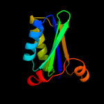
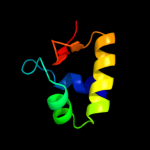
1 c3i6pF_
100.0
46
PDB header: structural proteinChain: F: PDB Molecule: ethanolamine utilization protein eutm;PDBTitle: ethanolamine utilization microcompartment shell subunit, eutm
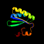
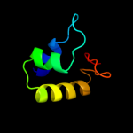
2 c3ngkA_
100.0
45
PDB header: unknown functionChain: A: PDB Molecule: propanediol utilization protein pdua;PDBTitle: pdua from salmonella enterica typhimurium
3 d2a10a1
100.0
29
Fold: Ferredoxin-likeSuperfamily: CcmK-likeFamily: CcmK-like4 d2a1ba1
100.0
34
Fold: Ferredoxin-likeSuperfamily: CcmK-likeFamily: CcmK-like5 d2ewha1
100.0
37
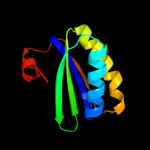
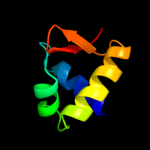
Fold: Ferredoxin-likeSuperfamily: CcmK-likeFamily: CcmK-like6 c3nwgA_
100.0
29
PDB header: structural proteinChain: A: PDB Molecule: microcompartments protein;PDBTitle: the crystal structure of a microcomparments protein from2 desulfitobacterium hafniense dcb
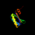
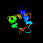
7 c3n79A_
100.0
28
PDB header: electron transportChain: A: PDB Molecule: pdut;PDBTitle: pdut c38s mutant from salmonella enterica typhimurium
8 c3i71B_
99.9
100
PDB header: unknown functionChain: B: PDB Molecule: ethanolamine utilization protein eutk;PDBTitle: ethanolamine utilization microcompartment shell subunit, eutk c-2 terminal domain
9 c3io0A_
99.8
22
PDB header: structural proteinChain: A: PDB Molecule: etub protein;PDBTitle: crystal structure of etub from clostridium kluyveri
10 c3i82A_
99.6
23
PDB header: structural proteinChain: A: PDB Molecule: ethanolamine utilization protein eutl;PDBTitle: ethanolamine utilization microcompartment shell subunit, eutl closed2 form
11 c3cgiD_
98.2
24
PDB header: unknown functionChain: D: PDB Molecule: propanediol utilization protein pduu;PDBTitle: crystal structure of the pduu shell protein from the pdu2 microcompartment
12 c3ia0c_
98.1
28
PDB header: structural proteinChain: C: PDB Molecule: ethanolamine utilization protein euts;PDBTitle: ethanolamine utilization microcompartment shell subunit,2 euts-g39v mutant
13 c3f56F_
96.2
23
PDB header: structural proteinChain: F: PDB Molecule: csos1d;PDBTitle: the structure of a previously undetected carboxysome shell2 protein: csos1d from prochlorococcus marinus med4
14 d1mkma1
83.9
25
Fold: DNA/RNA-binding 3-helical bundleSuperfamily: "Winged helix" DNA-binding domainFamily: Transcriptional regulator IclR, N-terminal domain15 c2g7uB_
82.7
24
PDB header: structural genomics, unknown functionChain: B: PDB Molecule: transcriptional regulator;PDBTitle: 2.3 a structure of putative catechol degradative operon regulator from2 rhodococcus sp. rha1
16 c3r4kD_
81.3
21
PDB header: dna binding proteinChain: D: PDB Molecule: transcriptional regulator, iclr family;PDBTitle: crystal structure of a putative iclr transcriptional regulator2 (tm1040_3717) from silicibacter sp. tm1040 at 2.46 a resolution
17 c2o0yB_
74.3
16
PDB header: transcriptionChain: B: PDB Molecule: transcriptional regulator;PDBTitle: crystal structure of putative transcriptional regulator rha1_ro069532 (iclr-family) from rhodococcus sp.
18 c2xroE_
64.9
19
PDB header: dna-binding protein/dnaChain: E: PDB Molecule: hth-type transcriptional regulator ttgv;PDBTitle: crystal structure of ttgv in complex with its dna operator
19 d1j5ya1
58.2
14
Fold: DNA/RNA-binding 3-helical bundleSuperfamily: "Winged helix" DNA-binding domainFamily: Biotin repressor-like20 c1mkmA_
56.7
21
PDB header: transcriptionChain: A: PDB Molecule: iclr transcriptional regulator;PDBTitle: crystal structure of the thermotoga maritima iclr
21 c3lstB_
not modelled
41.2
27
PDB header: transferaseChain: B: PDB Molecule: calo1 methyltransferase;PDBTitle: crystal structure of calo1, methyltransferase in calicheamicin2 biosynthesis, sah bound form
22 c2zmeA_
not modelled
37.2
35
PDB header: protein transportChain: A: PDB Molecule: vacuolar-sorting protein snf8;PDBTitle: integrated structural and functional model of the human escrt-ii2 complex
23 c3cuqA_
not modelled
37.0
35
PDB header: protein transportChain: A: PDB Molecule: vacuolar-sorting protein snf8;PDBTitle: integrated structural and functional model of the human escrt-ii2 complex
24 c3eqxB_
not modelled
36.0
18
PDB header: dna binding proteinChain: B: PDB Molecule: fic domain containing transcriptional regulator;PDBTitle: crystal structure of a fic family protein (so_4266) from shewanella2 oneidensis at 1.6 a resolution
25 c1f5tA_
not modelled
34.1
8
PDB header: transcription/dnaChain: A: PDB Molecule: diphtheria toxin repressor;PDBTitle: diphtheria tox repressor (c102d mutant) complexed with2 nickel and dtxr consensus binding sequence
26 c2ia2D_
not modelled
33.9
21
PDB header: transcriptionChain: D: PDB Molecule: putative transcriptional regulator;PDBTitle: the crystal structure of a putative transcriptional regulator rha061952 from rhodococcus sp. rha1
27 d1jhfa1
not modelled
32.6
26
Fold: DNA/RNA-binding 3-helical bundleSuperfamily: "Winged helix" DNA-binding domainFamily: LexA repressor, N-terminal DNA-binding domain28 c3mq0A_
not modelled
32.4
22
PDB header: transcription repressorChain: A: PDB Molecule: transcriptional repressor of the blcabc operon;PDBTitle: crystal structure of agobacterium tumefaciens repressor blcr
29 c1w7pD_
not modelled
29.5
16
PDB header: protein transportChain: D: PDB Molecule: vps36p, ylr417w;PDBTitle: the crystal structure of endosomal complex escrt-ii2 (vps22/vps25/vps36)
30 c2v9vA_
not modelled
26.9
24
PDB header: transcriptionChain: A: PDB Molecule: selenocysteine-specific elongation factor;PDBTitle: crystal structure of moorella thermoacetica selb(377-511)
31 c1u5tB_
not modelled
25.4
16
PDB header: transport proteinChain: B: PDB Molecule: defective in vacuolar protein sorting; vps36p;PDBTitle: structure of the escrt-ii endosomal trafficking complex
32 c2v79B_
not modelled
25.0
23
PDB header: dna-binding proteinChain: B: PDB Molecule: dna replication protein dnad;PDBTitle: crystal structure of the n-terminal domain of dnad from2 bacillus subtilis
33 c3cuoB_
not modelled
24.8
20
PDB header: transcription regulatorChain: B: PDB Molecule: uncharacterized hth-type transcriptional regulator ygav;PDBTitle: crystal structure of the predicted dna-binding transcriptional2 regulator from e. coli
34 c2vn2B_
not modelled
24.7
18
PDB header: replicationChain: B: PDB Molecule: chromosome replication initiation protein;PDBTitle: crystal structure of the n-terminal domain of dnad protein2 from geobacillus kaustophilus hta426
35 c2x4hA_
not modelled
24.7
9
PDB header: transcriptionChain: A: PDB Molecule: hypothetical protein sso2273;PDBTitle: crystal structure of the hypothetical protein sso2273 from2 sulfolobus solfataricus
36 c1u5tA_
not modelled
23.9
23
PDB header: transport proteinChain: A: PDB Molecule: appears to be functionally related to snf7;PDBTitle: structure of the escrt-ii endosomal trafficking complex
37 c2oqgA_
not modelled
21.6
22
PDB header: transcriptionChain: A: PDB Molecule: possible transcriptional regulator, arsr family protein;PDBTitle: arsr-like transcriptional regulator from rhodococcus sp. rha1
38 c2zmeB_
not modelled
21.6
26
PDB header: protein transportChain: B: PDB Molecule: vacuolar protein-sorting-associated protein 36;PDBTitle: integrated structural and functional model of the human escrt-ii2 complex
39 d2b0la1
not modelled
21.6
28
Fold: DNA/RNA-binding 3-helical bundleSuperfamily: "Winged helix" DNA-binding domainFamily: CodY HTH domain40 d1biaa1
not modelled
18.8
18
Fold: DNA/RNA-binding 3-helical bundleSuperfamily: "Winged helix" DNA-binding domainFamily: Biotin repressor-like41 c3f6vA_
not modelled
17.7
39
PDB header: transcription regulatorChain: A: PDB Molecule: possible transcriptional regulator, arsr familyPDBTitle: crystal structure of possible transcriptional regulator for2 arsenical resistance
42 c3f6oB_
not modelled
17.2
20
PDB header: transcription regulatorChain: B: PDB Molecule: probable transcriptional regulator, arsr familyPDBTitle: crystal structure of arsr family transcriptional regulator,2 rha00566
43 c3dp7B_
not modelled
16.8
28
PDB header: transferaseChain: B: PDB Molecule: sam-dependent methyltransferase;PDBTitle: crystal structure of sam-dependent methyltransferase from bacteroides2 vulgatus atcc 8482
44 c3mczB_
not modelled
16.4
37
PDB header: transferaseChain: B: PDB Molecule: o-methyltransferase;PDBTitle: the structure of an o-methyltransferase family protein from2 burkholderia thailandensis.
45 c1j5yA_
not modelled
16.0
15
PDB header: transcriptionChain: A: PDB Molecule: transcriptional regulator, biotin repressor family;PDBTitle: crystal structure of transcriptional regulator (tm1602) from2 thermotoga maritima at 2.3 a resolution
46 d1u2wa1
not modelled
15.8
22
Fold: DNA/RNA-binding 3-helical bundleSuperfamily: "Winged helix" DNA-binding domainFamily: ArsR-like transcriptional regulators47 d1tw3a1
not modelled
15.5
17
Fold: DNA/RNA-binding 3-helical bundleSuperfamily: "Winged helix" DNA-binding domainFamily: Plant O-methyltransferase, N-terminal domain48 d1u5ta2
not modelled
15.2
21
Fold: DNA/RNA-binding 3-helical bundleSuperfamily: "Winged helix" DNA-binding domainFamily: Vacuolar sorting protein domain49 c2h09A_
not modelled
14.5
19
PDB header: transcriptionChain: A: PDB Molecule: transcriptional regulator mntr;PDBTitle: crystal structure of diphtheria toxin repressor like protein2 from e. coli
50 d1wi9a_
not modelled
13.9
31
Fold: DNA/RNA-binding 3-helical bundleSuperfamily: "Winged helix" DNA-binding domainFamily: PCI domain (PINT motif)51 d1sfxa_
not modelled
12.9
23
Fold: DNA/RNA-binding 3-helical bundleSuperfamily: "Winged helix" DNA-binding domainFamily: TrmB-like52 c3hruA_
not modelled
11.6
19
PDB header: transcriptionChain: A: PDB Molecule: metalloregulator scar;PDBTitle: crystal structure of scar with bound zn2+
53 d2htja1
not modelled
10.9
27
Fold: DNA/RNA-binding 3-helical bundleSuperfamily: "Winged helix" DNA-binding domainFamily: FaeA-like54 c1r22B_
not modelled
10.8
25
PDB header: transcription repressorChain: B: PDB Molecule: transcriptional repressor smtb;PDBTitle: crystal structure of the cyanobacterial metallothionein2 repressor smtb (c14s/c61s/c121s mutant) in the zn2alpha5-3 form
55 c1x1aA_
not modelled
10.2
17
PDB header: transferaseChain: A: PDB Molecule: crtf-related protein;PDBTitle: crystal structure of bchu complexed with s-adenosyl-l-methionine
56 c2it0A_
not modelled
10.0
11
PDB header: transcription/dnaChain: A: PDB Molecule: iron-dependent repressor ider;PDBTitle: crystal structure of a two-domain ider-dna complex crystal2 form ii
57 c1fx7C_
not modelled
9.6
9
PDB header: signaling proteinChain: C: PDB Molecule: iron-dependent repressor ider;PDBTitle: crystal structure of the iron-dependent regulator (ider)2 from mycobacterium tuberculosis
58 d1ku9a_
not modelled
8.7
17
Fold: DNA/RNA-binding 3-helical bundleSuperfamily: "Winged helix" DNA-binding domainFamily: DNA-binding protein Mj22359 c3boqB_
not modelled
7.8
17
PDB header: transcription regulatorChain: B: PDB Molecule: transcriptional regulator, marr family;PDBTitle: crystal structure of marr family transcriptional regulator from2 silicibacter pomeroyi
60 c2eo4A_
not modelled
7.4
15
PDB header: hydrolaseChain: A: PDB Molecule: 150aa long hypothetical histidine triad nucleotide-bindingPDBTitle: crystal structure of hypothetical histidine triad nucleotide-binding2 protein st2152 from sulfolobus tokodaii strain7
61 c3r0aB_
not modelled
7.3
7
PDB header: transcription regulatorChain: B: PDB Molecule: putative transcriptional regulator;PDBTitle: possible transcriptional regulator from methanosarcina mazei go1 (gi2 21227196)
62 d2frha1
not modelled
6.7
11
Fold: DNA/RNA-binding 3-helical bundleSuperfamily: "Winged helix" DNA-binding domainFamily: MarR-like transcriptional regulators63 c3jthA_
not modelled
6.6
24
PDB header: transcriptionChain: A: PDB Molecule: transcription activator hlyu;PDBTitle: crystal structure of a transcriptional regulator hlyu from2 vibrio vulnificus cmcp6
64 c1dpuA_
not modelled
6.1
12
PDB header: dna binding proteinChain: A: PDB Molecule: replication protein a (rpa32) c-terminal domain;PDBTitle: solution structure of the c-terminal domain of human rpa322 complexed with ung2(73-88)
65 d1dpua_
not modelled
6.1
12
Fold: DNA/RNA-binding 3-helical bundleSuperfamily: "Winged helix" DNA-binding domainFamily: C-terminal domain of RPA3266 d2qn6b1
not modelled
6.1
15
Fold: Ferredoxin-likeSuperfamily: eIF-2-alpha, C-terminal domainFamily: eIF-2-alpha, C-terminal domain67 c3d5cX_
not modelled
6.1
16
PDB header: ribosomeChain: X: PDB Molecule: peptide chain release factor 1;PDBTitle: structural basis for translation termination on the 70s ribosome. this2 file contains the 30s subunit, release factor 1 (rf1), two trna, and3 mrna molecules of the second 70s ribosome. the entire crystal4 structure contains two 70s ribosomes as described in remark 400.
68 c3gwzB_
not modelled
5.9
20
PDB header: transferaseChain: B: PDB Molecule: mmcr;PDBTitle: structure of the mitomycin 7-o-methyltransferase mmcr
69 d1stza1
not modelled
5.7
16
Fold: DNA/RNA-binding 3-helical bundleSuperfamily: "Winged helix" DNA-binding domainFamily: Heat-inducible transcription repressor HrcA, N-terminal domain70 c3nzlA_
not modelled
5.7
43
PDB header: transcriptionChain: A: PDB Molecule: dna-binding protein satb1;PDBTitle: crystal structure of the n-terminal domain of dna-binding protein2 satb1 from homo sapiens, northeast structural genomics consortium3 target hr4435b
71 d1qzza1
not modelled
5.5
15
Fold: DNA/RNA-binding 3-helical bundleSuperfamily: "Winged helix" DNA-binding domainFamily: Plant O-methyltransferase, N-terminal domain72 c2ifoA_
not modelled
5.4
15
PDB header: virusChain: A: PDB Molecule: inovirus;PDBTitle: model-building studies of inovirus: genetic variations on a2 geometric theme
73 d2a61a1
not modelled
5.3
19
Fold: DNA/RNA-binding 3-helical bundleSuperfamily: "Winged helix" DNA-binding domainFamily: MarR-like transcriptional regulators