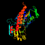
1 c2qa2A_
100.0
21
PDB header: oxidoreductaseChain: A: PDB Molecule: polyketide oxygenase cabe;PDBTitle: crystal structure of cabe, an aromatic hydroxylase from angucycline2 biosynthesis, determined to 2.7 a resolution
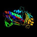
2 c3fmwC_
100.0
22
PDB header: oxidoreductaseChain: C: PDB Molecule: oxygenase;PDBTitle: the crystal structure of mtmoiv, a baeyer-villiger2 monooxygenase from the mithramycin biosynthetic pathway in3 streptomyces argillaceus.
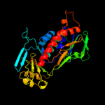
3 c2dkhA_
100.0
19
PDB header: oxidoreductaseChain: A: PDB Molecule: 3-hydroxybenzoate hydroxylase;PDBTitle: crystal structure of 3-hydroxybenzoate hydroxylase from comamonas2 testosteroni, in complex with the substrate
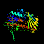
4 c1pn0A_
100.0
21
PDB header: oxidoreductaseChain: A: PDB Molecule: phenol 2-monooxygenase;PDBTitle: phenol hydroxylase from trichosporon cutaneum
5 c3ihgA_
100.0
20
PDB header: flavoprotein, oxidoreductaseChain: A: PDB Molecule: rdme;PDBTitle: crystal structure of a ternary complex of aklavinone-112 hydroxylase with fad and aklavinone
6 c1phhA_
100.0
16
PDB header: oxidoreductaseChain: A: PDB Molecule: p-hydroxybenzoate hydroxylase;PDBTitle: crystal structure of p-hydroxybenzoate hydroxylase complexed with its2 reaction product 3,4-dihydroxybenzoate
7 c3i3lA_
100.0
18
PDB header: hydrolaseChain: A: PDB Molecule: alkylhalidase cmls;PDBTitle: crystal structure of cmls, a flavin-dependent halogenase
8 c2r0gB_
100.0
21
PDB header: oxidoreductaseChain: B: PDB Molecule: rebc;PDBTitle: chromopyrrolic acid-soaked rebc with bound 7-carboxy-k252c
9 c3e1tA_
100.0
16
PDB header: flavoproteinChain: A: PDB Molecule: halogenase;PDBTitle: structure and action of the myxobacterial chondrochloren2 halogenase cndh, a new variant of fad-dependent halogenases
10 c3nixF_
100.0
14
PDB header: oxidoreductaseChain: F: PDB Molecule: flavoprotein/dehydrogenase;PDBTitle: crystal structure of flavoprotein/dehydrogenase from cytophaga2 hutchinsonii. northeast structural genomics consortium target chr43.
11 c2x3nA_
100.0
21
PDB header: oxidoreductaseChain: A: PDB Molecule: probable fad-dependent monooxygenase;PDBTitle: crystal structure of pqsl, a probable fad-dependent2 monooxygenase from pseudomonas aeruginosa
12 c3allA_
100.0
18
PDB header: oxidoreductaseChain: A: PDB Molecule: 2-methyl-3-hydroxypyridine-5-carboxylic acid oxygenase;PDBTitle: crystal structure of 2-methyl-3-hydroxypyridine-5-carboxylic acid2 oxygenase, mutant y270a
13 c3gmbB_
100.0
19
PDB header: oxidoreductaseChain: B: PDB Molecule: 2-methyl-3-hydroxypyridine-5-carboxylic acidPDBTitle: crystal structure of 2-methyl-3-hydroxypyridine-5-carboxylic2 acid oxygenase
14 c3c4aA_
100.0
16
PDB header: oxidoreductaseChain: A: PDB Molecule: probable tryptophan hydroxylase viod;PDBTitle: crystal structure of viod hydroxylase in complex with fad2 from chromobacterium violaceum. northeast structural3 genomics consortium target cvr158
15 c2rgjA_
100.0
17
PDB header: oxidoreductaseChain: A: PDB Molecule: flavin-containing monooxygenase;PDBTitle: crystal structure of flavin-containing monooxygenase phzs
16 c3atrA_
100.0
15
PDB header: oxidoreductaseChain: A: PDB Molecule: conserved archaeal protein;PDBTitle: geranylgeranyl reductase (ggr) from sulfolobus acidocaldarius co-2 crystallized with its ligand
17 d1k0ia1
100.0
23
Fold: FAD/NAD(P)-binding domainSuperfamily: FAD/NAD(P)-binding domainFamily: FAD-linked reductases, N-terminal domain18 c2xdoC_
100.0
18
PDB header: oxidoreductaseChain: C: PDB Molecule: tetx2 protein;PDBTitle: structure of the tetracycline degrading monooxygenase tetx2 from2 bacteroides thetaiotaomicron
19 c2vouA_
100.0
19
PDB header: oxidoreductaseChain: A: PDB Molecule: 2,6-dihydroxypyridine hydroxylase;PDBTitle: structure of 2,6-dihydroxypyridine-3-hydroxylase from2 arthrobacter nicotinovorans
20 c3cgvA_
100.0
15
PDB header: structural genomics, unknown functionChain: A: PDB Molecule: geranylgeranyl reductase related protein;PDBTitle: crystal structure of geranylgeranyl bacteriochlorophyll reductase-like2 fixc homolog (np_393992.1) from thermoplasma acidophilum at 1.60 a3 resolution
21 d1pn0a1
not modelled
100.0
18
Fold: FAD/NAD(P)-binding domainSuperfamily: FAD/NAD(P)-binding domainFamily: FAD-linked reductases, N-terminal domain22 c3ihmB_
not modelled
100.0
16
PDB header: oxidoreductaseChain: B: PDB Molecule: styrene monooxygenase a;PDBTitle: structure of the oxygenase component of a pseudomonas styrene2 monooxygenase
23 c2bryA_
not modelled
100.0
16
PDB header: transportChain: A: PDB Molecule: nedd9 interacting protein with calponin homologyPDBTitle: crystal structure of the native monooxygenase domain of2 mical at 1.45 a resolution
24 d3c96a1
not modelled
100.0
22
Fold: FAD/NAD(P)-binding domainSuperfamily: FAD/NAD(P)-binding domainFamily: FAD-linked reductases, N-terminal domain25 c2ardA_
not modelled
100.0
14
PDB header: biosynthetic proteinChain: A: PDB Molecule: tryptophan halogenase prna;PDBTitle: the structure of tryptophan 7-halogenase (prna) suggests a mechanism2 for regioselective chlorination
26 d2voua1
not modelled
100.0
24
Fold: FAD/NAD(P)-binding domainSuperfamily: FAD/NAD(P)-binding domainFamily: FAD-linked reductases, N-terminal domain27 c2weuD_
not modelled
100.0
15
PDB header: antifungal proteinChain: D: PDB Molecule: tryptophan 5-halogenase;PDBTitle: crystal structure of tryptophan 5-halogenase (pyrh) complex2 with substrate tryptophan
28 c2pyxA_
not modelled
100.0
17
PDB header: biosynthetic proteinChain: A: PDB Molecule: tryptophan halogenase;PDBTitle: crystal structure of tryptophan halogenase (yp_750003.1) from2 shewanella frigidimarina ncimb 400 at 1.50 a resolution
29 c2e4gB_
not modelled
100.0
15
PDB header: biosynthetic protein, flavoproteinChain: B: PDB Molecule: tryptophan halogenase;PDBTitle: rebh with bound l-trp
30 c2gmhA_
not modelled
100.0
14
PDB header: oxidoreductaseChain: A: PDB Molecule: electron transfer flavoprotein-ubiquinonePDBTitle: structure of porcine electron transfer flavoprotein-2 ubiquinone oxidoreductase in complexed with ubiquinone
31 d2gmha1
not modelled
99.9
14
Fold: FAD/NAD(P)-binding domainSuperfamily: FAD/NAD(P)-binding domainFamily: FAD-linked reductases, N-terminal domain32 c3nrnA_
not modelled
99.8
13
PDB header: structural genomics, unknown functionChain: A: PDB Molecule: uncharacterized protein pf1083;PDBTitle: crystal structure of pf1083 protein from pyrococcus furiosus,2 northeast structural genomics consortium target pfr223
33 c1yvvB_
not modelled
99.7
13
PDB header: oxidoreductaseChain: B: PDB Molecule: amine oxidase, flavin-containing;PDBTitle: x-ray structurure of p. syringae q888a4 oxidoreductase at2 resolution 2.5a. northeast structural genomics consortium3 target psr10.
34 c3da1A_
not modelled
99.7
13
PDB header: oxidoreductaseChain: A: PDB Molecule: glycerol-3-phosphate dehydrogenase;PDBTitle: x-ray structure of the glycerol-3-phosphate dehydrogenase2 from bacillus halodurans complexed with fad. northeast3 structural genomics consortium target bhr167.
35 c3qj4A_
not modelled
99.6
13
PDB header: oxidoreductaseChain: A: PDB Molecule: renalase;PDBTitle: crystal structure of human renalase (isoform 1)
36 c1y56B_
not modelled
99.6
11
PDB header: oxidoreductaseChain: B: PDB Molecule: sarcosine oxidase;PDBTitle: crystal structure of l-proline dehydrogenase from p.horikoshii
37 c3bhkA_
not modelled
99.6
12
PDB header: oxidoreductaseChain: A: PDB Molecule: monomeric sarcosine oxidase;PDBTitle: crystal structure of r49k mutant of monomeric sarcosine oxidase2 crystallized in phosphate as precipitant
38 c2r4jA_
not modelled
99.6
13
PDB header: oxidoreductaseChain: A: PDB Molecule: aerobic glycerol-3-phosphate dehydrogenase;PDBTitle: crystal structure of escherichia coli semet substituted2 glycerol-3-phosphate dehydrogenase in complex with dhap
39 c2olnA_
not modelled
99.6
16
PDB header: oxidoreductaseChain: A: PDB Molecule: nikd protein;PDBTitle: nikd, an unusual amino acid oxidase essential for2 nikkomycin biosynthesis: closed form at 1.15 a resolution
40 c3nyeA_
not modelled
99.6
14
PDB header: oxidoreductaseChain: A: PDB Molecule: d-arginine dehydrogenase;PDBTitle: crystal structure of pseudomonas aeruginosa d-arginine dehydrogenase2 in complex with imino-arginine
41 c2vvlD_
not modelled
99.5
12
PDB header: oxidoreductaseChain: D: PDB Molecule: monoamine oxidase n;PDBTitle: the structure of mao-n-d3, a variant of monoamine oxidase2 from aspergillus niger.
42 c1s3bB_
not modelled
99.5
16
PDB header: oxidoreductaseChain: B: PDB Molecule: amine oxidase [flavin-containing] b;PDBTitle: crystal structure of maob in complex with n-methyl-n-2 propargyl-1(r)-aminoindan
43 c1c0iA_
not modelled
99.5
13
PDB header: oxidoreductaseChain: A: PDB Molecule: d-amino acid oxidase;PDBTitle: crystal structure of d-amino acid oxidase in complex with2 two anthranylate molecules
44 c2zxiC_
not modelled
99.5
10
PDB header: fad-binding proteinChain: C: PDB Molecule: trna uridine 5-carboxymethylaminomethylPDBTitle: structure of aquifex aeolicus gida in the form ii crystal
45 c3ps9A_
not modelled
99.5
17
PDB header: transferaseChain: A: PDB Molecule: trna 5-methylaminomethyl-2-thiouridine biosynthesisPDBTitle: crystal structure of mnmc from e. coli
46 c3ka7A_
not modelled
99.5
15
PDB header: oxidoreductaseChain: A: PDB Molecule: oxidoreductase;PDBTitle: crystal structure of an oxidoreductase from methanosarcina2 mazei. northeast structural genomics consortium target id3 mar208
47 d1neka2
not modelled
99.5
12
Fold: FAD/NAD(P)-binding domainSuperfamily: FAD/NAD(P)-binding domainFamily: Succinate dehydrogenase/fumarate reductase flavoprotein N-terminal domain48 c2gahB_
not modelled
99.5
18
PDB header: oxidoreductaseChain: B: PDB Molecule: heterotetrameric sarcosine oxidase beta-subunit;PDBTitle: heterotetrameric sarcosine: structure of a diflavin2 metaloenzyme at 1.85 a resolution
49 c2ivdA_
not modelled
99.5
17
PDB header: oxidoreductaseChain: A: PDB Molecule: protoporphyrinogen oxidase;PDBTitle: structure of protoporphyrinogen oxidase from myxococcus2 xanthus with acifluorfen
50 c3f8rD_
not modelled
99.5
11
PDB header: oxidoreductaseChain: D: PDB Molecule: thioredoxin reductase (trxb-3);PDBTitle: crystal structure of sulfolobus solfataricus thioredoxin2 reductase b3 in complex with two nadp molecules
51 c1ryiB_
not modelled
99.5
11
PDB header: oxidoreductaseChain: B: PDB Molecule: glycine oxidase;PDBTitle: structure of glycine oxidase with bound inhibitor glycolate
52 c3dmeB_
not modelled
99.5
14
PDB header: structural genomics, unknown functionChain: B: PDB Molecule: conserved exported protein;PDBTitle: crystal structure of conserved exported protein from2 bordetella pertussis. northeast structural genomics target3 ber141
53 c3i6dA_
not modelled
99.4
13
PDB header: oxidoreductaseChain: A: PDB Molecule: protoporphyrinogen oxidase;PDBTitle: crystal structure of ppo from bacillus subtilis with af
54 c3ab1B_
not modelled
99.4
15
PDB header: oxidoreductaseChain: B: PDB Molecule: ferredoxin--nadp reductase;PDBTitle: crystal structure of ferredoxin nadp+ oxidoreductase
55 d2gqfa1
not modelled
99.4
19
Fold: FAD/NAD(P)-binding domainSuperfamily: FAD/NAD(P)-binding domainFamily: HI0933 N-terminal domain-like56 d1b5qa1
not modelled
99.4
10
Fold: FAD/NAD(P)-binding domainSuperfamily: FAD/NAD(P)-binding domainFamily: FAD-linked reductases, N-terminal domain57 c3k7tB_
not modelled
99.4
19
PDB header: oxidoreductaseChain: B: PDB Molecule: 6-hydroxy-l-nicotine oxidase;PDBTitle: crystal structure of apo-form 6-hydroxy-l-nicotine oxidase,2 crystal form p3121
58 d2bs2a2
not modelled
99.4
15
Fold: FAD/NAD(P)-binding domainSuperfamily: FAD/NAD(P)-binding domainFamily: Succinate dehydrogenase/fumarate reductase flavoprotein N-terminal domain59 c3pvcA_
not modelled
99.4
21
PDB header: oxidoreductase, transferaseChain: A: PDB Molecule: trna 5-methylaminomethyl-2-thiouridine biosynthesisPDBTitle: crystal structure of apo mnmc from yersinia pestis
60 c3rhaA_
not modelled
99.4
20
PDB header: oxidoreductaseChain: A: PDB Molecule: putrescine oxidase;PDBTitle: the crystal structure of oxidoreductase from arthrobacter aurescens
61 d2ivda1
not modelled
99.4
17
Fold: FAD/NAD(P)-binding domainSuperfamily: FAD/NAD(P)-binding domainFamily: FAD-linked reductases, N-terminal domain62 c3cesB_
not modelled
99.4
13
PDB header: rna binding proteinChain: B: PDB Molecule: trna uridine 5-carboxymethylaminomethyl modification enzymePDBTitle: crystal structure of e.coli mnmg (gida), a highly-conserved trna2 modifying enzyme
63 c2yg4B_
not modelled
99.4
21
PDB header: oxidoreductaseChain: B: PDB Molecule: putrescine oxidase;PDBTitle: structure-based redesign of cofactor binding in putrescine2 oxidase: wild type bound to putrescine
64 c3g05B_
not modelled
99.4
12
PDB header: rna binding proteinChain: B: PDB Molecule: trna uridine 5-carboxymethylaminomethyl modification enzymePDBTitle: crystal structure of n-terminal domain (2-550) of e.coli mnmg
65 d1jnra2
not modelled
99.3
15
Fold: FAD/NAD(P)-binding domainSuperfamily: FAD/NAD(P)-binding domainFamily: Succinate dehydrogenase/fumarate reductase flavoprotein N-terminal domain66 c1pj6A_
not modelled
99.3
14
PDB header: oxidoreductaseChain: A: PDB Molecule: n,n-dimethylglycine oxidase;PDBTitle: crystal structure of dimethylglycine oxidase of arthrobacter2 globiformis in complex with folic acid
67 c3cp8C_
not modelled
99.3
14
PDB header: oxidoreductaseChain: C: PDB Molecule: trna uridine 5-carboxymethylaminomethylPDBTitle: crystal structure of gida from chlorobium tepidum
68 c3djeA_
not modelled
99.3
12
PDB header: oxidoreductaseChain: A: PDB Molecule: fructosyl amine: oxygen oxidoreductase;PDBTitle: crystal structure of the deglycating enzyme fructosamine2 oxidase from aspergillus fumigatus (amadoriase ii) in3 complex with fsa
69 c2rghA_
not modelled
99.3
12
PDB header: oxidoreductaseChain: A: PDB Molecule: alpha-glycerophosphate oxidase;PDBTitle: structure of alpha-glycerophosphate oxidase from2 streptococcus sp.: a template for the mitochondrial alpha-3 glycerophosphate dehydrogenase
70 c2rgoA_
not modelled
99.3
12
PDB header: oxidoreductaseChain: A: PDB Molecule: alpha-glycerophosphate oxidase;PDBTitle: structure of alpha-glycerophosphate oxidase from2 streptococcus sp.: a template for the mitochondrial alpha-3 glycerophosphate dehydrogenase
71 d1kf6a2
not modelled
99.3
12
Fold: FAD/NAD(P)-binding domainSuperfamily: FAD/NAD(P)-binding domainFamily: Succinate dehydrogenase/fumarate reductase flavoprotein N-terminal domain72 d1reoa1
not modelled
99.3
13
Fold: FAD/NAD(P)-binding domainSuperfamily: FAD/NAD(P)-binding domainFamily: FAD-linked reductases, N-terminal domain73 d1qo8a2
not modelled
99.3
16
Fold: FAD/NAD(P)-binding domainSuperfamily: FAD/NAD(P)-binding domainFamily: Succinate dehydrogenase/fumarate reductase flavoprotein N-terminal domain74 d2i0za1
not modelled
99.2
14
Fold: FAD/NAD(P)-binding domainSuperfamily: FAD/NAD(P)-binding domainFamily: HI0933 N-terminal domain-like75 c3lovA_
not modelled
99.2
16
PDB header: oxidoreductaseChain: A: PDB Molecule: protoporphyrinogen oxidase;PDBTitle: crystal structure of putative protoporphyrinogen oxidase2 (yp_001813199.1) from exiguobacterium sp. 255-15 at 2.06 a resolution
76 c2q7vA_
not modelled
99.2
11
PDB header: oxidoreductaseChain: A: PDB Molecule: thioredoxin reductase;PDBTitle: crystal structure of deinococcus radiodurans thioredoxin2 reductase
77 c3jskN_
not modelled
99.2
18
PDB header: biosynthetic proteinChain: N: PDB Molecule: cypbp37 protein;PDBTitle: thiazole synthase from neurospora crassa
78 c2uzzD_
not modelled
99.2
12
PDB header: oxidoreductaseChain: D: PDB Molecule: n-methyl-l-tryptophan oxidase;PDBTitle: x-ray structure of n-methyl-l-tryptophan oxidase (mtox)
79 c1sezA_
not modelled
99.2
13
PDB header: oxidoreductaseChain: A: PDB Molecule: protoporphyrinogen oxidase, mitochondrial;PDBTitle: crystal structure of protoporphyrinogen ix oxidase
80 d1d4ca2
not modelled
99.2
18
Fold: FAD/NAD(P)-binding domainSuperfamily: FAD/NAD(P)-binding domainFamily: Succinate dehydrogenase/fumarate reductase flavoprotein N-terminal domain81 d1w4xa1
not modelled
99.2
23
Fold: FAD/NAD(P)-binding domainSuperfamily: FAD/NAD(P)-binding domainFamily: FAD/NAD-linked reductases, N-terminal and central domains82 c2a87A_
not modelled
99.2
11
PDB header: oxidoreductaseChain: A: PDB Molecule: thioredoxin reductase;PDBTitle: crystal structure of m. tuberculosis thioredoxin reductase
83 c2aczA_
not modelled
99.1
15
PDB header: oxidoreductase/electron transportChain: A: PDB Molecule: succinate dehydrogenase flavoprotein subunit;PDBTitle: complex ii (succinate dehydrogenase) from e. coli with atpenin a52 inhibitor co-crystallized at the ubiquinone binding site
84 d1ryia1
not modelled
99.1
14
Fold: FAD/NAD(P)-binding domainSuperfamily: FAD/NAD(P)-binding domainFamily: FAD-linked reductases, N-terminal domain85 c1yq4A_
not modelled
99.1
14
PDB header: oxidoreductaseChain: A: PDB Molecule: succinate dehydrogenase flavoprotein subunit;PDBTitle: avian respiratory complex ii with 3-nitropropionate and ubiquinone
86 c3h8lA_
not modelled
99.1
16
PDB header: oxidoreductaseChain: A: PDB Molecule: nadh oxidase;PDBTitle: the first x-ray structure of a sulfide:quinone2 oxidoreductase: insights into sulfide oxidation mechanism
87 c3fbsB_
not modelled
99.1
26
PDB header: oxidoreductaseChain: B: PDB Molecule: oxidoreductase;PDBTitle: the crystal structure of the oxidoreductase from agrobacterium2 tumefaciens
88 d2gjca1
not modelled
99.1
13
Fold: FAD/NAD(P)-binding domainSuperfamily: FAD/NAD(P)-binding domainFamily: Thi4-like89 d2cula1
not modelled
99.0
18
Fold: FAD/NAD(P)-binding domainSuperfamily: FAD/NAD(P)-binding domainFamily: GidA-like90 d2gf3a1
not modelled
99.0
15
Fold: FAD/NAD(P)-binding domainSuperfamily: FAD/NAD(P)-binding domainFamily: FAD-linked reductases, N-terminal domain91 c1bwcA_
not modelled
99.0
39
PDB header: oxidoreductaseChain: A: PDB Molecule: protein (glutathione reductase);PDBTitle: structure of human glutathione reductase complexed with ajoene2 inhibitor and subversive substrate
92 c2eq8E_
not modelled
99.0
28
PDB header: oxidoreductaseChain: E: PDB Molecule: pyruvate dehydrogenase complex, dihydrolipoamidePDBTitle: crystal structure of lipoamide dehydrogenase from thermus thermophilus2 hb8 with psbdp
93 d1rp0a1
not modelled
99.0
18
Fold: FAD/NAD(P)-binding domainSuperfamily: FAD/NAD(P)-binding domainFamily: Thi4-like94 d1pj5a2
not modelled
99.0
12
Fold: FAD/NAD(P)-binding domainSuperfamily: FAD/NAD(P)-binding domainFamily: FAD-linked reductases, N-terminal domain95 c3hdqI_
not modelled
99.0
34
PDB header: isomeraseChain: I: PDB Molecule: udp-galactopyranose mutase;PDBTitle: crystal structure of udp-galactopyranose mutase (oxidized2 form) in complex with substrate
96 d1d5ta1
not modelled
99.0
18
Fold: FAD/NAD(P)-binding domainSuperfamily: FAD/NAD(P)-binding domainFamily: GDI-like N domain97 c2bs3A_
not modelled
99.0
18
PDB header: oxidoreductaseChain: A: PDB Molecule: quinol-fumarate reductase flavoprotein subunit a;PDBTitle: glu c180 -> gln variant quinol:fumarate reductase from2 wolinella succinogenes
98 d1o5wa1
not modelled
99.0
30
Fold: FAD/NAD(P)-binding domainSuperfamily: FAD/NAD(P)-binding domainFamily: FAD-linked reductases, N-terminal domain99 d3grsa1
not modelled
99.0
39
Fold: FAD/NAD(P)-binding domainSuperfamily: FAD/NAD(P)-binding domainFamily: FAD/NAD-linked reductases, N-terminal and central domains100 d2bcgg1
not modelled
99.0
18
Fold: FAD/NAD(P)-binding domainSuperfamily: FAD/NAD(P)-binding domainFamily: GDI-like N domain101 d1c0pa1
not modelled
98.9
18
Fold: Nucleotide-binding domainSuperfamily: Nucleotide-binding domainFamily: D-aminoacid oxidase, N-terminal domain102 c1ltxR_
not modelled
98.9
23
PDB header: transferase/protein bindingChain: R: PDB Molecule: rab escort protein 1;PDBTitle: structure of rab escort protein-1 in complex with rab2 geranylgeranyl transferase and isoprenoid
103 d2iida1
not modelled
98.9
29
Fold: FAD/NAD(P)-binding domainSuperfamily: FAD/NAD(P)-binding domainFamily: FAD-linked reductases, N-terminal domain104 c3ctyA_
not modelled
98.9
40
PDB header: oxidoreductaseChain: A: PDB Molecule: thioredoxin reductase;PDBTitle: crystal structure of t. acidophilum thioredoxin reductase
105 c1qo8A_
not modelled
98.9
17
PDB header: oxidoreductaseChain: A: PDB Molecule: flavocytochrome c3 fumarate reductase;PDBTitle: the structure of the open conformation of a flavocytochrome2 c3 fumarate reductase
106 c1jrxA_
not modelled
98.9
21
PDB header: oxidoreductaseChain: A: PDB Molecule: flavocytochrome c;PDBTitle: crystal structure of arg402ala mutant flavocytochrome c32 from shewanella frigidimarina
107 c1kifE_
not modelled
98.9
11
PDB header: flavoproteinChain: E: PDB Molecule: d-amino acid oxidase;PDBTitle: d-amino acid oxidase from pig kidney
108 c3p4rM_
not modelled
98.9
16
PDB header: oxidoreductaseChain: M: PDB Molecule: fumarate reductase flavoprotein subunit;PDBTitle: crystal structure of menaquinol:fumarate oxidoreductase in complex2 with glutarate
109 c1w4xA_
not modelled
98.9
20
PDB header: oxygenaseChain: A: PDB Molecule: phenylacetone monooxygenase;PDBTitle: phenylacetone monooxygenase, a baeyer-villiger2 monooxygenase
110 c2i0zA_
not modelled
98.9
34
PDB header: oxidoreductaseChain: A: PDB Molecule: nad(fad)-utilizing dehydrogenases;PDBTitle: crystal structure of a fad binding protein from bacillus2 cereus, a putative nad(fad)-utilizing dehydrogenases
111 c2gqfA_
not modelled
98.9
29
PDB header: structural genomics, unknown functionChain: A: PDB Molecule: hypothetical protein hi0933;PDBTitle: crystal structure of flavoprotein hi0933 from haemophilus influenzae2 rd
112 c3gwdA_
not modelled
98.9
15
PDB header: oxidoreductaseChain: A: PDB Molecule: cyclohexanone monooxygenase;PDBTitle: closed crystal structure of cyclohexanone monooxygenase
113 c3lzxB_
not modelled
98.9
32
PDB header: oxidoreductaseChain: B: PDB Molecule: ferredoxin--nadp reductase 2;PDBTitle: crystal structure of ferredoxin-nadp+ oxidoreductase from bacillus2 subtilis (form ii)
114 d3lada1
not modelled
98.9
29
Fold: FAD/NAD(P)-binding domainSuperfamily: FAD/NAD(P)-binding domainFamily: FAD/NAD-linked reductases, N-terminal and central domains115 d1v59a1
not modelled
98.9
33
Fold: FAD/NAD(P)-binding domainSuperfamily: FAD/NAD(P)-binding domainFamily: FAD/NAD-linked reductases, N-terminal and central domains116 d2v5za1
not modelled
98.9
37
Fold: FAD/NAD(P)-binding domainSuperfamily: FAD/NAD(P)-binding domainFamily: FAD-linked reductases, N-terminal domain117 c2zbwA_
not modelled
98.9
26
PDB header: oxidoreductaseChain: A: PDB Molecule: thioredoxin reductase;PDBTitle: crystal structure of thioredoxin reductase-like protein from thermus2 thermophilus hb8
118 c2hqmB_
not modelled
98.9
26
PDB header: oxidoreductaseChain: B: PDB Molecule: glutathione reductase;PDBTitle: crystal structure of glutathione reductase glr1 from the yeast2 saccharomyces cerevisiae
119 d1lpfa1
not modelled
98.9
28
Fold: FAD/NAD(P)-binding domainSuperfamily: FAD/NAD(P)-binding domainFamily: FAD/NAD-linked reductases, N-terminal and central domains120 c1v59B_
not modelled
98.9
33
PDB header: oxidoreductaseChain: B: PDB Molecule: dihydrolipoamide dehydrogenase;PDBTitle: crystal structure of yeast lipoamide dehydrogenase2 complexed with nad+