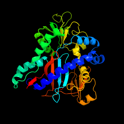
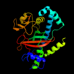
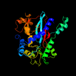
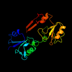
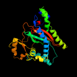
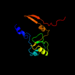
| 1 | c2ioaA_
|
|
 |
100.0 |
28 |
PDB header:ligase, hydrolase
Chain: A: PDB Molecule:bifunctional glutathionylspermidine
PDBTitle: e. coli bifunctional glutathionylspermidine2 synthetase/amidase incomplex with mg2+ and adp and3 phosphinate inhibitor
|
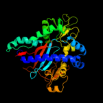
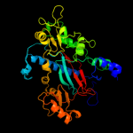
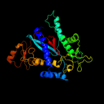
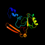
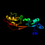
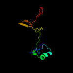
| 2 | c2vpmB_
|
|
 |
100.0 |
29 |
PDB header:ligase
Chain: B: PDB Molecule:trypanothione synthetase;
PDBTitle: trypanothione synthetase
|
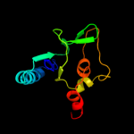
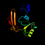
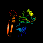
| 3 | d2io8a3
|
|
 |
100.0 |
33 |
Fold:ATP-grasp
Superfamily:Glutathione synthetase ATP-binding domain-like
Family:Glutathionylspermidine synthase ATP-binding domain-like |
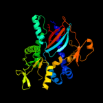
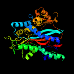
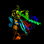
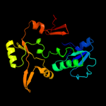
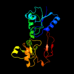
| 4 | c3n6xA_
|
|
 |
100.0 |
13 |
PDB header:ligase
Chain: A: PDB Molecule:putative glutathionylspermidine synthase;
PDBTitle: crystal structure of a putative glutathionylspermidine synthase2 (mfla_0391) from methylobacillus flagellatus kt at 2.35 a resolution
|
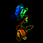
| 5 | d2io8a1
|
|
 |
99.8 |
20 |
Fold:PreATP-grasp domain
Superfamily:PreATP-grasp domain
Family:Glutathionylspermidine synthase substrate-binding domain-like |
| 6 | c2hgsA_
|
|
 |
99.5 |
15 |
PDB header:amine/carboxylate ligase
Chain: A: PDB Molecule:protein (glutathione synthetase);
PDBTitle: human glutathione synthetase
|
| 7 | c3kalB_
|
|
 |
99.5 |
17 |
PDB header:ligase
Chain: B: PDB Molecule:homoglutathione synthetase;
PDBTitle: structure of homoglutathione synthetase from glycine max in2 closed conformation with homoglutathione, adp, a sulfate3 ion, and three magnesium ions bound
|
| 8 | c1m0tB_
|
|
 |
99.3 |
16 |
PDB header:ligase
Chain: B: PDB Molecule:glutathione synthetase;
PDBTitle: yeast glutathione synthase
|
| 9 | c2wyoC_
|
|
 |
99.3 |
17 |
PDB header:ligase
Chain: C: PDB Molecule:glutathione synthetase;
PDBTitle: trypanosoma brucei glutathione synthetase
|
| 10 | c1gshA_
|
|
 |
99.1 |
14 |
PDB header:glutathione biosynthesis ligase
Chain: A: PDB Molecule:glutathione biosynthetic ligase;
PDBTitle: structure of escherichia coli glutathione synthetase at ph 7.5
|
| 11 | c2p0aA_
|
|
 |
98.5 |
13 |
PDB header:neuropeptide
Chain: A: PDB Molecule:synapsin-3;
PDBTitle: the crystal structure of human synapsin iii (syn3) in complex with2 amppnp
|
| 12 | c1i7nA_
|
|
 |
98.3 |
13 |
PDB header:neuropeptide
Chain: A: PDB Molecule:synapsin ii;
PDBTitle: crystal structure analysis of the c domain of synapsin ii2 from rat brain
|
| 13 | c1pk8D_
|
|
 |
98.2 |
13 |
PDB header:membrane protein
Chain: D: PDB Molecule:rat synapsin i;
PDBTitle: crystal structure of rat synapsin i c domain complexed to2 ca.atp
|
| 14 | c1uc8B_
|
|
 |
98.2 |
13 |
PDB header:biosynthetic protein
Chain: B: PDB Molecule:lysine biosynthesis enzyme;
PDBTitle: crystal structure of a lysine biosynthesis enzyme, lysx,2 from thermus thermophilus hb8
|
| 15 | d2hgsa4
|
|
 |
98.0 |
16 |
Fold:ATP-grasp
Superfamily:Glutathione synthetase ATP-binding domain-like
Family:Eukaryotic glutathione synthetase ATP-binding domain |
| 16 | c2qb5B_
|
|
 |
98.0 |
13 |
PDB header:transferase
Chain: B: PDB Molecule:inositol-tetrakisphosphate 1-kinase;
PDBTitle: crystal structure of human inositol 1,3,4-trisphosphate 5/6-kinase2 (itpk1) in complex with adp and mn2+
|
| 17 | d1pk8a2
|
|
 |
97.9 |
8 |
Fold:ATP-grasp
Superfamily:Glutathione synthetase ATP-binding domain-like
Family:Synapsin C-terminal domain |
| 18 | c2dlnA_
|
|
 |
97.6 |
12 |
PDB header:ligase(peptidoglycan synthesis)
Chain: A: PDB Molecule:d-alanine--d-alanine ligase;
PDBTitle: vancomycin resistance: structure of d-alanine:d-alanine2 ligase at 2.3 angstroms resolution
|
| 19 | c3ln6A_
|
|
 |
97.4 |
20 |
PDB header:ligase
Chain: A: PDB Molecule:glutathione biosynthesis bifunctional protein gshab;
PDBTitle: crystal structure of a bifunctional glutathione synthetase from2 streptococcus agalactiae
|
| 20 | c3ln7A_
|
|
 |
97.4 |
17 |
PDB header:ligase
Chain: A: PDB Molecule:glutathione biosynthesis bifunctional protein gshab;
PDBTitle: crystal structure of a bifunctional glutathione synthetase from2 pasteurella multocida
|
| 21 | d1i7na2 |
|
not modelled |
97.3 |
10 |
Fold:ATP-grasp
Superfamily:Glutathione synthetase ATP-binding domain-like
Family:Synapsin C-terminal domain |
| 22 | c3t9aA_ |
|
not modelled |
97.2 |
16 |
PDB header:transferase
Chain: A: PDB Molecule:inositol pyrophosphate kinase;
PDBTitle: crystal structure of the catalytic domain of human diphosphoinositol2 pentakisphosphate kinase 2 (ppip5k2) in complex with amppnp at ph 7.0
|
| 23 | c1kjjA_ |
|
not modelled |
97.1 |
14 |
PDB header:transferase
Chain: A: PDB Molecule:phosphoribosylglycinamide formyltransferase 2;
PDBTitle: crystal structure of glycniamide ribonucleotide2 transformylase in complex with mg-atp-gamma-s
|
| 24 | c2yyaB_ |
|
not modelled |
97.1 |
14 |
PDB header:ligase
Chain: B: PDB Molecule:phosphoribosylamine--glycine ligase;
PDBTitle: crystal structure of gar synthetase from aquifex aeolicus
|
| 25 | c2xd4A_ |
|
not modelled |
97.0 |
13 |
PDB header:ligase
Chain: A: PDB Molecule:phosphoribosylamine--glycine ligase;
PDBTitle: nucleotide-bound structures of bacillus subtilis glycinamide2 ribonucleotide synthetase
|
| 26 | c3tqtB_ |
|
not modelled |
97.0 |
13 |
PDB header:ligase
Chain: B: PDB Molecule:d-alanine--d-alanine ligase;
PDBTitle: structure of the d-alanine-d-alanine ligase from coxiella burnetii
|
| 27 | c3i12A_ |
|
not modelled |
97.0 |
13 |
PDB header:ligase
Chain: A: PDB Molecule:d-alanine-d-alanine ligase a;
PDBTitle: the crystal structure of the d-alanyl-alanine synthetase a from2 salmonella enterica subsp. enterica serovar typhimurium str. lt2
|
| 28 | d1gsaa2 |
|
not modelled |
96.8 |
18 |
Fold:ATP-grasp
Superfamily:Glutathione synthetase ATP-binding domain-like
Family:ATP-binding domain of peptide synthetases |
| 29 | d1m0wa2 |
|
not modelled |
96.7 |
17 |
Fold:ATP-grasp
Superfamily:Glutathione synthetase ATP-binding domain-like
Family:Eukaryotic glutathione synthetase ATP-binding domain |
| 30 | c1e4eB_ |
|
not modelled |
96.7 |
9 |
PDB header:ligase
Chain: B: PDB Molecule:vancomycin/teicoplanin a-type resistance protein vana;
PDBTitle: d-alanyl-d-lacate ligase
|
| 31 | c2pvpB_ |
|
not modelled |
96.7 |
12 |
PDB header:ligase
Chain: B: PDB Molecule:d-alanine-d-alanine ligase;
PDBTitle: crystal structure of d-alanine-d-alanine ligase from helicobacter2 pylori
|
| 32 | c3lp8A_ |
|
not modelled |
96.4 |
17 |
PDB header:ligase
Chain: A: PDB Molecule:phosphoribosylamine-glycine ligase;
PDBTitle: crystal structure of phosphoribosylamine-glycine ligase from2 ehrlichia chaffeensis
|
| 33 | c3lwbA_ |
|
not modelled |
96.3 |
13 |
PDB header:ligase
Chain: A: PDB Molecule:d-alanine--d-alanine ligase;
PDBTitle: crystal structure of apo d-alanine:d-alanine ligase (ddl) from2 mycobacterium tuberculosis
|
| 34 | c3bg5C_ |
|
not modelled |
96.1 |
16 |
PDB header:ligase
Chain: C: PDB Molecule:pyruvate carboxylase;
PDBTitle: crystal structure of staphylococcus aureus pyruvate2 carboxylase
|
| 35 | c2hjwA_ |
|
not modelled |
95.9 |
14 |
PDB header:ligase
Chain: A: PDB Molecule:acetyl-coa carboxylase 2;
PDBTitle: crystal structure of the bc domain of acc2
|
| 36 | d1gsaa1 |
|
not modelled |
95.8 |
9 |
Fold:PreATP-grasp domain
Superfamily:PreATP-grasp domain
Family:Prokaryotic glutathione synthetase, N-terminal domain |
| 37 | c2ip4A_ |
|
not modelled |
95.8 |
19 |
PDB header:ligase
Chain: A: PDB Molecule:phosphoribosylamine--glycine ligase;
PDBTitle: crystal structure of glycinamide ribonucleotide synthetase from2 thermus thermophilus hb8
|
| 38 | c2zdqA_ |
|
not modelled |
95.7 |
18 |
PDB header:ligase
Chain: A: PDB Molecule:d-alanine--d-alanine ligase;
PDBTitle: crystal structure of d-alanine:d-alanine ligase with atp2 and d-alanine:d-alanine from thermus thermophius hb8
|
| 39 | c1vkzA_ |
|
not modelled |
95.5 |
14 |
PDB header:ligase
Chain: A: PDB Molecule:phosphoribosylamine--glycine ligase;
PDBTitle: crystal structure of phosphoribosylamine--glycine ligase (tm1250) from2 thermotoga maritima at 2.30 a resolution
|
| 40 | c1z2pX_ |
|
not modelled |
95.5 |
15 |
PDB header:transferase
Chain: X: PDB Molecule:inositol 1,3,4-trisphosphate 5/6-kinase;
PDBTitle: inositol 1,3,4-trisphosphate 5/6-kinase in complex with mg2+/amp-2 pcp/ins(1,3,4)p3
|
| 41 | c3tinA_ |
|
not modelled |
95.4 |
15 |
PDB header:ligase
Chain: A: PDB Molecule:ttl protein;
PDBTitle: tubulin tyrosine ligase
|
| 42 | c2dzdB_ |
|
not modelled |
95.2 |
11 |
PDB header:ligase
Chain: B: PDB Molecule:pyruvate carboxylase;
PDBTitle: crystal structure of the biotin carboxylase domain of2 pyruvate carboxylase
|
| 43 | c1ehiB_ |
|
not modelled |
95.2 |
9 |
PDB header:ligase
Chain: B: PDB Molecule:d-alanine:d-lactate ligase;
PDBTitle: d-alanine:d-lactate ligase (lmddl2) of vancomycin-resistant2 leuconostoc mesenteroides
|
| 44 | c1w96B_ |
|
not modelled |
95.1 |
11 |
PDB header:ligase
Chain: B: PDB Molecule:acetyl-coenzyme a carboxylase;
PDBTitle: crystal structure of biotin carboxylase domain of acetyl-2 coenzyme a carboxylase from saccharomyces cerevisiae in3 complex with soraphen a
|
| 45 | c3g8cB_ |
|
not modelled |
95.0 |
11 |
PDB header:ligase
Chain: B: PDB Molecule:biotin carboxylase;
PDBTitle: crystal stucture of biotin carboxylase in complex with2 biotin, bicarbonate, adp and mg ion
|
| 46 | c3ouzA_ |
|
not modelled |
94.9 |
15 |
PDB header:ligase
Chain: A: PDB Molecule:biotin carboxylase;
PDBTitle: crystal structure of biotin carboxylase-adp complex from campylobacter2 jejuni
|
| 47 | c2cqyA_ |
|
not modelled |
94.8 |
13 |
PDB header:ligase
Chain: A: PDB Molecule:propionyl-coa carboxylase alpha chain,
PDBTitle: solution structure of b domain from human propionyl-coa2 carboxylase alpha subunit
|
| 48 | d1uc8a2 |
|
not modelled |
94.4 |
15 |
Fold:ATP-grasp
Superfamily:Glutathione synthetase ATP-binding domain-like
Family:Lysine biosynthesis enzyme LysX ATP-binding domain |
| 49 | c3df7A_ |
|
not modelled |
94.4 |
21 |
PDB header:structural genomics, unknown function
Chain: A: PDB Molecule:putative atp-grasp superfamily protein;
PDBTitle: crystal structure of a putative atp-grasp superfamily2 protein from archaeoglobus fulgidus
|
| 50 | c1ulzA_ |
|
not modelled |
94.3 |
18 |
PDB header:ligase
Chain: A: PDB Molecule:pyruvate carboxylase n-terminal domain;
PDBTitle: crystal structure of the biotin carboxylase subunit of pyruvate2 carboxylase
|
| 51 | c2vpqA_ |
|
not modelled |
94.0 |
14 |
PDB header:ligase
Chain: A: PDB Molecule:acetyl-coa carboxylase;
PDBTitle: crystal structure of biotin carboxylase from s. aureus2 complexed with amppnp
|
| 52 | c3orqA_ |
|
not modelled |
93.9 |
9 |
PDB header:ligase,biosynthetic protein
Chain: A: PDB Molecule:n5-carboxyaminoimidazole ribonucleotide synthetase;
PDBTitle: crystal structure of n5-carboxyaminoimidazole synthetase from2 staphylococcus aureus complexed with adp
|
| 53 | c3gidB_ |
|
not modelled |
93.9 |
11 |
PDB header:ligase
Chain: B: PDB Molecule:acetyl-coa carboxylase 2;
PDBTitle: the biotin carboxylase (bc) domain of human acetyl-coa2 carboxylase 2 (acc2) in complex with soraphen a
|
| 54 | c3uvzB_ |
|
not modelled |
93.8 |
9 |
PDB header:lyase
Chain: B: PDB Molecule:phosphoribosylaminoimidazole carboxylase, atpase subunit;
PDBTitle: crystal structure of phosphoribosylaminoimidazole carboxylase, atpase2 subunit from burkholderia ambifaria
|
| 55 | c3k5iB_ |
|
not modelled |
93.7 |
9 |
PDB header:lyase
Chain: B: PDB Molecule:phosphoribosyl-aminoimidazole carboxylase;
PDBTitle: crystal structure of n5-carboxyaminoimidazole synthase from2 aspergillus clavatus in complex with adp and 5-3 aminoimadazole ribonucleotide
|
| 56 | d1w96a3 |
|
not modelled |
92.9 |
10 |
Fold:ATP-grasp
Superfamily:Glutathione synthetase ATP-binding domain-like
Family:BC ATP-binding domain-like |
| 57 | d1e4ea2 |
|
not modelled |
91.8 |
23 |
Fold:ATP-grasp
Superfamily:Glutathione synthetase ATP-binding domain-like
Family:ATP-binding domain of peptide synthetases |
| 58 | c2i80B_ |
|
not modelled |
91.1 |
24 |
PDB header:ligase
Chain: B: PDB Molecule:d-alanine-d-alanine ligase;
PDBTitle: allosteric inhibition of staphylococcus aureus d-alanine:d-alanine2 ligase revealed by crystallographic studies
|
| 59 | c3se7A_ |
|
not modelled |
90.9 |
11 |
PDB header:ligase
Chain: A: PDB Molecule:vana;
PDBTitle: ancient vana
|
| 60 | d1a9xa5 |
|
not modelled |
90.8 |
12 |
Fold:ATP-grasp
Superfamily:Glutathione synthetase ATP-binding domain-like
Family:BC ATP-binding domain-like |
| 61 | d1gsoa3 |
|
not modelled |
90.4 |
32 |
Fold:ATP-grasp
Superfamily:Glutathione synthetase ATP-binding domain-like
Family:BC ATP-binding domain-like |
| 62 | c2ys6A_ |
|
not modelled |
90.0 |
13 |
PDB header:ligase
Chain: A: PDB Molecule:phosphoribosylglycinamide synthetase;
PDBTitle: crystal structure of gar synthetase from geobacillus kaustophilus
|
| 63 | d2r85a2 |
|
not modelled |
89.4 |
10 |
Fold:ATP-grasp
Superfamily:Glutathione synthetase ATP-binding domain-like
Family:PurP ATP-binding domain-like |
| 64 | d1ehia2 |
|
not modelled |
88.8 |
33 |
Fold:ATP-grasp
Superfamily:Glutathione synthetase ATP-binding domain-like
Family:ATP-binding domain of peptide synthetases |
| 65 | c3e5nA_ |
|
not modelled |
88.8 |
23 |
PDB header:ligase
Chain: A: PDB Molecule:d-alanine-d-alanine ligase a;
PDBTitle: crystal strucutre of d-alanine-d-alanine ligase from2 xanthomonas oryzae pv. oryzae kacc10331
|
| 66 | d2r7ka2 |
|
not modelled |
88.7 |
15 |
Fold:ATP-grasp
Superfamily:Glutathione synthetase ATP-binding domain-like
Family:PurP ATP-binding domain-like |
| 67 | d1vkza3 |
|
not modelled |
87.8 |
14 |
Fold:ATP-grasp
Superfamily:Glutathione synthetase ATP-binding domain-like
Family:BC ATP-binding domain-like |
| 68 | d1iowa2 |
|
not modelled |
87.7 |
36 |
Fold:ATP-grasp
Superfamily:Glutathione synthetase ATP-binding domain-like
Family:ATP-binding domain of peptide synthetases |
| 69 | c3k3pA_ |
|
not modelled |
86.8 |
33 |
PDB header:ligase
Chain: A: PDB Molecule:d-alanine--d-alanine ligase;
PDBTitle: crystal structure of the apo form of d-alanine:d-alanine ligase (ddl)2 from streptococcus mutans
|
| 70 | c1m6vE_ |
|
not modelled |
85.5 |
14 |
PDB header:ligase
Chain: E: PDB Molecule:carbamoyl phosphate synthetase large chain;
PDBTitle: crystal structure of the g359f (small subunit) point mutant of2 carbamoyl phosphate synthetase
|
| 71 | c3q2oB_ |
|
not modelled |
83.7 |
10 |
PDB header:lyase
Chain: B: PDB Molecule:phosphoribosylaminoimidazole carboxylase, atpase subunit;
PDBTitle: crystal structure of purk: n5-carboxyaminoimidazole ribonucleotide2 synthetase
|
| 72 | c3r23B_ |
|
not modelled |
83.4 |
9 |
PDB header:ligase
Chain: B: PDB Molecule:d-alanine--d-alanine ligase;
PDBTitle: crystal structure of d-alanine--d-alanine ligase from bacillus2 anthracis
|
| 73 | d1kjqa3 |
|
not modelled |
79.0 |
15 |
Fold:ATP-grasp
Superfamily:Glutathione synthetase ATP-binding domain-like
Family:BC ATP-binding domain-like |
| 74 | c1gsoA_ |
|
not modelled |
75.7 |
32 |
PDB header:ligase
Chain: A: PDB Molecule:protein (glycinamide ribonucleotide synthetase);
PDBTitle: glycinamide ribonucleotide synthetase (gar-syn) from e.2 coli.
|
| 75 | c2z04A_ |
|
not modelled |
75.5 |
8 |
PDB header:lyase
Chain: A: PDB Molecule:phosphoribosylaminoimidazole carboxylase atpase
PDBTitle: crystal structure of phosphoribosylaminoimidazole2 carboxylase atpase subunit from aquifex aeolicus
|
| 76 | c3u9sE_ |
|
not modelled |
74.4 |
18 |
PDB header:ligase
Chain: E: PDB Molecule:methylcrotonyl-coa carboxylase, alpha-subunit;
PDBTitle: crystal structure of p. aeruginosa 3-methylcrotonyl-coa carboxylase2 (mcc) 750 kd holoenzyme, coa complex
|
| 77 | d3etja3 |
|
not modelled |
73.9 |
7 |
Fold:ATP-grasp
Superfamily:Glutathione synthetase ATP-binding domain-like
Family:BC ATP-binding domain-like |
| 78 | d1ulza3 |
|
not modelled |
72.6 |
10 |
Fold:ATP-grasp
Superfamily:Glutathione synthetase ATP-binding domain-like
Family:BC ATP-binding domain-like |
| 79 | c2r85B_ |
|
not modelled |
72.0 |
15 |
PDB header:unknown function
Chain: B: PDB Molecule:purp protein pf1517;
PDBTitle: crystal structure of purp from pyrococcus furiosus complexed with amp
|
| 80 | c3n6rK_ |
|
not modelled |
70.9 |
23 |
PDB header:ligase
Chain: K: PDB Molecule:propionyl-coa carboxylase, alpha subunit;
PDBTitle: crystal structure of the holoenzyme of propionyl-coa carboxylase (pcc)
|
| 81 | c2pn1A_ |
|
not modelled |
70.1 |
13 |
PDB header:ligase
Chain: A: PDB Molecule:carbamoylphosphate synthase large subunit;
PDBTitle: crystal structure of carbamoylphosphate synthase large subunit (split2 gene in mj) (zp_00538348.1) from exiguobacterium sp. 255-15 at 2.00 a3 resolution
|
| 82 | c2dwcB_ |
|
not modelled |
67.0 |
8 |
PDB header:transferase
Chain: B: PDB Molecule:433aa long hypothetical phosphoribosylglycinamide formyl
PDBTitle: crystal structure of probable phosphoribosylglycinamide formyl2 transferase from pyrococcus horikoshii ot3 complexed with adp
|
| 83 | c2qk4A_ |
|
not modelled |
63.0 |
12 |
PDB header:ligase
Chain: A: PDB Molecule:trifunctional purine biosynthetic protein adenosine-3;
PDBTitle: human glycinamide ribonucleotide synthetase
|
| 84 | c3etjB_ |
|
not modelled |
59.0 |
12 |
PDB header:lyase
Chain: B: PDB Molecule:phosphoribosylaminoimidazole carboxylase atpase
PDBTitle: crystal structure e. coli purk in complex with mg, adp, and2 pi
|
| 85 | c2gpwC_ |
|
not modelled |
56.7 |
14 |
PDB header:ligase
Chain: C: PDB Molecule:biotin carboxylase;
PDBTitle: crystal structure of the biotin carboxylase subunit, f363a2 mutant, of acetyl-coa carboxylase from escherichia coli.
|
| 86 | d2j9ga3 |
|
not modelled |
55.5 |
9 |
Fold:ATP-grasp
Superfamily:Glutathione synthetase ATP-binding domain-like
Family:BC ATP-binding domain-like |
| 87 | d1a9xa6 |
|
not modelled |
47.5 |
15 |
Fold:ATP-grasp
Superfamily:Glutathione synthetase ATP-binding domain-like
Family:BC ATP-binding domain-like |
| 88 | c3k1tA_ |
|
not modelled |
44.2 |
15 |
PDB header:ligase
Chain: A: PDB Molecule:glutamate--cysteine ligase gsha;
PDBTitle: crystal structure of putative gamma-glutamylcysteine synthetase2 (yp_546622.1) from methylobacillus flagellatus kt at 1.90 a3 resolution
|
| 89 | c3l4eA_ |
|
not modelled |
27.9 |
15 |
PDB header:hydrolase
Chain: A: PDB Molecule:uncharacterized peptidase lmo0363;
PDBTitle: 1.5a crystal structure of a putative peptidase e protein from listeria2 monocytogenes egd-e
|
| 90 | c3oetF_ |
|
not modelled |
23.7 |
25 |
PDB header:oxidoreductase
Chain: F: PDB Molecule:erythronate-4-phosphate dehydrogenase;
PDBTitle: d-erythronate-4-phosphate dehydrogenase complexed with nad
|
| 91 | c3d3kD_ |
|
not modelled |
22.1 |
7 |
PDB header:protein binding
Chain: D: PDB Molecule:enhancer of mrna-decapping protein 3;
PDBTitle: crystal structure of human edc3p
|
| 92 | d1v65a_ |
|
not modelled |
20.5 |
14 |
Fold:KRAB domain (Kruppel-associated box)
Superfamily:KRAB domain (Kruppel-associated box)
Family:KRAB domain (Kruppel-associated box) |
| 93 | c2o4cB_ |
|
not modelled |
20.3 |
19 |
PDB header:oxidoreductase
Chain: B: PDB Molecule:erythronate-4-phosphate dehydrogenase;
PDBTitle: crystal structure of d-erythronate-4-phosphate dehydrogenase complexed2 with nad
|
| 94 | d2q22a1 |
|
not modelled |
18.1 |
11 |
Fold:Ava3019-like
Superfamily:Ava3019-like
Family:Ava3019-like |
| 95 | c3e90C_ |
|
not modelled |
17.6 |
33 |
PDB header:hydrolase
Chain: C: PDB Molecule:ns2b cofactor;
PDBTitle: west nile vi rus ns2b-ns3protease in complexed with2 inhibitor naph-kkr-h
|
| 96 | d2oo3a1 |
|
not modelled |
17.4 |
21 |
Fold:S-adenosyl-L-methionine-dependent methyltransferases
Superfamily:S-adenosyl-L-methionine-dependent methyltransferases
Family:LPG1296-like |
| 97 | c2ijoA_ |
|
not modelled |
16.4 |
33 |
PDB header:hydrolase/hydrolase inhibitor
Chain: A: PDB Molecule:polyprotein;
PDBTitle: crystal structure of the west nile virus ns2b-ns3 protease2 complexed with bovine pancreatic trypsin inhibitor
|
| 98 | c1f13A_ |
|
not modelled |
15.5 |
14 |
PDB header:coagulation factor
Chain: A: PDB Molecule:cellular coagulation factor xiii zymogen;
PDBTitle: recombinant human cellular coagulation factor xiii
|
| 99 | c2r7mA_ |
|
not modelled |
14.0 |
12 |
PDB header:ligase
Chain: A: PDB Molecule:5-formaminoimidazole-4-carboxamide-1-(beta)-d-
PDBTitle: crystal structure of faicar synthetase (purp) from m.2 jannaschii complexed with amp
|