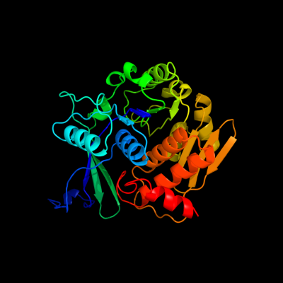
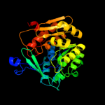
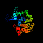
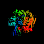
1 c3in1A_
100.0
98
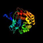
PDB header: transferaseChain: A: PDB Molecule: uncharacterized sugar kinase ydjh;PDBTitle: crystal structure of a putative ribokinase in complex with2 adp from e.coli
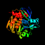
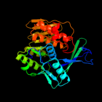
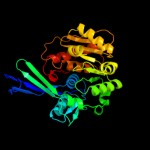
2 c3iq0B_
100.0
20
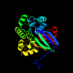
PDB header: transferaseChain: B: PDB Molecule: putative ribokinase ii;PDBTitle: crystal structure of a putative ribokinase ii in complex2 with atp and mg+2 from e.coli
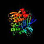
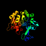
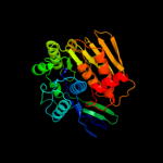
3 c3pl2D_
100.0
23
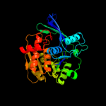
PDB header: transferaseChain: D: PDB Molecule: sugar kinase, ribokinase family;PDBTitle: crystal structure of a 5-keto-2-deoxygluconokinase (ncgl0155, cgl0158)2 from corynebacterium glutamicum atcc 13032 kitasato at 1.89 a3 resolution
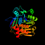
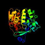
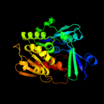
4 c2pkkA_
100.0
19
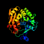
PDB header: transferaseChain: A: PDB Molecule: adenosine kinase;PDBTitle: crystal structure of m tuberculosis adenosine kinase complexed with 2-2 fluro adenosine
5 c2rbcA_
100.0
20
PDB header: transferaseChain: A: PDB Molecule: sugar kinase;PDBTitle: crystal structure of a putative ribokinase from agrobacterium2 tumefaciens
6 c2qcvA_
100.0
22
PDB header: transferaseChain: A: PDB Molecule: putative 5-dehydro-2-deoxygluconokinase;PDBTitle: crystal structure of a putative 5-dehydro-2-deoxygluconokinase (iolc)2 from bacillus halodurans c-125 at 1.90 a resolution
7 c2varB_
100.0
20
PDB header: transferaseChain: B: PDB Molecule: fructokinase;PDBTitle: crystal structure of sulfolobus solfataricus 2-keto-3-2 deoxygluconate kinase complexed with 2-keto-3-3 deoxygluconate
8 d1rkda_
100.0
27
Fold: Ribokinase-likeSuperfamily: Ribokinase-likeFamily: Ribokinase-like9 c3b1qD_
100.0
19
PDB header: transferaseChain: D: PDB Molecule: ribokinase, putative;PDBTitle: structure of burkholderia thailandensis nucleoside kinase (bthnk) in2 complex with inosine
10 c2c49A_
100.0
21
PDB header: transferaseChain: A: PDB Molecule: sugar kinase mj0406;PDBTitle: crystal structure of methanocaldococcus jannaschii2 nucleoside kinase - an archaeal member of the ribokinase3 family
11 c3kzhA_
100.0
23
PDB header: transferaseChain: A: PDB Molecule: probable sugar kinase;PDBTitle: crystal structure of a putative sugar kinase from2 clostridium perfringens
12 d1v19a_
100.0
25
Fold: Ribokinase-likeSuperfamily: Ribokinase-likeFamily: Ribokinase-like13 c2nwhA_
100.0
25
PDB header: signaling protein,transferaseChain: A: PDB Molecule: carbohydrate kinase;PDBTitle: carbohydrate kinase from agrobacterium tumefaciens
14 d2dcna1
100.0
18
Fold: Ribokinase-likeSuperfamily: Ribokinase-likeFamily: Ribokinase-like15 d1vm7a_
100.0
28
Fold: Ribokinase-likeSuperfamily: Ribokinase-likeFamily: Ribokinase-like16 d2fv7a1
100.0
22
Fold: Ribokinase-likeSuperfamily: Ribokinase-likeFamily: Ribokinase-like17 c3go6B_
100.0
18
PDB header: transferaseChain: B: PDB Molecule: ribokinase rbsk;PDBTitle: crystal structure of m. tuberculosis ribokinase (rv2436) in2 complex with ribose and amp-pnp
18 d1bx4a_
100.0
19
Fold: Ribokinase-likeSuperfamily: Ribokinase-likeFamily: Ribokinase-like19 c3gbuD_
100.0
22
PDB header: transferaseChain: D: PDB Molecule: uncharacterized sugar kinase ph1459;PDBTitle: crystal structure of an uncharacterized sugar kinase ph1459 from2 pyrococcus horikoshii in complex with atp
20 c3lkiA_
100.0
18
PDB header: transferaseChain: A: PDB Molecule: fructokinase;PDBTitle: crystal structure of fructokinase with bound atp from2 xylella fastidiosa
21 d1tyya_
not modelled
100.0
22
Fold: Ribokinase-likeSuperfamily: Ribokinase-likeFamily: Ribokinase-like22 c2absA_
not modelled
100.0
24
PDB header: signaling protein,transferaseChain: A: PDB Molecule: adenosine kinase;PDBTitle: crystal structure of t. gondii adenosine kinase complexed2 with amp-pcp
23 d2absa1
not modelled
100.0
24
Fold: Ribokinase-likeSuperfamily: Ribokinase-likeFamily: Ribokinase-like24 c3ktnA_
not modelled
100.0
17
PDB header: transferaseChain: A: PDB Molecule: carbohydrate kinase, pfkb family;PDBTitle: crystal structure of a putative 2-keto-3-deoxygluconate2 kinase from enterococcus faecalis
25 d2afba1
not modelled
100.0
17
Fold: Ribokinase-likeSuperfamily: Ribokinase-likeFamily: Ribokinase-like26 c3i3yB_
not modelled
100.0
24
PDB header: transferaseChain: B: PDB Molecule: carbohydrate kinase;PDBTitle: crystal structure of ribokinase in complex with d-ribose from2 klebsiella pneumoniae
27 c3cqdB_
not modelled
100.0
17
PDB header: transferaseChain: B: PDB Molecule: 6-phosphofructokinase isozyme 2;PDBTitle: structure of the tetrameric inhibited form of2 phosphofructokinase-2 from escherichia coli
28 c3looC_
not modelled
100.0
20
PDB header: transferaseChain: C: PDB Molecule: anopheles gambiae adenosine kinase;PDBTitle: crystal structure of anopheles gambiae adenosine kinase in complex2 with p1,p4-di(adenosine-5) tetraphosphate
29 d2f02a1
not modelled
100.0
19
Fold: Ribokinase-likeSuperfamily: Ribokinase-likeFamily: Ribokinase-like30 d2abqa1
not modelled
100.0
22
Fold: Ribokinase-likeSuperfamily: Ribokinase-likeFamily: Ribokinase-like31 c2xtbA_
not modelled
100.0
21
PDB header: transferaseChain: A: PDB Molecule: adenosine kinase;PDBTitle: crystal structure of trypanosoma brucei rhodesiense2 adenosine kinase complexed with activator
32 c3kd6B_
not modelled
100.0
23
PDB header: transferaseChain: B: PDB Molecule: carbohydrate kinase, pfkb family;PDBTitle: crystal structure of nucleoside kinase from chlorobium tepidum in2 complex with amp
33 c2jg1C_
not modelled
100.0
18
PDB header: transferaseChain: C: PDB Molecule: tagatose-6-phosphate kinase;PDBTitle: structure of staphylococcus aureus d-tagatose-6-phosphate2 kinase with cofactor and substrate
34 c2jg5B_
not modelled
100.0
15
PDB header: transferaseChain: B: PDB Molecule: fructose 1-phosphate kinase;PDBTitle: crystal structure of a putative phosphofructokinase from2 staphylococcus aureus
35 c1tz6B_
not modelled
100.0
22
PDB header: transferaseChain: B: PDB Molecule: putative sugar kinase;PDBTitle: crystal structure of aminoimidazole riboside kinase from2 salmonella enterica complexed with aminoimidazole riboside3 and atp analog
36 c3b3lC_
not modelled
100.0
19
PDB header: transferaseChain: C: PDB Molecule: ketohexokinase;PDBTitle: crystal structures of alternatively-spliced isoforms of human2 ketohexokinase
37 c3lhxA_
not modelled
100.0
21
PDB header: transferaseChain: A: PDB Molecule: ketodeoxygluconokinase;PDBTitle: crystal structure of a ketodeoxygluconokinase (kdgk) from2 shigella flexneri
38 c2qhpA_
not modelled
100.0
18
PDB header: transferaseChain: A: PDB Molecule: fructokinase;PDBTitle: crystal structure of fructokinase (np_810670.1) from bacteroides2 thetaiotaomicron vpi-5482 at 1.80 a resolution
39 d2ajra1
not modelled
100.0
15
Fold: Ribokinase-likeSuperfamily: Ribokinase-likeFamily: Ribokinase-like40 c3bf5A_
not modelled
100.0
16
PDB header: transferaseChain: A: PDB Molecule: ribokinase related protein;PDBTitle: crystal structure of putative ribokinase (10640157) from thermoplasma2 acidophilum at 1.91 a resolution
41 c3julA_
not modelled
100.0
17
PDB header: transferaseChain: A: PDB Molecule: lin2199 protein;PDBTitle: crystal structure of listeria innocua d-tagatose-6-phosphate2 kinase bound with substrate
42 c3hj6B_
not modelled
100.0
28
PDB header: transferaseChain: B: PDB Molecule: fructokinase;PDBTitle: structure of halothermothrix orenii fructokinase (frk)
43 d1vk4a_
not modelled
100.0
17
Fold: Ribokinase-likeSuperfamily: Ribokinase-likeFamily: Ribokinase-like44 c2ddmA_
not modelled
99.8
19
PDB header: transferaseChain: A: PDB Molecule: pyridoxine kinase;PDBTitle: crystal structure of pyridoxal kinase from the escherichia2 coli pdxk gene at 2.1 a resolution
45 c3mbjA_
not modelled
99.7
15
PDB header: transferaseChain: A: PDB Molecule: putative phosphomethylpyrimidine kinase;PDBTitle: crystal structure of a putative phosphomethylpyrimidine kinase2 (bt_4458) from bacteroides thetaiotaomicron vpi-5482 at 2.10 a3 resolution (rhombohedral form)
46 c2i5bC_
not modelled
99.6
16
PDB header: transferaseChain: C: PDB Molecule: phosphomethylpyrimidine kinase;PDBTitle: the crystal structure of an adp complex of bacillus2 subtilis pyridoxal kinase provides evidence for the3 parralel emergence of enzyme activity during evolution
47 c3ibqA_
not modelled
99.5
20
PDB header: transferaseChain: A: PDB Molecule: pyridoxal kinase;PDBTitle: crystal structure of pyridoxal kinase from lactobacillus2 plantarum in complex with atp
48 d1vi9a_
not modelled
99.5
16
Fold: Ribokinase-likeSuperfamily: Ribokinase-likeFamily: PfkB-like kinase49 d1ub0a_
not modelled
99.5
19
Fold: Ribokinase-likeSuperfamily: Ribokinase-likeFamily: Thiamin biosynthesis kinases50 d1lhpa_
not modelled
99.5
19
Fold: Ribokinase-likeSuperfamily: Ribokinase-likeFamily: PfkB-like kinase51 d1jxha_
not modelled
99.3
17
Fold: Ribokinase-likeSuperfamily: Ribokinase-likeFamily: Thiamin biosynthesis kinases52 c3rm5B_
not modelled
99.3
17
PDB header: transferaseChain: B: PDB Molecule: hydroxymethylpyrimidine/phosphomethylpyrimidine kinasePDBTitle: structure of trifunctional thi20 from yeast
53 c3dzvB_
not modelled
98.9
15
PDB header: transferaseChain: B: PDB Molecule: 4-methyl-5-(beta-hydroxyethyl)thiazole kinase;PDBTitle: crystal structure of 4-methyl-5-(beta-hydroxyethyl)thiazole2 kinase (np_816404.1) from enterococcus faecalis v583 at3 2.57 a resolution
54 d1kyha_
not modelled
98.8
17
Fold: Ribokinase-likeSuperfamily: Ribokinase-likeFamily: YjeF C-terminal domain-like55 d1v8aa_
not modelled
98.6
16
Fold: Ribokinase-likeSuperfamily: Ribokinase-likeFamily: Thiamin biosynthesis kinases56 d1gc5a_
not modelled
98.6
16
Fold: Ribokinase-likeSuperfamily: Ribokinase-likeFamily: ADP-specific Phosphofructokinase/Glucokinase57 d1l2la_
not modelled
98.4
13
Fold: Ribokinase-likeSuperfamily: Ribokinase-likeFamily: ADP-specific Phosphofructokinase/Glucokinase58 d1ua4a_
not modelled
98.4
14
Fold: Ribokinase-likeSuperfamily: Ribokinase-likeFamily: ADP-specific Phosphofructokinase/Glucokinase59 d1ekqa_
not modelled
98.4
11
Fold: Ribokinase-likeSuperfamily: Ribokinase-likeFamily: Thiamin biosynthesis kinases60 c2r3bA_
not modelled
98.3
15
PDB header: transferaseChain: A: PDB Molecule: yjef-related protein;PDBTitle: crystal structure of a ribokinase-like superfamily protein (ef1790)2 from enterococcus faecalis v583 at 1.80 a resolution
61 d2ax3a1
not modelled
98.1
13
Fold: Ribokinase-likeSuperfamily: Ribokinase-likeFamily: YjeF C-terminal domain-like62 c3nm3D_
not modelled
98.0
12
PDB header: transferaseChain: D: PDB Molecule: thiamine biosynthetic bifunctional enzyme;PDBTitle: the crystal structure of candida glabrata thi6, a bifunctional enzyme2 involved in thiamin biosyhthesis of eukaryotes
63 d1u2xa_
not modelled
97.8
18
Fold: Ribokinase-likeSuperfamily: Ribokinase-likeFamily: ADP-specific Phosphofructokinase/Glucokinase64 c3drwA_
not modelled
97.8
16
PDB header: transferaseChain: A: PDB Molecule: adp-specific phosphofructokinase;PDBTitle: crystal structure of a phosphofructokinase from pyrococcus2 horikoshii ot3 with amp
65 c3k5wA_
not modelled
97.8
19
PDB header: transferaseChain: A: PDB Molecule: carbohydrate kinase;PDBTitle: crystal structure of a carbohydrate kinase (yjef family)from2 helicobacter pylori
66 c2ax3A_
not modelled
97.8
12
PDB header: transferaseChain: A: PDB Molecule: hypothetical protein tm0922;PDBTitle: crystal structure of a putative carbohydrate kinase (tm0922) from2 thermotoga maritima msb8 at 2.25 a resolution
67 c3bgkA_
not modelled
97.5
13
PDB header: unknown functionChain: A: PDB Molecule: putative uncharacterized protein;PDBTitle: the crystal structure of hypothetic protein smu.573 from2 streptococcus mutans
68 d3grsa1
not modelled
87.0
28
Fold: FAD/NAD(P)-binding domainSuperfamily: FAD/NAD(P)-binding domainFamily: FAD/NAD-linked reductases, N-terminal and central domains69 c2zxiC_
not modelled
86.0
38
PDB header: fad-binding proteinChain: C: PDB Molecule: trna uridine 5-carboxymethylaminomethylPDBTitle: structure of aquifex aeolicus gida in the form ii crystal
70 c2eq8E_
not modelled
85.9
34
PDB header: oxidoreductaseChain: E: PDB Molecule: pyruvate dehydrogenase complex, dihydrolipoamidePDBTitle: crystal structure of lipoamide dehydrogenase from thermus thermophilus2 hb8 with psbdp
71 c2xdoC_
not modelled
85.6
19
PDB header: oxidoreductaseChain: C: PDB Molecule: tetx2 protein;PDBTitle: structure of the tetracycline degrading monooxygenase tetx2 from2 bacteroides thetaiotaomicron
72 c1bwcA_
not modelled
82.9
28
PDB header: oxidoreductaseChain: A: PDB Molecule: protein (glutathione reductase);PDBTitle: structure of human glutathione reductase complexed with ajoene2 inhibitor and subversive substrate
73 c3fbsB_
not modelled
79.4
29
PDB header: oxidoreductaseChain: B: PDB Molecule: oxidoreductase;PDBTitle: the crystal structure of the oxidoreductase from agrobacterium2 tumefaciens
74 c2aczA_
not modelled
76.4
44
PDB header: oxidoreductase/electron transportChain: A: PDB Molecule: succinate dehydrogenase flavoprotein subunit;PDBTitle: complex ii (succinate dehydrogenase) from e. coli with atpenin a52 inhibitor co-crystallized at the ubiquinone binding site
75 c2i0zA_
not modelled
72.7
32
PDB header: oxidoreductaseChain: A: PDB Molecule: nad(fad)-utilizing dehydrogenases;PDBTitle: crystal structure of a fad binding protein from bacillus2 cereus, a putative nad(fad)-utilizing dehydrogenases
76 c3ctyA_
not modelled
72.2
35
PDB header: oxidoreductaseChain: A: PDB Molecule: thioredoxin reductase;PDBTitle: crystal structure of t. acidophilum thioredoxin reductase
77 c3d8xB_
not modelled
71.8
35
PDB header: oxidoreductaseChain: B: PDB Molecule: thioredoxin reductase 1;PDBTitle: crystal structure of saccharomyces cerevisiae nadph dependent2 thioredoxin reductase 1
78 d3lada1
not modelled
69.2
48
Fold: FAD/NAD(P)-binding domainSuperfamily: FAD/NAD(P)-binding domainFamily: FAD/NAD-linked reductases, N-terminal and central domains79 c3ic9D_
not modelled
68.6
28
PDB header: oxidoreductaseChain: D: PDB Molecule: dihydrolipoamide dehydrogenase;PDBTitle: the structure of dihydrolipoamide dehydrogenase from colwellia2 psychrerythraea 34h.
80 c3i3lA_
not modelled
68.3
25
PDB header: hydrolaseChain: A: PDB Molecule: alkylhalidase cmls;PDBTitle: crystal structure of cmls, a flavin-dependent halogenase
81 c3l8kB_
not modelled
67.6
32
PDB header: oxidoreductaseChain: B: PDB Molecule: dihydrolipoyl dehydrogenase;PDBTitle: crystal structure of a dihydrolipoyl dehydrogenase from2 sulfolobus solfataricus
82 d1neka2
not modelled
67.4
44
Fold: FAD/NAD(P)-binding domainSuperfamily: FAD/NAD(P)-binding domainFamily: Succinate dehydrogenase/fumarate reductase flavoprotein N-terminal domain83 c1v59B_
not modelled
64.9
31
PDB header: oxidoreductaseChain: B: PDB Molecule: dihydrolipoamide dehydrogenase;PDBTitle: crystal structure of yeast lipoamide dehydrogenase2 complexed with nad+
84 c2gqfA_
not modelled
64.6
32
PDB header: structural genomics, unknown functionChain: A: PDB Molecule: hypothetical protein hi0933;PDBTitle: crystal structure of flavoprotein hi0933 from haemophilus influenzae2 rd
85 c3atrA_
not modelled
63.9
38
PDB header: oxidoreductaseChain: A: PDB Molecule: conserved archaeal protein;PDBTitle: geranylgeranyl reductase (ggr) from sulfolobus acidocaldarius co-2 crystallized with its ligand
86 d1dxla1
not modelled
62.4
32
Fold: FAD/NAD(P)-binding domainSuperfamily: FAD/NAD(P)-binding domainFamily: FAD/NAD-linked reductases, N-terminal and central domains87 d1v59a1
not modelled
61.9
32
Fold: FAD/NAD(P)-binding domainSuperfamily: FAD/NAD(P)-binding domainFamily: FAD/NAD-linked reductases, N-terminal and central domains88 c3f8rD_
not modelled
61.2
35
PDB header: oxidoreductaseChain: D: PDB Molecule: thioredoxin reductase (trxb-3);PDBTitle: crystal structure of sulfolobus solfataricus thioredoxin2 reductase b3 in complex with two nadp molecules
89 d1gesa1
not modelled
59.7
29
Fold: FAD/NAD(P)-binding domainSuperfamily: FAD/NAD(P)-binding domainFamily: FAD/NAD-linked reductases, N-terminal and central domains90 c3rhaA_
not modelled
59.6
38
PDB header: oxidoreductaseChain: A: PDB Molecule: putrescine oxidase;PDBTitle: the crystal structure of oxidoreductase from arthrobacter aurescens
91 d1vdca1
not modelled
58.9
29
Fold: FAD/NAD(P)-binding domainSuperfamily: FAD/NAD(P)-binding domainFamily: FAD/NAD-linked reductases, N-terminal and central domains92 d1ebda1
not modelled
58.6
26
Fold: FAD/NAD(P)-binding domainSuperfamily: FAD/NAD(P)-binding domainFamily: FAD/NAD-linked reductases, N-terminal and central domains93 d1lpfa1
not modelled
58.4
42
Fold: FAD/NAD(P)-binding domainSuperfamily: FAD/NAD(P)-binding domainFamily: FAD/NAD-linked reductases, N-terminal and central domains94 c2hqmB_
not modelled
57.9
26
PDB header: oxidoreductaseChain: B: PDB Molecule: glutathione reductase;PDBTitle: crystal structure of glutathione reductase glr1 from the yeast2 saccharomyces cerevisiae
95 c3crcB_
not modelled
56.2
16
PDB header: hydrolaseChain: B: PDB Molecule: protein mazg;PDBTitle: crystal structure of escherichia coli mazg, the regulator2 of nutritional stress response
96 d1h6va1
not modelled
56.2
20
Fold: FAD/NAD(P)-binding domainSuperfamily: FAD/NAD(P)-binding domainFamily: FAD/NAD-linked reductases, N-terminal and central domains97 c1ojtA_
not modelled
56.1
23
PDB header: oxidoreductaseChain: A: PDB Molecule: surface protein;PDBTitle: structure of dihydrolipoamide dehydrogenase
98 c3dgzA_
not modelled
55.9
26
PDB header: oxidoreductaseChain: A: PDB Molecule: thioredoxin reductase 2;PDBTitle: crystal structure of mouse mitochondrial thioredoxin reductase, c-2 terminal 3-residue truncation
99 c1zkqA_
not modelled
55.9
26
PDB header: oxidoreductaseChain: A: PDB Molecule: thioredoxin reductase 2, mitochondrial;PDBTitle: crystal structure of mouse thioredoxin reductase type 2
100 c1lvlA_
not modelled
54.4
26
PDB header: oxidoreductaseChain: A: PDB Molecule: dihydrolipoamide dehydrogenase;PDBTitle: the refined structure of pseudomonas putida lipoamide dehydrogenase2 complexed with nad+ at 2.45 angstroms resolution
101 c1ebdB_
not modelled
54.2
26
PDB header: complex (oxidoreductase/transferase)Chain: B: PDB Molecule: dihydrolipoamide dehydrogenase;PDBTitle: dihydrolipoamide dehydrogenase complexed with the binding2 domain of the dihydrolipoamide acetylase
102 d2i0za1
not modelled
54.1
32
Fold: FAD/NAD(P)-binding domainSuperfamily: FAD/NAD(P)-binding domainFamily: HI0933 N-terminal domain-like103 c2r4jA_
not modelled
53.9
25
PDB header: oxidoreductaseChain: A: PDB Molecule: aerobic glycerol-3-phosphate dehydrogenase;PDBTitle: crystal structure of escherichia coli semet substituted2 glycerol-3-phosphate dehydrogenase in complex with dhap
104 c1geuA_
not modelled
53.5
29
PDB header: oxidoreductase(flavoenzyme)Chain: A: PDB Molecule: glutathione reductase;PDBTitle: anatomy of an engineered nad-binding site
105 d1ryia1
not modelled
52.3
25
Fold: FAD/NAD(P)-binding domainSuperfamily: FAD/NAD(P)-binding domainFamily: FAD-linked reductases, N-terminal domain106 c3g05B_
not modelled
51.6
39
PDB header: rna binding proteinChain: B: PDB Molecule: trna uridine 5-carboxymethylaminomethyl modification enzymePDBTitle: crystal structure of n-terminal domain (2-550) of e.coli mnmg
107 c1yq4A_
not modelled
49.6
36
PDB header: oxidoreductaseChain: A: PDB Molecule: succinate dehydrogenase flavoprotein subunit;PDBTitle: avian respiratory complex ii with 3-nitropropionate and ubiquinone
108 c1qo8A_
not modelled
48.8
29
PDB header: oxidoreductaseChain: A: PDB Molecule: flavocytochrome c3 fumarate reductase;PDBTitle: the structure of the open conformation of a flavocytochrome2 c3 fumarate reductase
109 c1ryiB_
not modelled
48.0
25
PDB header: oxidoreductaseChain: B: PDB Molecule: glycine oxidase;PDBTitle: structure of glycine oxidase with bound inhibitor glycolate
110 c2r9zB_
not modelled
47.7
32
PDB header: oxidoreductaseChain: B: PDB Molecule: glutathione amide reductase;PDBTitle: glutathione amide reductase from chromatium gracile
111 c3lzxB_
not modelled
45.9
19
PDB header: oxidoreductaseChain: B: PDB Molecule: ferredoxin--nadp reductase 2;PDBTitle: crystal structure of ferredoxin-nadp+ oxidoreductase from bacillus2 subtilis (form ii)
112 c3o0hA_
not modelled
45.8
39
PDB header: oxidoreductaseChain: A: PDB Molecule: glutathione reductase;PDBTitle: crystal structure of glutathione reductase from bartonella henselae
113 c2bs3A_
not modelled
45.8
25
PDB header: oxidoreductaseChain: A: PDB Molecule: quinol-fumarate reductase flavoprotein subunit a;PDBTitle: glu c180 -> gln variant quinol:fumarate reductase from2 wolinella succinogenes
114 c1vdcA_
not modelled
45.7
28
PDB header: oxidoreductaseChain: A: PDB Molecule: nadph dependent thioredoxin reductase;PDBTitle: structure of nadph dependent thioredoxin reductase
115 c1f6mF_
not modelled
45.6
23
PDB header: oxidoreductaseChain: F: PDB Molecule: thioredoxin reductase;PDBTitle: crystal structure of a complex between thioredoxin2 reductase, thioredoxin, and the nadp+ analog, aadp+
116 d1kf6a2
not modelled
45.2
34
Fold: FAD/NAD(P)-binding domainSuperfamily: FAD/NAD(P)-binding domainFamily: Succinate dehydrogenase/fumarate reductase flavoprotein N-terminal domain117 c1kf6A_
not modelled
43.8
35
PDB header: oxidoreductaseChain: A: PDB Molecule: fumarate reductase flavoprotein;PDBTitle: e. coli quinol-fumarate reductase with bound inhibitor hqno
118 c2a87A_
not modelled
43.3
33
PDB header: oxidoreductaseChain: A: PDB Molecule: thioredoxin reductase;PDBTitle: crystal structure of m. tuberculosis thioredoxin reductase
119 c3allA_
not modelled
43.3
16
PDB header: oxidoreductaseChain: A: PDB Molecule: 2-methyl-3-hydroxypyridine-5-carboxylic acid oxygenase;PDBTitle: crystal structure of 2-methyl-3-hydroxypyridine-5-carboxylic acid2 oxygenase, mutant y270a
120 d1k0ia1
not modelled
42.7
29
Fold: FAD/NAD(P)-binding domainSuperfamily: FAD/NAD(P)-binding domainFamily: FAD-linked reductases, N-terminal domain