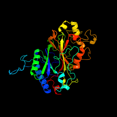
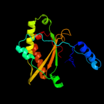
Phyre Home Retrieve Phyre Job Id
Emaill.a.kelley@imperial.ac.uk
DescriptionP31545
DateThu Jan 5 11:48:05 GMT 2012
Unique Job IDce39836f0622b3bc

Summary 

Top model
Image coloured by rainbow N → C terminus
Model (left) based on template c2wx6B_
Top template information
PDB header:oxidoreductase
Chain: B: PDB Molecule:peroxidase ycdb;
PDBTitle: x-ray crystallographic structure of e. coli apo-efeb
Confidence and coverage
Confidence:100.0% Coverage: 87%
368 residues ( 87% of your sequence) have been modelled with 100.0% confidence by the single highest scoring template.
3D viewing
Interactive 3D view in Jmol

Sequence analysis 

Secondary structure and disorder prediction 

   1........10.........20.........30.........40.........50.........60
Sequence  MQYKDENGVNEPSRRRLLKVIGALALAGSCPVAHAQKTQSAPGTLSPDARNEKQPFYGEH
Secondary structure 


SS confidence 


Disorder  ?????????????


????????????????????


Disorder confidence 


   .........70.........80.........90.........100.........110.........120
Sequence  QAGILTPQQAAMMLVAFDVLASDKADLERLFRLLTQRFAFLTQGGAAPETPNPRLPPLDS
Secondary structure 


SS confidence 


Disorder 


????????????


Disorder confidence 


   .........130.........140.........150.........160.........170.........180
Sequence  GILGGYIAPDNLTITLSVGHSLFDERFGLAPQMPKKLQKMTRFPNDSLDAALCHGDVLLQ
Secondary structure 


SS confidence 


Disorder 
??????


?


?


?

?????


Disorder confidence 


   .........190.........200.........210.........220.........230.........240
Sequence  ICANTQDTVIHALRDIIKHTPDLLSVRWKREGFISDHAARSKGKETPINLLGFKDGTANP
Secondary structure 


SS confidence 


Disorder 


?????????????


?

Disorder confidence 


   .........250.........260.........270.........280.........290.........300
Sequence  DSQNDKLMQKVVWVTADQQEPAWTIGGSYQAVRLIQFRVEFWDRTPLKEQQTIFGRDKQT
Secondary structure 


SS confidence 


Disorder  ?????


???


Disorder confidence 


   .........310.........320.........330.........340.........350.........360
Sequence  GAPLGMQHEHDVPDYASDPEGKVIALDSHIRLANPRTAESESSLMLRRGYSYSLGVTNSG
Secondary structure 


SS confidence 


Disorder 
?

????


?


??????????


???????
Disorder confidence 


   .........370.........380.........390.........400.........410.........420
Sequence  QLDMGLLFVCYQHDLEKGFLTVQKRLNGEALEEYVKPIGGGYFFALPGVKDANDYFGSAL
Secondary structure 


SS confidence 


Disorder  ?


?


Disorder confidence 


   ...
Sequence  LRV
Secondary structure 

SS confidence 


Disorder  ???
Disorder confidence 


Confidence Key
High(9)                    Low (0)
?Disordered
Alpha helix
Beta strand

Domain analysis 

Hover over an aligned region to see model and summary info

Please note, only up to the top 20 hits are modelled to reduce computer load

RankAligned region

PDB 2wx6 chain B

3D model

Region: 48 - 423
Aligned: 368
Modelled: 376
Confidence: 100.0%
Identity: 100%
PDB header:oxidoreductase
Chain: B: PDB Molecule:peroxidase ycdb;
PDBTitle: x-ray crystallographic structure of e. coli apo-efeb

Phyre2

PDB 2iiz chain A domain 1

3D model

Region: 59 - 415
Aligned: 296
Modelled: 316
Confidence: 100.0%
Identity: 24%
Fold: Ferredoxin-like
Superfamily: Dimeric alpha+beta barrel
Family: Dyp-type peroxidase-like

Phyre2

PDB 2gvk chain A domain 1

3D model

Region: 56 - 407
Aligned: 292
Modelled: 315
Confidence: 100.0%
Identity: 20%
Fold: Ferredoxin-like
Superfamily: Dimeric alpha+beta barrel
Family: Dyp-type peroxidase-like

Phyre2

PDB 3qns chain A

3D model

Region: 60 - 407
Aligned: 290
Modelled: 302
Confidence: 100.0%
Identity: 23%
PDB header:oxidoreductase
Chain: A: PDB Molecule:dyp peroxidase;
PDBTitle: dypb from rhodococcus jostii rha1, crystal form 2

Phyre2

PDB 2d3q chain A domain 1

3D model

Region: 57 - 418
Aligned: 332
Modelled: 341
Confidence: 100.0%
Identity: 25%
Fold: Ferredoxin-like
Superfamily: Dimeric alpha+beta barrel
Family: Dyp-type peroxidase-like

Phyre2

PDB 2e76 chain D

3D model

Region: 8 - 63
Aligned: 56
Modelled: 56
Confidence: 89.5%
Identity: 11%
PDB header:photosynthesis
Chain: D: PDB Molecule:cytochrome b6-f complex iron-sulfur subunit;
PDBTitle: crystal structure of the cytochrome b6f complex with tridecyl-2 stigmatellin (tds) from m.laminosus

Phyre2

PDB 3nn4 chain C

3D model

Region: 172 - 403
Aligned: 160
Modelled: 181
Confidence: 73.8%
Identity: 19%
PDB header:oxidoreductase
Chain: C: PDB Molecule:chlorite dismutase;
PDBTitle: structure of chlorite dismutase from candidatus nitrospira defluvii2 r173k mutant

Phyre2

PDB 1p84 chain E

3D model

Region: 6 - 35
Aligned: 30
Modelled: 30
Confidence: 69.1%
Identity: 10%
PDB header:oxidoreductase
Chain: E: PDB Molecule:ubiquinol-cytochrome c reductase iron-sulfur
PDBTitle: hdbt inhibited yeast cytochrome bc1 complex

Phyre2

PDB 2fyn chain O

3D model

Region: 13 - 63
Aligned: 51
Modelled: 51
Confidence: 69.1%
Identity: 18%
PDB header:oxidoreductase
Chain: O: PDB Molecule:ubiquinol-cytochrome c reductase iron-sulfur
PDBTitle: crystal structure analysis of the double mutant rhodobacter2 sphaeroides bc1 complex

Phyre2

PDB 2fyu chain E

3D model

Region: 1 - 33
Aligned: 33
Modelled: 33
Confidence: 68.7%
Identity: 12%
PDB header:oxidoreductase
Chain: E: PDB Molecule:ubiquinol-cytochrome c reductase iron-sulfur subunit,
PDBTitle: crystal structure of bovine heart mitochondrial bc1 with jg1442 inhibitor

Phyre2

PDB 2pq4 chain B

3D model

Region: 13 - 40
Aligned: 28
Modelled: 28
Confidence: 53.6%
Identity: 21%
PDB header:chaperone/oxidoreductase
Chain: B: PDB Molecule:periplasmic nitrate reductase precursor;
PDBTitle: nmr solution structure of napd in complex with napa1-352 signal peptide

Phyre2

PDB 2vxh chain F

3D model

Region: 172 - 386
Aligned: 145
Modelled: 161
Confidence: 47.4%
Identity: 17%
PDB header:oxidoreductase
Chain: F: PDB Molecule:chlorite dismutase;
PDBTitle: the crystal structure of chlorite dismutase: a detox enzyme2 producing molecular oxygen

Phyre2

PDB 1t0t chain V

3D model

Region: 175 - 212
Aligned: 38
Modelled: 38
Confidence: 36.3%
Identity: 21%
Fold: Ferredoxin-like
Superfamily: Dimeric alpha+beta barrel
Family: Chlorite dismutase-like

Phyre2

PDB 2hpg chain B

3D model

Region: 231 - 293
Aligned: 56
Modelled: 63
Confidence: 33.4%
Identity: 13%
PDB header:ligand binding protein
Chain: B: PDB Molecule:abc transporter, periplasmic substrate-binding
PDBTitle: the crystal structure of a thermophilic trap periplasmic2 binding protein

Phyre2

PDB 3b0v chain D

3D model

Region: 177 - 206
Aligned: 30
Modelled: 30
Confidence: 33.3%
Identity: 17%
PDB header:oxidoreductase/rna
Chain: D: PDB Molecule:trna-dihydrouridine synthase;
PDBTitle: trna-dihydrouridine synthase from thermus thermophilus in complex with2 trna

Phyre2

PDB 2hzk chain B

3D model

Region: 232 - 293
Aligned: 58
Modelled: 62
Confidence: 25.0%
Identity: 14%
PDB header:ligand binding, transport protein
Chain: B: PDB Molecule:trap-t family sorbitol/mannitol transporter, periplasmic
PDBTitle: crystal structures of a sodium-alpha-keto acid binding subunit from a2 trap transporter in its open form

Phyre2

PDB 3b50 chain A

3D model

Region: 232 - 293
Aligned: 55
Modelled: 62
Confidence: 24.3%
Identity: 16%
PDB header:transport protein
Chain: A: PDB Molecule:sialic acid-binding periplasmic protein siap;
PDBTitle: structure of h. influenzae sialic acid binding protein2 bound to neu5ac.

Phyre2

PDB 2pfy chain A

3D model

Region: 232 - 293
Aligned: 55
Modelled: 62
Confidence: 24.1%
Identity: 15%
PDB header:transport protein
Chain: A: PDB Molecule:putative exported protein;
PDBTitle: crystal structure of dctp7, a bordetella pertussis2 extracytoplasmic solute receptor binding pyroglutamic acid

Phyre2

PDB 3gyy chain C

3D model

Region: 232 - 293
Aligned: 55
Modelled: 62
Confidence: 18.1%
Identity: 18%
PDB header:transport protein
Chain: C: PDB Molecule:periplasmic substrate binding protein;
PDBTitle: the ectoine binding protein of the teaabc trap transporter teaa in the2 apo-state

Phyre2

PDB 1vdh chain A

3D model

Region: 175 - 209
Aligned: 35
Modelled: 35
Confidence: 18.0%
Identity: 14%
Fold: Ferredoxin-like
Superfamily: Dimeric alpha+beta barrel
Family: Chlorite dismutase-like

Phyre2
1

c2wx6B_
2

d2iiza1
3

d2gvka1
4

c3qnsA_
5

d2d3qa1
6

c2e76D_
7

c3nn4C_
8

c1p84E_
9

c2fynO_
10

c2fyuE_
11

c2pq4B_
12

c2vxhF_
13

d1t0tv_
14

c2hpgB_
15

c3b0vD_
16

c2hzkB_
17

c3b50A_
18

c2pfyA_
19

c3gyyC_
20

d1vdha_
21


22


23


24


25


26


27


28


29


30


31


32


33


34


35


36


37


38


39


40


41


42


43


44


45


46


47


48


49


50


51


52


53


54


55


56


57


58


59


60


61


62


63


Detailed template information 

#
Template Alignment Coverage3D Model Confidence
% i.d. Template Information
1c2wx6B_


100.0 100 PDB header:oxidoreductase
Chain: B: PDB Molecule:peroxidase ycdb;
PDBTitle: x-ray crystallographic structure of e. coli apo-efeb
2d2iiza1


100.0 24 Fold:Ferredoxin-like
Superfamily:Dimeric alpha+beta barrel
Family:Dyp-type peroxidase-like
3d2gvka1


100.0 20 Fold:Ferredoxin-like
Superfamily:Dimeric alpha+beta barrel
Family:Dyp-type peroxidase-like
4c3qnsA_


100.0 23 PDB header:oxidoreductase
Chain: A: PDB Molecule:dyp peroxidase;
PDBTitle: dypb from rhodococcus jostii rha1, crystal form 2
5d2d3qa1


100.0 25 Fold:Ferredoxin-like
Superfamily:Dimeric alpha+beta barrel
Family:Dyp-type peroxidase-like
6c2e76D_


89.5 11 PDB header:photosynthesis
Chain: D: PDB Molecule:cytochrome b6-f complex iron-sulfur subunit;
PDBTitle: crystal structure of the cytochrome b6f complex with tridecyl-2 stigmatellin (tds) from m.laminosus
7c3nn4C_


73.8 19 PDB header:oxidoreductase
Chain: C: PDB Molecule:chlorite dismutase;
PDBTitle: structure of chlorite dismutase from candidatus nitrospira defluvii2 r173k mutant
8c1p84E_


69.1 10 PDB header:oxidoreductase
Chain: E: PDB Molecule:ubiquinol-cytochrome c reductase iron-sulfur
PDBTitle: hdbt inhibited yeast cytochrome bc1 complex
9c2fynO_


69.1 18 PDB header:oxidoreductase
Chain: O: PDB Molecule:ubiquinol-cytochrome c reductase iron-sulfur
PDBTitle: crystal structure analysis of the double mutant rhodobacter2 sphaeroides bc1 complex
10c2fyuE_


68.7 12 PDB header:oxidoreductase
Chain: E: PDB Molecule:ubiquinol-cytochrome c reductase iron-sulfur subunit,
PDBTitle: crystal structure of bovine heart mitochondrial bc1 with jg1442 inhibitor
11c2pq4B_


53.6 21 PDB header:chaperone/oxidoreductase
Chain: B: PDB Molecule:periplasmic nitrate reductase precursor;
PDBTitle: nmr solution structure of napd in complex with napa1-352 signal peptide
12c2vxhF_


47.4 17 PDB header:oxidoreductase
Chain: F: PDB Molecule:chlorite dismutase;
PDBTitle: the crystal structure of chlorite dismutase: a detox enzyme2 producing molecular oxygen
13d1t0tv_


36.3 21 Fold:Ferredoxin-like
Superfamily:Dimeric alpha+beta barrel
Family:Chlorite dismutase-like
14c2hpgB_


33.4 13 PDB header:ligand binding protein
Chain: B: PDB Molecule:abc transporter, periplasmic substrate-binding
PDBTitle: the crystal structure of a thermophilic trap periplasmic2 binding protein
15c3b0vD_


33.3 17 PDB header:oxidoreductase/rna
Chain: D: PDB Molecule:trna-dihydrouridine synthase;
PDBTitle: trna-dihydrouridine synthase from thermus thermophilus in complex with2 trna
16c2hzkB_


25.0 14 PDB header:ligand binding, transport protein
Chain: B: PDB Molecule:trap-t family sorbitol/mannitol transporter, periplasmic
PDBTitle: crystal structures of a sodium-alpha-keto acid binding subunit from a2 trap transporter in its open form
17c3b50A_


24.3 16 PDB header:transport protein
Chain: A: PDB Molecule:sialic acid-binding periplasmic protein siap;
PDBTitle: structure of h. influenzae sialic acid binding protein2 bound to neu5ac.
18c2pfyA_


24.1 15 PDB header:transport protein
Chain: A: PDB Molecule:putative exported protein;
PDBTitle: crystal structure of dctp7, a bordetella pertussis2 extracytoplasmic solute receptor binding pyroglutamic acid
19c3gyyC_


18.1 18 PDB header:transport protein
Chain: C: PDB Molecule:periplasmic substrate binding protein;
PDBTitle: the ectoine binding protein of the teaabc trap transporter teaa in the2 apo-state
20d1vdha_


18.0 14 Fold:Ferredoxin-like
Superfamily:Dimeric alpha+beta barrel
Family:Chlorite dismutase-like
21c3fxbB_


not modelled 17.3 16 PDB header:transport protein
Chain: B: PDB Molecule:trap dicarboxylate transporter, dctp subunit;
PDBTitle: crystal structure of the ectoine-binding protein ueha
22d1vjja1


not modelled 15.5 35 Fold:Immunoglobulin-like beta-sandwich
Superfamily:E set domains
Family:Transglutaminase N-terminal domain
23d1g0da1


not modelled 15.3 26 Fold:Immunoglobulin-like beta-sandwich
Superfamily:E set domains
Family:Transglutaminase N-terminal domain
24c3njqB_


not modelled 14.8 25 PDB header:viral protein/inhibitor
Chain: B: PDB Molecule:orf 17;
PDBTitle: crystal structure of kaposi's sarcoma-associated herpesvirus protease2 in complex with dimer disruptor
25d1kfia4


not modelled 14.8 33 Fold:TBP-like
Superfamily:Phosphoglucomutase, C-terminal domain
Family:Phosphoglucomutase, C-terminal domain
26d2q3za1


not modelled 14.6 17 Fold:Immunoglobulin-like beta-sandwich
Superfamily:E set domains
Family:Transglutaminase N-terminal domain
27d3pmga4


not modelled 14.1 24 Fold:TBP-like
Superfamily:Phosphoglucomutase, C-terminal domain
Family:Phosphoglucomutase, C-terminal domain
28c2r8kB_


not modelled 12.9 6 PDB header:replication, transferase/dna
Chain: B: PDB Molecule:dna polymerase eta;
PDBTitle: structure of the eukaryotic dna polymerase eta in complex with 1,2-2 d(gpg)-cisplatin containing dna
29d1nmla2


not modelled 12.9 33 Fold:Cytochrome c
Superfamily:Cytochrome c
Family:Di-heme cytochrome c peroxidase
30d1ex0a1


not modelled 12.6 23 Fold:Immunoglobulin-like beta-sandwich
Superfamily:E set domains
Family:Transglutaminase N-terminal domain
31d1jiha2


not modelled 12.1 6 Fold:DNA/RNA polymerases
Superfamily:DNA/RNA polymerases
Family:Lesion bypass DNA polymerase (Y-family), catalytic domain
32c2pfzA_


not modelled 11.9 11 PDB header:transport protein
Chain: A: PDB Molecule:putative exported protein;
PDBTitle: crystal structure of dctp6, a bordetella pertussis2 extracytoplasmic solute receptor binding pyroglutamic acid
33d1uoua3


not modelled 11.8 11 Fold:alpha/beta-Hammerhead
Superfamily:Pyrimidine nucleoside phosphorylase C-terminal domain
Family:Pyrimidine nucleoside phosphorylase C-terminal domain
34c1jihA_


not modelled 11.7 6 PDB header:translation
Chain: A: PDB Molecule:dna polymerase eta;
PDBTitle: yeast dna polymerase eta
35d2pbka1


not modelled 11.7 25 Fold:Herpes virus serine proteinase, assemblin
Superfamily:Herpes virus serine proteinase, assemblin
Family:Herpes virus serine proteinase, assemblin
36c3hq7A_


not modelled 11.6 23 PDB header:oxidoreductase
Chain: A: PDB Molecule:cytochrome c551 peroxidase;
PDBTitle: ccpa from g. sulfurreducens, g94k/k97q/r100i variant
37c1yqmA_


not modelled 11.3 17 PDB header:hydrolase/dna
Chain: A: PDB Molecule:n-glycosylase/dna lyase;
PDBTitle: catalytically inactive human 8-oxoguanine glycosylase2 crosslinked to 7-deazaguanine containing dna
38c2x29A_


not modelled 11.0 25 PDB header:cell adhesion
Chain: A: PDB Molecule:tumor necrosis factor ligand superfamily member
PDBTitle: crystal structure of human4-1bb ligand ectodomain
39c2aq4A_


not modelled 10.8 23 PDB header:transferase
Chain: A: PDB Molecule:dna repair protein rev1;
PDBTitle: ternary complex of the catalytic core of rev1 with dna and dctp.
40c2oh2B_


not modelled 10.5 9 PDB header:transferase/dna
Chain: B: PDB Molecule:dna polymerase kappa;
PDBTitle: ternary complex of human dna polymerase
41d1tdja3


not modelled 9.4 27 Fold:Ferredoxin-like
Superfamily:ACT-like
Family:Allosteric threonine deaminase C-terminal domain
42d1na6a1


not modelled 9.4 40 Fold:DNA-binding pseudobarrel domain
Superfamily:DNA-binding pseudobarrel domain
Family:Type II restriction endonuclease effector domain
43c3eq1A_


not modelled 9.0 25 PDB header:transferase
Chain: A: PDB Molecule:porphobilinogen deaminase;
PDBTitle: the crystal structure of human porphobilinogen deaminase at2 2.8a resolution
44d1at3a_


not modelled 8.4 19 Fold:Herpes virus serine proteinase, assemblin
Superfamily:Herpes virus serine proteinase, assemblin
Family:Herpes virus serine proteinase, assemblin
45d2i09a2


not modelled 7.9 21 Fold:DeoB insert domain-like
Superfamily:DeoB insert domain-like
Family:DeoB insert domain-like
46c3gqcB_


not modelled 7.8 9 PDB header:transferase/dna
Chain: B: PDB Molecule:dna repair protein rev1;
PDBTitle: structure of human rev1-dna-dntp ternary complex
47c2p5xB_


not modelled 7.8 18 PDB header:structural genomics, unknown function
Chain: B: PDB Molecule:n-acetylserotonin o-methyltransferase-like protein;
PDBTitle: crystal structure of maf domain of human n-acetylserotonin o-2 methyltransferase-like protein
48c3ps9A_


not modelled 7.7 23 PDB header:transferase
Chain: A: PDB Molecule:trna 5-methylaminomethyl-2-thiouridine biosynthesis
PDBTitle: crystal structure of mnmc from e. coli
49d1o6ea_


not modelled 7.2 16 Fold:Herpes virus serine proteinase, assemblin
Superfamily:Herpes virus serine proteinase, assemblin
Family:Herpes virus serine proteinase, assemblin
50c3mr2A_


not modelled 7.1 6 PDB header:transferase/dna
Chain: A: PDB Molecule:dna polymerase eta;
PDBTitle: human dna polymerase eta in complex with normal dna and incoming2 nucleotide (nrm)
51c2vfyA_


not modelled 6.9 20 PDB header:hydrolase
Chain: A: PDB Molecule:akap18 delta;
PDBTitle: akap18 delta central domain
52c1nmlA_


not modelled 6.9 19 PDB header:oxidoreductase
Chain: A: PDB Molecule:di-haem cytochrome c peroxidase;
PDBTitle: di-haemic cytochrome c peroxidase from pseudomonas nautica 617, form2 in (ph 4.0)
53c2c1uB_


not modelled 6.8 23 PDB header:oxidoreductase
Chain: B: PDB Molecule:di-haem cytochrome c peroxidase;
PDBTitle: crystal structure of the di-haem cytochrome c peroxidase2 from paracoccus pantotrophus - oxidised form
54d1eb7a2


not modelled 6.7 16 Fold:Cytochrome c
Superfamily:Cytochrome c
Family:Di-heme cytochrome c peroxidase
55c3pt1A_


not modelled 6.7 11 PDB header:hydrolase
Chain: A: PDB Molecule:upf0364 protein ymr027w;
PDBTitle: structure of duf89 from saccharomyces cerevisiae co-crystallized with2 f6p.
56c2e1aD_


not modelled 6.6 11 PDB header:transcription
Chain: D: PDB Molecule:75aa long hypothetical regulatory protein asnc;
PDBTitle: crystal structure of ffrp-dm1
57c1ko9A_


not modelled 5.8 16 PDB header:hydrolase
Chain: A: PDB Molecule:8-oxoguanine dna glycosylase;
PDBTitle: native structure of the human 8-oxoguanine dna glycosylase2 hogg1
58c2qdrA_


not modelled 5.7 12 PDB header:unknown function
Chain: A: PDB Molecule:uncharacterized protein;
PDBTitle: crystal structure of a putative dioxygenase (npun_f5605) from nostoc2 punctiforme pcc 73102 at 2.60 a resolution
59c3dtzB_


not modelled 5.7 14 PDB header:structural genomics, unknown function
Chain: B: PDB Molecule:putative chlorite dismutase ta0507;
PDBTitle: crystal structure of putative chlorite dismutase ta0507
60c1cq0A_


not modelled 5.6 78 PDB header:de novo protein
Chain: A: PDB Molecule:protein (new hypothalamic neuropeptide/orexin-
PDBTitle: solution structure of a human hypocretin-2/orexin-2 b'solution structure of a human hypocretin-2/orexin-b '
61d1iega_


not modelled 5.6 20 Fold:Herpes virus serine proteinase, assemblin
Superfamily:Herpes virus serine proteinase, assemblin
Family:Herpes virus serine proteinase, assemblin
62d2cg4a2


not modelled 5.5 17 Fold:Ferredoxin-like
Superfamily:Dimeric alpha+beta barrel
Family:Lrp/AsnC-like transcriptional regulator C-terminal domain
63c1t94B_


not modelled 5.3 9 PDB header:replication
Chain: B: PDB Molecule:polymerase (dna directed) kappa;
PDBTitle: crystal structure of the catalytic core of human dna2 polymerase kappa

Binding site prediction 

Due to computational demand, binding site predictions are not run for batch jobs

If you want to predict binding sites, please manually submit your model of choice to 3DLigandSite


Phyre is for academic use only

Please cite: Protein structure prediction on the web: a case study using the Phyre server
Kelley LA and Sternberg MJE. Nature Protocols 4, 363 - 371 (2009) [pdf] [Import into BibTeX]
 
If you use the binding site predictions from 3DLigandSite, please also cite:
3DLigandSite: predicting ligand-binding sites using similar structures.
Wass MN, Kelley LA and Sternberg MJ Nucleic Acids Research 38, W469-73 (2010) [PubMed]
 
© Structural Bioinformatics Group
Imperial College London
Lawrence Kelley, Benjamin Jefferys
Disclaimer
Terms and Conditions
Component software
Template detection: HHpred 1.51
Secondary structure prediction: Psi-pred 2.5
Disorder prediction: Disopred 2.4
Transmembrane prediction: Memsat_SVM
Multi-template modelling and ab initio: Poing 1.0