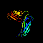
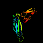
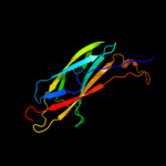
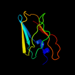
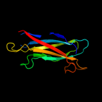
| 1 | c1qunA_
|
|
 |
100.0 |
48 |
PDB header:chaperone/structural protein
Chain: A: PDB Molecule:papd-like chaperone fimc;
PDBTitle: x-ray structure of the fimc-fimh chaperone adhesin complex2 from uropathogenic e.coli
|
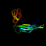
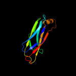
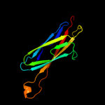
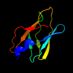
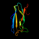
| 2 | c1z9sA_
|
|
 |
100.0 |
32 |
PDB header:chaperone/immune system
Chain: A: PDB Molecule:chaperone protein caf1m;
PDBTitle: crystal structure of the native chaperone:subunit:subunit2 caf1m:caf1:caf1 complex
|
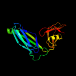
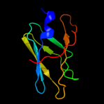
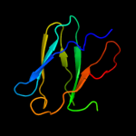
| 3 | c2co7B_
|
|
 |
100.0 |
29 |
PDB header:fibril protein
Chain: B: PDB Molecule:putative fimbriae assembly chaperone;
PDBTitle: salmonella enterica safa pilin in complex with the safb2 chaperone (type ii)
|
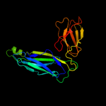
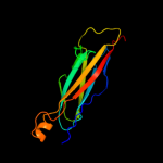
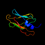
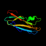
| 4 | c1l4iA_
|
|
 |
100.0 |
45 |
PDB header:chaperone
Chain: A: PDB Molecule:sfae protein;
PDBTitle: crystal structure of the periplasmic chaperone sfae
|
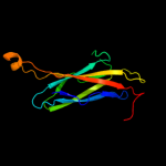
| 5 | c1qpxA_
|
|
 |
100.0 |
29 |
PDB header:chaperone
Chain: A: PDB Molecule:papd chaperone;
PDBTitle: crystal structures of self-capping papd chaperone homodimers
|
| 6 | c3f6iB_
|
|
 |
100.0 |
27 |
PDB header:chaperone
Chain: B: PDB Molecule:chaperone protein faee;
PDBTitle: structure of the semet labeled f4 fibrial chaperone faee
|
| 7 | c3q48B_
|
|
 |
100.0 |
29 |
PDB header:chaperone
Chain: B: PDB Molecule:chaperone cupb2;
PDBTitle: crystal structure of pseudomonas aeruginosa cupb2 chaperone
|
| 8 | d3bwuc1
|
|
 |
100.0 |
58 |
Fold:Immunoglobulin-like beta-sandwich
Superfamily:PapD-like
Family:Pilus chaperone |
| 9 | d2co7b1
|
|
 |
100.0 |
32 |
Fold:Immunoglobulin-like beta-sandwich
Superfamily:PapD-like
Family:Pilus chaperone |
| 10 | d2j2za1
|
|
 |
100.0 |
34 |
Fold:Immunoglobulin-like beta-sandwich
Superfamily:PapD-like
Family:Pilus chaperone |
| 11 | d1p5va1
|
|
 |
100.0 |
38 |
Fold:Immunoglobulin-like beta-sandwich
Superfamily:PapD-like
Family:Pilus chaperone |
| 12 | d1l4ia1
|
|
 |
100.0 |
54 |
Fold:Immunoglobulin-like beta-sandwich
Superfamily:PapD-like
Family:Pilus chaperone |
| 13 | d3bwuc2
|
|
 |
99.8 |
35 |
Fold:C2 domain-like
Superfamily:Periplasmic chaperone C-domain
Family:Periplasmic chaperone C-domain |
| 14 | d1p5va2
|
|
 |
99.8 |
23 |
Fold:C2 domain-like
Superfamily:Periplasmic chaperone C-domain
Family:Periplasmic chaperone C-domain |
| 15 | d2co7b2
|
|
 |
99.8 |
23 |
Fold:C2 domain-like
Superfamily:Periplasmic chaperone C-domain
Family:Periplasmic chaperone C-domain |
| 16 | d1l4ia2
|
|
 |
99.7 |
33 |
Fold:C2 domain-like
Superfamily:Periplasmic chaperone C-domain
Family:Periplasmic chaperone C-domain |
| 17 | d2j2za2
|
|
 |
99.6 |
23 |
Fold:C2 domain-like
Superfamily:Periplasmic chaperone C-domain
Family:Periplasmic chaperone C-domain |
| 18 | d1m1sa_
|
|
 |
97.2 |
14 |
Fold:Immunoglobulin-like beta-sandwich
Superfamily:PapD-like
Family:MSP-like |
| 19 | d1grwa_
|
|
 |
96.5 |
19 |
Fold:Immunoglobulin-like beta-sandwich
Superfamily:PapD-like
Family:MSP-like |
| 20 | d1rowa_
|
|
 |
96.5 |
14 |
Fold:Immunoglobulin-like beta-sandwich
Superfamily:PapD-like
Family:MSP-like |
| 21 | c2qsvA_ |
|
not modelled |
95.4 |
10 |
PDB header:structural genomics, unknown function
Chain: A: PDB Molecule:uncharacterized protein;
PDBTitle: crystal structure of protein of unknown function from porphyromonas2 gingivalis w83
|
| 22 | d1mspa_ |
|
not modelled |
95.2 |
16 |
Fold:Immunoglobulin-like beta-sandwich
Superfamily:PapD-like
Family:MSP-like |
| 23 | c2e6jA_ |
|
not modelled |
94.3 |
16 |
PDB header:structural genomics, unknown function
Chain: A: PDB Molecule:hydin protein;
PDBTitle: solution structure of the c-terminal papd-like domain from2 human hydin protein
|
| 24 | c1z9oB_ |
|
not modelled |
93.2 |
15 |
PDB header:protein binding/lipid binding protein
Chain: B: PDB Molecule:vesicle-associated membrane protein-associated protein a;
PDBTitle: 1.9 angstrom crystal structure of the rat vap-a msp homology domain in2 complex with the rat orp1 ffat motif
|
| 25 | c3o0lB_ |
|
not modelled |
92.9 |
7 |
PDB header:structural genomics, unknown function
Chain: B: PDB Molecule:uncharacterized protein;
PDBTitle: crystal structure of a pfam duf1425 family member (shew_1734) from2 shewanella sp. pv-4 at 1.81 a resolution
|
| 26 | c3qisA_ |
|
not modelled |
91.8 |
12 |
PDB header:hydrolase/protein binding
Chain: A: PDB Molecule:inositol polyphosphate 5-phosphatase ocrl-1;
PDBTitle: recognition of the f&h motif by the lowe syndrome protein ocrl
|
| 27 | c3qbtH_ |
|
not modelled |
90.5 |
12 |
PDB header:protein transport/hydrolase
Chain: H: PDB Molecule:inositol polyphosphate 5-phosphatase ocrl-1;
PDBTitle: crystal structure of ocrl1 540-678 in complex with rab8a:gppnhp
|
| 28 | c2ys4A_ |
|
not modelled |
88.0 |
12 |
PDB header:structural genomics, unknown function
Chain: A: PDB Molecule:hydrocephalus-inducing protein homolog;
PDBTitle: solution structure of the n-terminal papd-like domain of2 hydin protein from human
|
| 29 | d2vzsa2 |
|
not modelled |
82.1 |
18 |
Fold:Immunoglobulin-like beta-sandwich
Superfamily:beta-Galactosidase/glucuronidase domain
Family:beta-Galactosidase/glucuronidase domain |
| 30 | d1wica_ |
|
not modelled |
78.1 |
9 |
Fold:Immunoglobulin-like beta-sandwich
Superfamily:PapD-like
Family:MSP-like |
| 31 | c3ginB_ |
|
not modelled |
71.9 |
11 |
PDB header:metal binding protein
Chain: B: PDB Molecule:sodium/calcium exchanger 1;
PDBTitle: crystal structure of e454k-cbd1
|
| 32 | d1ufga_ |
|
not modelled |
71.8 |
19 |
Fold:Immunoglobulin-like beta-sandwich
Superfamily:Lamin A/C globular tail domain
Family:Lamin A/C globular tail domain |
| 33 | d2dpka1 |
|
not modelled |
69.4 |
13 |
Fold:Immunoglobulin-like beta-sandwich
Superfamily:CalX-like
Family:CalX-beta domain |
| 34 | d4ubpb_ |
|
not modelled |
68.0 |
22 |
Fold:beta-clip
Superfamily:Urease, beta-subunit
Family:Urease, beta-subunit |
| 35 | d1ejxb_ |
|
not modelled |
65.7 |
14 |
Fold:beta-clip
Superfamily:Urease, beta-subunit
Family:Urease, beta-subunit |
| 36 | d1kyaa2 |
|
not modelled |
63.5 |
6 |
Fold:Cupredoxin-like
Superfamily:Cupredoxins
Family:Multidomain cupredoxins |
| 37 | d1aoza2 |
|
not modelled |
63.0 |
26 |
Fold:Cupredoxin-like
Superfamily:Cupredoxins
Family:Multidomain cupredoxins |
| 38 | d1e9ya1 |
|
not modelled |
62.1 |
21 |
Fold:beta-clip
Superfamily:Urease, beta-subunit
Family:Urease, beta-subunit |
| 39 | d1hfua2 |
|
not modelled |
60.3 |
10 |
Fold:Cupredoxin-like
Superfamily:Cupredoxins
Family:Multidomain cupredoxins |
| 40 | c2lllA_ |
|
not modelled |
55.9 |
18 |
PDB header:structural protein
Chain: A: PDB Molecule:lamin-b2;
PDBTitle: solution nmr structure of c-terminal globular domain of human lamin-2 b2, northeast structural genomics consortium target hr8546a
|
| 41 | c3h6aB_ |
|
not modelled |
54.1 |
8 |
PDB header:cell adhesion
Chain: B: PDB Molecule:integrin beta-4;
PDBTitle: structure of the calx-beta domain of integrin beta42 crystallized in the presence of calcium
|
| 42 | d1ifra_ |
|
not modelled |
53.9 |
19 |
Fold:Immunoglobulin-like beta-sandwich
Superfamily:Lamin A/C globular tail domain
Family:Lamin A/C globular tail domain |
| 43 | d2q9oa2 |
|
not modelled |
53.3 |
23 |
Fold:Cupredoxin-like
Superfamily:Cupredoxins
Family:Multidomain cupredoxins |
| 44 | c3jt0B_ |
|
not modelled |
52.8 |
11 |
PDB header:structural protein
Chain: B: PDB Molecule:lamin-b1;
PDBTitle: crystal structure of the c-terminal fragment (426-558)2 lamin-b1 from homo sapiens, northeast structural genomics3 consortium target hr5546a
|
| 45 | c1e9zA_ |
|
not modelled |
50.6 |
21 |
PDB header:hydrolase
Chain: A: PDB Molecule:urease subunit alpha;
PDBTitle: crystal structure of helicobacter pylori urease
|
| 46 | d1k3ra1 |
|
not modelled |
50.4 |
18 |
Fold:OB-fold
Superfamily:Nucleic acid-binding proteins
Family:Hypothetical protein MTH1 (MT0001), insert domain |
| 47 | c2je8B_ |
|
not modelled |
50.2 |
7 |
PDB header:hydrolase
Chain: B: PDB Molecule:beta-mannosidase;
PDBTitle: structure of a beta-mannosidase from bacteroides2 thetaiotaomicron
|
| 48 | d1v10a2 |
|
not modelled |
48.6 |
10 |
Fold:Cupredoxin-like
Superfamily:Cupredoxins
Family:Multidomain cupredoxins |
| 49 | c2qvkA_ |
|
not modelled |
47.4 |
10 |
PDB header:metal binding protein
Chain: A: PDB Molecule:sodium/calcium exchanger 1;
PDBTitle: the second ca2+-binding domain of the na+-ca2+ exchanger is2 essential for regulation: crystal structures and3 mutational analysis
|
| 50 | c3qgaD_ |
|
not modelled |
46.2 |
17 |
PDB header:hydrolase
Chain: D: PDB Molecule:fusion of urease beta and gamma subunits;
PDBTitle: 3.0 a model of iron containing urease urea2b2 from helicobacter2 mustelae
|
| 51 | c3eujB_ |
|
not modelled |
44.9 |
15 |
PDB header:cell cycle
Chain: B: PDB Molecule:chromosome partition protein mukf;
PDBTitle: crystal structure of muke-mukf(residues 292-443)-mukb(head2 domain)-atpgammas complex, symmetric dimer
|
| 52 | d1e42a1 |
|
not modelled |
44.7 |
20 |
Fold:Immunoglobulin-like beta-sandwich
Superfamily:Clathrin adaptor appendage domain
Family:Alpha-adaptin ear subdomain-like |
| 53 | c3e9uA_ |
|
not modelled |
40.5 |
12 |
PDB header:membrane protein
Chain: A: PDB Molecule:na/ca exchange protein;
PDBTitle: crystal structure of calx cbd2 domain
|
| 54 | c3rb7E_ |
|
not modelled |
37.4 |
12 |
PDB header:metal binding protein
Chain: E: PDB Molecule:na/ca exchange protein;
PDBTitle: crystal structure of cbd12 from calx1.2
|
| 55 | c3ac0B_ |
|
not modelled |
33.0 |
13 |
PDB header:hydrolase
Chain: B: PDB Molecule:beta-glucosidase i;
PDBTitle: crystal structure of beta-glucosidase from kluyveromyces marxianus in2 complex with glucose
|
| 56 | c1yycA_ |
|
not modelled |
31.9 |
13 |
PDB header:structural genomics, unknown function
Chain: A: PDB Molecule:putative late embryogenesis abundant protein;
PDBTitle: solution structure of a putative late embryogenesis2 abundant (lea) protein at2g46140.1
|
| 57 | d1gyca2 |
|
not modelled |
30.3 |
3 |
Fold:Cupredoxin-like
Superfamily:Cupredoxins
Family:Multidomain cupredoxins |
| 58 | d1ivta_ |
|
not modelled |
27.5 |
22 |
Fold:Immunoglobulin-like beta-sandwich
Superfamily:Lamin A/C globular tail domain
Family:Lamin A/C globular tail domain |
| 59 | d1xo8a_ |
|
not modelled |
23.7 |
13 |
Fold:Immunoglobulin-like beta-sandwich
Superfamily:LEA14-like
Family:LEA14-like |
| 60 | c2vzvB_ |
|
not modelled |
22.5 |
20 |
PDB header:hydrolase
Chain: B: PDB Molecule:exo-beta-d-glucosaminidase;
PDBTitle: substrate complex of amycolatopsis orientalis exo-2 chitosanase csxa e541a with chitosan
|
| 61 | c3butA_ |
|
not modelled |
21.3 |
10 |
PDB header:structural genomics, unknown function
Chain: A: PDB Molecule:uncharacterized protein af_0446;
PDBTitle: crystal structure of protein af_0446 from archaeoglobus fulgidus
|
| 62 | c1a65A_ |
|
not modelled |
21.1 |
9 |
PDB header:oxidoreductase
Chain: A: PDB Molecule:laccase;
PDBTitle: type-2 cu-depleted laccase from coprinus cinereus
|
| 63 | d1hmja_ |
|
not modelled |
20.9 |
26 |
Fold:RPB5-like RNA polymerase subunit
Superfamily:RPB5-like RNA polymerase subunit
Family:RPB5 |
| 64 | c2x41A_ |
|
not modelled |
20.7 |
9 |
PDB header:hydrolase
Chain: A: PDB Molecule:beta-glucosidase;
PDBTitle: structure of beta-glucosidase 3b from thermotoga neapolitana2 in complex with glucose
|
| 65 | c2l02B_ |
|
not modelled |
19.4 |
16 |
PDB header:structural genomics, unknown function
Chain: B: PDB Molecule:uncharacterized protein;
PDBTitle: solution nmr structure of protein bt2368 from bacteroides2 thetaiotaomicron, northeast structural genomics consortium target3 btr375
|
| 66 | d1w8oa1 |
|
not modelled |
16.3 |
13 |
Fold:Immunoglobulin-like beta-sandwich
Superfamily:E set domains
Family:E-set domains of sugar-utilizing enzymes |
| 67 | d1r7aa1 |
|
not modelled |
15.9 |
13 |
Fold:Glycosyl hydrolase domain
Superfamily:Glycosyl hydrolase domain
Family:alpha-Amylases, C-terminal beta-sheet domain |
| 68 | c3ppsD_ |
|
not modelled |
15.7 |
21 |
PDB header:oxidoreductase
Chain: D: PDB Molecule:laccase;
PDBTitle: crystal structure of an ascomycete fungal laccase from thielavia2 arenaria
|
| 69 | c1asqB_ |
|
not modelled |
15.4 |
27 |
PDB header:oxidoreductase
Chain: B: PDB Molecule:ascorbate oxidase;
PDBTitle: x-ray structures and mechanistic implications of three functional2 derivatives of ascorbate oxidase from zucchini: reduced-, peroxide-,3 and azide-forms
|
| 70 | c2q9oA_ |
|
not modelled |
15.4 |
25 |
PDB header:oxidoreductase
Chain: A: PDB Molecule:laccase-1;
PDBTitle: near-atomic resolution structure of a melanocarpus albomyces laccase
|
| 71 | d2fwua1 |
|
not modelled |
14.6 |
8 |
Fold:Immunoglobulin-like beta-sandwich
Superfamily:CalX-like
Family:CalX-beta domain |
| 72 | c1v10A_ |
|
not modelled |
14.6 |
9 |
PDB header:oxidase
Chain: A: PDB Molecule:laccase;
PDBTitle: structure of rigidoporus lignosus laccase from hemihedrally2 twinned crystals
|
| 73 | c1wkwB_ |
|
not modelled |
14.0 |
67 |
PDB header:translation/protein binding
Chain: B: PDB Molecule:eukaryotic translation initiation factor 4e
PDBTitle: crystal structure of the ternary complex of eif4e-m7gpppa-2 4ebp1 peptide
|
| 74 | c1gycA_ |
|
not modelled |
12.1 |
3 |
PDB header:oxidoreductase
Chain: A: PDB Molecule:laccase 2;
PDBTitle: crystal structure determination at room temperature of a2 laccase from trametes versicolor in its oxidised form3 containing a full complement of copper ions
|
| 75 | c3of6D_ |
|
not modelled |
12.0 |
12 |
PDB header:immune system
Chain: D: PDB Molecule:pre t-cell antigen receptor alpha;
PDBTitle: human pre-t cell receptor crystal structure
|
| 76 | d1yq2a1 |
|
not modelled |
11.6 |
13 |
Fold:Immunoglobulin-like beta-sandwich
Superfamily:beta-Galactosidase/glucuronidase domain
Family:beta-Galactosidase/glucuronidase domain |
| 77 | c3e9tD_ |
|
not modelled |
11.5 |
11 |
PDB header:membrane protein
Chain: D: PDB Molecule:na/ca exchange protein;
PDBTitle: crystal structure of apo-form calx cbd1 domain
|
| 78 | c2f7fA_ |
|
not modelled |
11.5 |
16 |
PDB header:transferase
Chain: A: PDB Molecule:nicotinate phosphoribosyltransferase, putative;
PDBTitle: crystal structure of enterococcus faecalis putative nicotinate2 phosphoribosyltransferase, new york structural genomics consortium
|
| 79 | c1e42A_ |
|
not modelled |
11.0 |
16 |
PDB header:endocytosis
Chain: A: PDB Molecule:ap-2 complex subunit beta;
PDBTitle: beta2-adaptin appendage domain, from clathrin adaptor ap2
|
| 80 | d1eu3a1 |
|
not modelled |
10.6 |
24 |
Fold:OB-fold
Superfamily:Bacterial enterotoxins
Family:Superantigen toxins, N-terminal domain |
| 81 | c3pe9D_ |
|
not modelled |
10.3 |
16 |
PDB header:unknown function
Chain: D: PDB Molecule:fibronectin(iii)-like module;
PDBTitle: structures of clostridium thermocellum cbha fibronectin(iii)-like2 modules
|
| 82 | c2vqiA_ |
|
not modelled |
9.9 |
19 |
PDB header:transport
Chain: A: PDB Molecule:outer membrane usher protein papc;
PDBTitle: structure of the p pilus usher (papc) translocation pore
|
| 83 | d1jz8a2 |
|
not modelled |
9.5 |
25 |
Fold:Immunoglobulin-like beta-sandwich
Superfamily:beta-Galactosidase/glucuronidase domain
Family:beta-Galactosidase/glucuronidase domain |
| 84 | c3k6sB_ |
|
not modelled |
9.4 |
11 |
PDB header:cell adhesion
Chain: B: PDB Molecule:integrin beta-2;
PDBTitle: structure of integrin alphaxbeta2 ectodomain
|
| 85 | c2pheC_ |
|
not modelled |
9.2 |
33 |
PDB header:transcription
Chain: C: PDB Molecule:alpha trans-inducing protein;
PDBTitle: model for vp16 binding to pc4
|
| 86 | c2phgB_ |
|
not modelled |
9.1 |
33 |
PDB header:transcription
Chain: B: PDB Molecule:alpha trans-inducing protein;
PDBTitle: model for vp16 binding to tfiib
|
| 87 | c2aenH_ |
|
not modelled |
7.9 |
10 |
PDB header:viral protein
Chain: H: PDB Molecule:outer capsid protein vp4, vp8* core;
PDBTitle: crystal structure of the rotavirus strain ds-1 vp8* core
|
| 88 | c2r2yA_ |
|
not modelled |
7.6 |
20 |
PDB header:protein binding
Chain: A: PDB Molecule:protein adrm1;
PDBTitle: crystal structure of the proteasomal rpn13 pru-domain
|
| 89 | c2z59A_ |
|
not modelled |
7.6 |
20 |
PDB header:protein transport
Chain: A: PDB Molecule:protein adrm1;
PDBTitle: complex structures of mouse rpn13 (22-130aa) and ubiquitin
|
| 90 | c2dv6F_ |
|
not modelled |
7.2 |
10 |
PDB header:oxidoreductase
Chain: F: PDB Molecule:nitrite reductase;
PDBTitle: crystal structure of nitrite reductase from hyphomicrobium2 denitrificans
|
| 91 | c2z4dA_ |
|
not modelled |
6.7 |
17 |
PDB header:nuclear protein
Chain: A: PDB Molecule:26s proteasome regulatory subunit rpn13;
PDBTitle: nmr structures of yeast proteasome component rpn13
|
| 92 | c3g7mA_ |
|
not modelled |
6.2 |
18 |
PDB header:hydrolase inhibitor
Chain: A: PDB Molecule:xylanase inhibitor tl-xi;
PDBTitle: structure of the thaumatin-like xylanase inhibitor tlxi
|
| 93 | c2kr0A_ |
|
not modelled |
6.1 |
20 |
PDB header:protein binding
Chain: A: PDB Molecule:proteasomal ubiquitin receptor adrm1;
PDBTitle: a proteasome protein
|
| 94 | d1t4za_ |
|
not modelled |
6.0 |
29 |
Fold:Thioredoxin fold
Superfamily:Thioredoxin-like
Family:KaiB-like |
| 95 | c1u8cB_ |
|
not modelled |
6.0 |
13 |
PDB header:cell adhesion
Chain: B: PDB Molecule:integrin beta-3;
PDBTitle: a novel adaptation of the integrin psi domain revealed from its2 crystal structure
|
| 96 | d1lm8v_ |
|
not modelled |
5.9 |
21 |
Fold:Prealbumin-like
Superfamily:VHL
Family:VHL |
| 97 | d1kqra_ |
|
not modelled |
5.9 |
9 |
Fold:Concanavalin A-like lectins/glucanases
Superfamily:Concanavalin A-like lectins/glucanases
Family:vp4 sialic acid binding domain |
| 98 | c1kriA_ |
|
not modelled |
5.9 |
9 |
PDB header:viral protein
Chain: A: PDB Molecule:vp4;
PDBTitle: nmr solution structures of the rhesus rotavirus vp4 sialic2 acid binding domain without ligand
|
| 99 | d2plta_ |
|
not modelled |
5.7 |
14 |
Fold:Cupredoxin-like
Superfamily:Cupredoxins
Family:Plastocyanin/azurin-like |