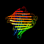
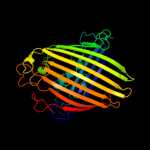
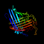
1 d1af6a_
100.0
28
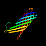
Fold: Transmembrane beta-barrelsSuperfamily: PorinsFamily: Maltoporin-like2 d2mpra_
100.0
26
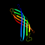
Fold: Transmembrane beta-barrelsSuperfamily: PorinsFamily: Maltoporin-like3 d1a0tp_
100.0
22
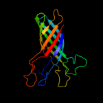
Fold: Transmembrane beta-barrelsSuperfamily: PorinsFamily: Maltoporin-like4 d2zfga1
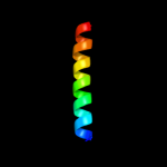
94.5
13
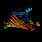
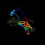
Fold: Transmembrane beta-barrelsSuperfamily: PorinsFamily: Porin5 c3qraA_
93.7
16
PDB header: cell invasionChain: A: PDB Molecule: attachment invasion locus protein;PDBTitle: the crystal structure of ail, the attachment invasion locus protein of2 yersinia pestis
6 d1qj8a_
91.4
7
Fold: Transmembrane beta-barrelsSuperfamily: OMPA-likeFamily: Outer membrane protein7 d1phoa_
90.5
12
Fold: Transmembrane beta-barrelsSuperfamily: PorinsFamily: Porin8 c2k0lA_
88.2
10
PDB header: membrane proteinChain: A: PDB Molecule: outer membrane protein a;PDBTitle: nmr structure of the transmembrane domain of the outer2 membrane protein a from klebsiella pneumoniae in dhpc3 micelles.
9 d1g90a_
85.0
9
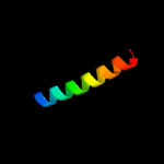
Fold: Transmembrane beta-barrelsSuperfamily: OMPA-likeFamily: Outer membrane protein10 c1cosB_
81.1
24
PDB header: alpha-helical bundleChain: B: PDB Molecule: coiled serine;PDBTitle: crystal structure of a synthetic triple-stranded alpha-2 helical bundle
11 c1cosA_
81.1
24
PDB header: alpha-helical bundleChain: A: PDB Molecule: coiled serine;PDBTitle: crystal structure of a synthetic triple-stranded alpha-2 helical bundle
12 c1cosC_
80.7
24
PDB header: alpha-helical bundleChain: C: PDB Molecule: coiled serine;PDBTitle: crystal structure of a synthetic triple-stranded alpha-2 helical bundle
13 c3nsgA_
79.9
13
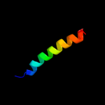
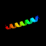
PDB header: membrane proteinChain: A: PDB Molecule: outer membrane protein f;PDBTitle: crystal structure of ompf, an outer membrane protein from salmonella2 typhi
14 c2x6pC_
76.8
24
PDB header: de novo proteinChain: C: PDB Molecule: coil ser l19c;PDBTitle: crystal structure of coil ser l19c
15 c2x6pA_
76.8
24
PDB header: de novo proteinChain: A: PDB Molecule: coil ser l19c;PDBTitle: crystal structure of coil ser l19c
16 c2x6pB_
76.8
24
PDB header: de novo proteinChain: B: PDB Molecule: coil ser l19c;PDBTitle: crystal structure of coil ser l19c
17 c3n4xB_
76.2
15
PDB header: replicationChain: B: PDB Molecule: monopolin complex subunit csm1;PDBTitle: structure of csm1 full-length
18 c3mkxC_
74.4
9
PDB header: antiviral proteinChain: C: PDB Molecule: bone marrow stromal antigen 2;PDBTitle: crystal structure of bst2/tetherin
19 c1zxaB_
73.0
37
PDB header: transferaseChain: B: PDB Molecule: cgmp-dependent protein kinase 1, alpha isozyme;PDBTitle: solution structure of the coiled-coil domain of cgmp-2 dependent protein kinase ia
20 c1aq5C_
72.6
29
PDB header: coiled-coilChain: C: PDB Molecule: cartilage matrix protein;PDBTitle: high-resolution solution nmr structure of the trimeric coiled-coil2 domain of chicken cartilage matrix protein, 20 structures
21 c3u1aC_
not modelled
66.1
15
PDB header: contractile proteinChain: C: PDB Molecule: smooth muscle tropomyosin alpha;PDBTitle: n-terminal 81-aa fragment of smooth muscle tropomyosin alpha
22 c2a3dA_
not modelled
65.9
32
PDB header: three-helix bundleChain: A: PDB Molecule: protein (de novo three-helix bundle);PDBTitle: solution structure of a de novo designed single chain three-2 helix bundle (a3d)
23 c1y4cA_
not modelled
63.7
10
PDB header: de novo proteinChain: A: PDB Molecule: maltose binding protein fused with designedPDBTitle: designed helical protein fusion mbp
24 c2x7aB_
not modelled
63.3
9
PDB header: immune systemChain: B: PDB Molecule: bone marrow stromal antigen 2;PDBTitle: structural basis of hiv-1 tethering to membranes by the2 bst2-tetherin ectodomain
25 c3m9bK_
not modelled
60.5
11
PDB header: chaperoneChain: K: PDB Molecule: proteasome-associated atpase;PDBTitle: crystal structure of the amino terminal coiled coil domain and the2 inter domain of the mycobacterium tuberculosis proteasomal atpase mpa
26 c2xzrA_
not modelled
59.8
10
PDB header: cell adhesionChain: A: PDB Molecule: immunoglobulin-binding protein eibd;PDBTitle: escherichia coli immunoglobulin-binding protein eibd 391-438 fused2 to gcn4 adaptors
27 c3cveC_
not modelled
59.3
16
PDB header: signaling proteinChain: C: PDB Molecule: homer protein homolog 1;PDBTitle: crystal structure of the carboxy terminus of homer1
28 c2jgoB_
not modelled
58.8
20
PDB header: de novo proteinChain: B: PDB Molecule: coil ser l9c;PDBTitle: stucture of the arsenated de novo designed peptide coil ser2 l9c
29 c3ljmA_
not modelled
58.8
20
PDB header: de novo proteinChain: A: PDB Molecule: coil ser l9c;PDBTitle: structure of de novo designed apo peptide coil ser l9c
30 c2jgoC_
not modelled
58.8
20
PDB header: de novo proteinChain: C: PDB Molecule: coil ser l9c;PDBTitle: stucture of the arsenated de novo designed peptide coil ser2 l9c
31 c3ljmB_
not modelled
58.8
20
PDB header: de novo proteinChain: B: PDB Molecule: coil ser l9c;PDBTitle: structure of de novo designed apo peptide coil ser l9c
32 c3ljmC_
not modelled
58.8
20
PDB header: de novo proteinChain: C: PDB Molecule: coil ser l9c;PDBTitle: structure of de novo designed apo peptide coil ser l9c
33 c2jgoA_
not modelled
58.8
20
PDB header: de novo proteinChain: A: PDB Molecule: coil ser l9c;PDBTitle: stucture of the arsenated de novo designed peptide coil ser2 l9c
34 c2wjqA_
not modelled
58.2
16
PDB header: transport proteinChain: A: PDB Molecule: probable n-acetylneuraminic acid outer membrane channelPDBTitle: nanc porin structure in hexagonal crystal form.
35 c3cvfA_
not modelled
57.9
24
PDB header: signaling proteinChain: A: PDB Molecule: homer protein homolog 3;PDBTitle: crystal structure of the carboxy terminus of homer3
36 c1ca9D_
not modelled
57.8
6
PDB header: tnf signalingChain: D: PDB Molecule: protein (tnf receptor associated factor 2);PDBTitle: structure of tnf receptor associated factor 2 in complex2 with a peptide from tnf-r2
37 c2zvfG_
not modelled
56.3
12
PDB header: ligaseChain: G: PDB Molecule: alanyl-trna synthetase;PDBTitle: crystal structure of archaeoglobus fulgidus alanyl-trna2 synthetase c-terminal dimerization domain
38 c3ni0A_
not modelled
56.2
19
PDB header: immune systemChain: A: PDB Molecule: bone marrow stromal antigen 2;PDBTitle: crystal structure of mouse bst-2/tetherin ectodomain
39 c3e98B_
not modelled
54.1
21
PDB header: unknown functionChain: B: PDB Molecule: gaf domain of unknown function;PDBTitle: crystal structure of a gaf domain containing protein that belongs to2 pfam duf484 family (pa5279) from pseudomonas aeruginosa at 2.43 a3 resolution
40 c1u0iA_
not modelled
51.9
24
PDB header: de novo proteinChain: A: PDB Molecule: iaal-e3;PDBTitle: iaal-e3/k3 heterodimer
41 c3nb3C_
not modelled
51.7
11
PDB header: virusChain: C: PDB Molecule: outer membrane protein a;PDBTitle: the host outer membrane proteins ompa and ompc are packed at specific2 sites in the shigella phage sf6 virion as structural components
42 c1kzzA_
not modelled
48.7
16
PDB header: signaling proteinChain: A: PDB Molecule: tnf receptor associated factor 3;PDBTitle: downstream regulator tank binds to the cd40 recognition2 site on traf3
43 c3iynR_
not modelled
48.1
14
PDB header: virusChain: R: PDB Molecule: hexon-associated protein;PDBTitle: 3.6-angstrom cryoem structure of human adenovirus type 5
44 c1coiA_
not modelled
47.4
12
PDB header: alpha-helical bundleChain: A: PDB Molecule: coil-vald;PDBTitle: designed trimeric coiled coil-vald
45 c1abzA_
not modelled
46.6
30
PDB header: de novo designChain: A: PDB Molecule: alpha-t-alpha;PDBTitle: alpha-t-alpha, a de novo designed peptide, nmr, 232 structures
46 c1g6uB_
not modelled
46.1
26
PDB header: de novo proteinChain: B: PDB Molecule: domain swapped dimer;PDBTitle: crystal structure of a domain swapped dimer
47 d1i78a_
not modelled
46.1
18
Fold: Transmembrane beta-barrelsSuperfamily: OMPT-likeFamily: Outer membrane protease OMPT48 d1vp7b_
not modelled
46.0
15
Fold: Spectrin repeat-likeSuperfamily: XseB-likeFamily: XseB-like49 c1kwwC_
not modelled
44.4
21
PDB header: immune system, sugar binding proteinChain: C: PDB Molecule: mannose-binding protein a;PDBTitle: rat mannose protein a complexed with a-me-fuc.
50 c2xdjF_
not modelled
43.4
18
PDB header: unknown functionChain: F: PDB Molecule: uncharacterized protein ybgf;PDBTitle: crystal structure of the n-terminal domain of e.coli ybgf
51 c1ei3E_
not modelled
39.6
9
PDB header: PDB COMPND: 52 d1osma_
not modelled
39.5
15
Fold: Transmembrane beta-barrelsSuperfamily: PorinsFamily: Porin53 c2iwvD_
not modelled
39.0
9
PDB header: ion channelChain: D: PDB Molecule: outer membrane protein g;PDBTitle: structure of the monomeric outer membrane porin ompg in the2 open and closed conformation
54 c2x27X_
not modelled
38.8
10
PDB header: membrane proteinChain: X: PDB Molecule: outer membrane protein oprg;PDBTitle: crystal structure of the outer membrane protein oprg from2 pseudomonas aeruginosa
55 c3efgA_
not modelled
38.3
14
PDB header: structural genomics, unknown functionChain: A: PDB Molecule: protein slyx homolog;PDBTitle: structure of slyx protein from xanthomonas campestris pv. campestris2 str. atcc 33913
56 c1rb1A_
not modelled
36.8
19
PDB header: dna binding proteinChain: A: PDB Molecule: general control protein gcn4;PDBTitle: gcn4-leucine zipper core mutant as n16a trigonal automatic2 solution
57 c3k7zA_
not modelled
36.8
19
PDB header: dna binding proteinChain: A: PDB Molecule: general control protein gcn4;PDBTitle: gcn4-leucine zipper core mutant as n16a trigonal automatic2 solution
58 c1rb1B_
not modelled
36.8
19
PDB header: dna binding proteinChain: B: PDB Molecule: general control protein gcn4;PDBTitle: gcn4-leucine zipper core mutant as n16a trigonal automatic2 solution
59 c3k7zB_
not modelled
36.8
19
PDB header: dna binding proteinChain: B: PDB Molecule: general control protein gcn4;PDBTitle: gcn4-leucine zipper core mutant as n16a trigonal automatic2 solution
60 c1swiA_
not modelled
36.8
19
PDB header: leucine zipperChain: A: PDB Molecule: gcn4p1;PDBTitle: gcn4-leucine zipper core mutant as n16a complexed with2 benzene
61 c1rb6C_
not modelled
36.8
19
PDB header: dna binding proteinChain: C: PDB Molecule: general control protein gcn4;PDBTitle: antiparallel trimer of gcn4-leucine zipper core mutant as2 n16a tetragonal form
62 c2yy0D_
not modelled
36.7
15
PDB header: transcriptionChain: D: PDB Molecule: c-myc-binding protein;PDBTitle: crystal structure of ms0802, c-myc-1 binding protein domain2 from homo sapiens
63 d1t3ua_
not modelled
35.4
15
Fold: Cell division protein ZapA-likeSuperfamily: Cell division protein ZapA-likeFamily: Cell division protein ZapA-like64 d1fxka_
not modelled
34.5
9
Fold: Long alpha-hairpinSuperfamily: PrefoldinFamily: Prefoldin65 c2zdiA_
not modelled
34.4
18
PDB header: chaperoneChain: A: PDB Molecule: prefoldin subunit beta;PDBTitle: crystal structure of prefoldin from pyrococcus horikoshii2 ot3
66 c2ykqC_
not modelled
34.0
16
PDB header: rna-binding proteinChain: C: PDB Molecule: line-1 orf1p;PDBTitle: structure of the human line-1 orf1p trimer
67 c1ztaA_
not modelled
32.8
22
PDB header: dna-binding motifChain: A: PDB Molecule: leucine zipper monomer;PDBTitle: the solution structure of a leucine-zipper motif peptide
68 d1seda_
not modelled
32.7
40
Fold: Hypothetical protein YhaISuperfamily: Hypothetical protein YhaIFamily: Hypothetical protein YhaI69 c1deqF_
not modelled
32.6
15
PDB header: PDB COMPND: 70 d1ykhb1
not modelled
32.2
14
Fold: Mediator hinge subcomplex-likeSuperfamily: Mediator hinge subcomplex-likeFamily: CSE2-like71 c3ipkA_
not modelled
32.0
10
PDB header: cell adhesionChain: A: PDB Molecule: agi/ii;PDBTitle: crystal structure of a3vp1 of agi/ii of streptococcus mutans
72 c1ce0B_
not modelled
31.2
25
PDB header: hiv-1 envelope proteinChain: B: PDB Molecule: protein (leucine zipper model h38-p1);PDBTitle: trimerization specificity in hiv-1 gp41: analysis with a2 gcn4 leucine zipper model
73 c2f1tB_
not modelled
31.2
9
PDB header: membrane proteinChain: B: PDB Molecule: outer membrane protein w;PDBTitle: outer membrane protein ompw
74 c2o7hF_
not modelled
30.4
16
PDB header: transcriptionChain: F: PDB Molecule: general control protein gcn4;PDBTitle: crystal structure of trimeric coiled coil gcn4 leucine zipper
75 c2l8aA_
not modelled
30.0
14
PDB header: hydrolaseChain: A: PDB Molecule: endoglucanase;PDBTitle: structure of a novel cbm3 lacking the calcium-binding site
76 c2x4mD_
not modelled
29.4
12
PDB header: hydrolaseChain: D: PDB Molecule: coagulase/fibrinolysin;PDBTitle: yersinia pestis plasminogen activator pla
77 c2zv4O_
not modelled
28.8
10
PDB header: structural proteinChain: O: PDB Molecule: major vault protein;PDBTitle: the structure of rat liver vault at 3.5 angstrom resolution
78 c1l6nA_
not modelled
28.3
20
PDB header: viral proteinChain: A: PDB Molecule: gag polyprotein;PDBTitle: structure of the n-terminal 283-residue fragment of the hiv-2 1 gag polyprotein
79 c1d7mA_
not modelled
27.6
26
PDB header: contractile proteinChain: A: PDB Molecule: cortexillin i;PDBTitle: coiled-coil dimerization domain from cortexillin i
80 c3dbzB_
not modelled
27.4
24
PDB header: sugar binding proteinChain: B: PDB Molecule: pulmonary surfactant-associated protein d;PDBTitle: human surfactant protein d
81 c1ij2C_
not modelled
27.4
19
PDB header: transcriptionChain: C: PDB Molecule: general control protein gcn4;PDBTitle: gcn4-pvtl coiled-coil trimer with threonine at the a(16)2 position
82 c1n73C_
not modelled
27.1
8
PDB header: blood clottingChain: C: PDB Molecule: fibrin gamma chain;PDBTitle: fibrin d-dimer, lamprey complexed with the peptide ligand: gly-his-2 arg-pro-amide
83 c1ij3B_
not modelled
27.0
19
PDB header: transcriptionChain: B: PDB Molecule: general control protein gcn4;PDBTitle: gcn4-pvsl coiled-coil trimer with serine at the a(16)2 position
84 c1ij3C_
not modelled
27.0
19
PDB header: transcriptionChain: C: PDB Molecule: general control protein gcn4;PDBTitle: gcn4-pvsl coiled-coil trimer with serine at the a(16)2 position
85 c3bvhE_
not modelled
26.9
15
PDB header: blood clottingChain: E: PDB Molecule: fibrinogen beta chain;PDBTitle: crystal structure of recombinant gammad364a fibrinogen fragment d with2 the peptide ligand gly-pro-arg-pro-amide
86 c3kqgC_
not modelled
26.6
18
PDB header: immune systemChain: C: PDB Molecule: c-type lectin domain family 4 member k;PDBTitle: trimeric structure of langerin
87 c2z5hB_
not modelled
26.1
21
PDB header: contractile proteinChain: B: PDB Molecule: general control protein gcn4 and tropomyosinPDBTitle: crystal structure of the head-to-tail junction of2 tropomyosin complexed with a fragment of tnt
88 c2zdiC_
not modelled
25.9
15
PDB header: chaperoneChain: C: PDB Molecule: prefoldin subunit alpha;PDBTitle: crystal structure of prefoldin from pyrococcus horikoshii2 ot3
89 c1ij2B_
not modelled
25.7
19
PDB header: transcriptionChain: B: PDB Molecule: general control protein gcn4;PDBTitle: gcn4-pvtl coiled-coil trimer with threonine at the a(16)2 position
90 d1ivsa1
not modelled
25.6
23
Fold: Long alpha-hairpinSuperfamily: tRNA-binding armFamily: Valyl-tRNA synthetase (ValRS) C-terminal domain91 c2zshB_
not modelled
24.4
40
PDB header: hormone receptorChain: B: PDB Molecule: della protein gai;PDBTitle: structural basis of gibberellin(ga3)-induced della2 recognition by the gibberellin receptor
92 d1qjpa_
not modelled
24.4
11
Fold: Transmembrane beta-barrelsSuperfamily: OMPA-likeFamily: Outer membrane protein93 c2wamB_
not modelled
24.0
17
PDB header: unknown functionChain: B: PDB Molecule: conserved hypothetical alanine and leucine richPDBTitle: crystal structure of mycobacterium tuberculosis unknown2 function protein rv2714
94 c3h5gB_
not modelled
23.8
21
PDB header: de novo proteinChain: B: PDB Molecule: coil ser l16d-pen;PDBTitle: switching the chirality of the metal environment alters the2 coordination mode in designed peptides.
95 c3h5fA_
not modelled
23.8
21
PDB header: de novo proteinChain: A: PDB Molecule: coil ser l16l-pen;PDBTitle: switching the chirality of the metal environment alters the2 coordination mode in designed peptides.
96 c3h5gC_
not modelled
23.8
21
PDB header: de novo proteinChain: C: PDB Molecule: coil ser l16d-pen;PDBTitle: switching the chirality of the metal environment alters the2 coordination mode in designed peptides.
97 c3h5fB_
not modelled
23.8
21
PDB header: de novo proteinChain: B: PDB Molecule: coil ser l16l-pen;PDBTitle: switching the chirality of the metal environment alters the2 coordination mode in designed peptides.
98 c3h5fC_
not modelled
23.8
21
PDB header: de novo proteinChain: C: PDB Molecule: coil ser l16l-pen;PDBTitle: switching the chirality of the metal environment alters the2 coordination mode in designed peptides.
99 c3h5gA_
not modelled
23.8
21
PDB header: de novo proteinChain: A: PDB Molecule: coil ser l16d-pen;PDBTitle: switching the chirality of the metal environment alters the2 coordination mode in designed peptides.
100 c2rjzA_
not modelled
23.6
9
PDB header: structural genomics, unknown functionChain: A: PDB Molecule: pilo protein;PDBTitle: crystal structure of the type 4 fimbrial biogenesis protein pilo from2 pseudomonas aeruginosa
101 d2ao9a1
not modelled
23.4
22
Fold: DNA/RNA-binding 3-helical bundleSuperfamily: Homeodomain-likeFamily: Nanomeric phage protein-like102 c2hpcH_
not modelled
21.6
15
PDB header: blood clottingChain: H: PDB Molecule: fibrinogen beta chain;PDBTitle: crystal structure of fragment d from human fibrinogen complexed with2 gly-pro-arg-pro-amide.
103 c2hpcF_
not modelled
20.6
9
PDB header: blood clottingChain: F: PDB Molecule: fibrinogen, gamma polypeptide;PDBTitle: crystal structure of fragment d from human fibrinogen complexed with2 gly-pro-arg-pro-amide.
104 c3ojaB_
not modelled
20.6
8
PDB header: protein bindingChain: B: PDB Molecule: anopheles plasmodium-responsive leucine-rich repeat proteinPDBTitle: crystal structure of lrim1/apl1c complex