1 c3cp8C_
100.0
48
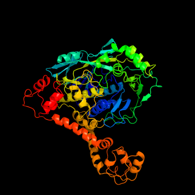
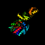
PDB header: oxidoreductaseChain: C: PDB Molecule: trna uridine 5-carboxymethylaminomethylPDBTitle: crystal structure of gida from chlorobium tepidum
2 c2zxiC_
100.0
52
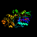
PDB header: fad-binding proteinChain: C: PDB Molecule: trna uridine 5-carboxymethylaminomethylPDBTitle: structure of aquifex aeolicus gida in the form ii crystal
3 c3g05B_
100.0
100
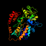
PDB header: rna binding proteinChain: B: PDB Molecule: trna uridine 5-carboxymethylaminomethyl modification enzymePDBTitle: crystal structure of n-terminal domain (2-550) of e.coli mnmg
4 c3cesB_
100.0
100
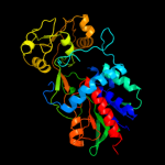
PDB header: rna binding proteinChain: B: PDB Molecule: trna uridine 5-carboxymethylaminomethyl modification enzymePDBTitle: crystal structure of e.coli mnmg (gida), a highly-conserved trna2 modifying enzyme
5 c3g5rA_
100.0
28
PDB header: transferaseChain: A: PDB Molecule: methylenetetrahydrofolate--trna-(uracil-5-)-PDBTitle: crystal structure of thermus thermophilus trmfo in complex with2 tetrahydrofolate
6 d2cula1
100.0
25
Fold: FAD/NAD(P)-binding domainSuperfamily: FAD/NAD(P)-binding domainFamily: GidA-like7 c2i0zA_
100.0
19
PDB header: oxidoreductaseChain: A: PDB Molecule: nad(fad)-utilizing dehydrogenases;PDBTitle: crystal structure of a fad binding protein from bacillus2 cereus, a putative nad(fad)-utilizing dehydrogenases
8 c1jrxA_
100.0
18
PDB header: oxidoreductaseChain: A: PDB Molecule: flavocytochrome c;PDBTitle: crystal structure of arg402ala mutant flavocytochrome c32 from shewanella frigidimarina
9 c3p4rM_
99.9
20
PDB header: oxidoreductaseChain: M: PDB Molecule: fumarate reductase flavoprotein subunit;PDBTitle: crystal structure of menaquinol:fumarate oxidoreductase in complex2 with glutarate
10 c1kf6A_
99.9
19
PDB header: oxidoreductaseChain: A: PDB Molecule: fumarate reductase flavoprotein;PDBTitle: e. coli quinol-fumarate reductase with bound inhibitor hqno
11 c2fjaC_
99.9
16
PDB header: oxidoreductaseChain: C: PDB Molecule: adenylylsulfate reductase, subunit a;PDBTitle: adenosine 5'-phosphosulfate reductase in complex with2 substrate
12 d2gqfa1
99.9
23
Fold: FAD/NAD(P)-binding domainSuperfamily: FAD/NAD(P)-binding domainFamily: HI0933 N-terminal domain-like13 c1d4cB_
99.9
16
PDB header: oxidoreductaseChain: B: PDB Molecule: flavocytochrome c fumarate reductase;PDBTitle: crystal structure of the uncomplexed form of the2 flavocytochrome c fumarate reductase of shewanella3 putrefaciens strain mr-1
14 c1yq4A_
99.9
17
PDB header: oxidoreductaseChain: A: PDB Molecule: succinate dehydrogenase flavoprotein subunit;PDBTitle: avian respiratory complex ii with 3-nitropropionate and ubiquinone
15 c1qo8A_
99.9
18
PDB header: oxidoreductaseChain: A: PDB Molecule: flavocytochrome c3 fumarate reductase;PDBTitle: the structure of the open conformation of a flavocytochrome2 c3 fumarate reductase
16 c2gqfA_
99.9
15
PDB header: structural genomics, unknown functionChain: A: PDB Molecule: hypothetical protein hi0933;PDBTitle: crystal structure of flavoprotein hi0933 from haemophilus influenzae2 rd
17 c3nlcA_
99.9
17
PDB header: structural genomics, unknown functionChain: A: PDB Molecule: uncharacterized protein vp0956;PDBTitle: crystal structure of the vp0956 protein from vibrio parahaemolyticus.2 northeast structural genomics consortium target vpr147
18 c2aczA_
99.9
16
PDB header: oxidoreductase/electron transportChain: A: PDB Molecule: succinate dehydrogenase flavoprotein subunit;PDBTitle: complex ii (succinate dehydrogenase) from e. coli with atpenin a52 inhibitor co-crystallized at the ubiquinone binding site
19 c3v76A_
99.9
16
PDB header: flavoproteinChain: A: PDB Molecule: flavoprotein;PDBTitle: the crystal structure of a flavoprotein from sinorhizobium meliloti
20 c2bs3A_
99.9
17
PDB header: oxidoreductaseChain: A: PDB Molecule: quinol-fumarate reductase flavoprotein subunit a;PDBTitle: glu c180 -> gln variant quinol:fumarate reductase from2 wolinella succinogenes
21 c3gyxA_
not modelled
99.9
17
PDB header: oxidoreductaseChain: A: PDB Molecule: adenylylsulfate reductase;PDBTitle: crystal structure of adenylylsulfate reductase from2 desulfovibrio gigas
22 c1chuA_
not modelled
99.9
20
PDB header: flavoenzymeChain: A: PDB Molecule: protein (l-aspartate oxidase);PDBTitle: structure of l-aspartate oxidase: implications for the2 succinate dehydrogenase/ fumarate reducatse family
23 c2e5vA_
not modelled
99.8
18
PDB header: oxidoreductaseChain: A: PDB Molecule: l-aspartate oxidase;PDBTitle: crystal structure of l-aspartate oxidase from2 hyperthermophilic archaeon sulfolobus tokodaii
24 d1qo8a2
not modelled
99.8
24
Fold: FAD/NAD(P)-binding domainSuperfamily: FAD/NAD(P)-binding domainFamily: Succinate dehydrogenase/fumarate reductase flavoprotein N-terminal domain25 d1y0pa2
not modelled
99.8
24
Fold: FAD/NAD(P)-binding domainSuperfamily: FAD/NAD(P)-binding domainFamily: Succinate dehydrogenase/fumarate reductase flavoprotein N-terminal domain26 d1d4ca2
not modelled
99.8
26
Fold: FAD/NAD(P)-binding domainSuperfamily: FAD/NAD(P)-binding domainFamily: Succinate dehydrogenase/fumarate reductase flavoprotein N-terminal domain27 c1x31A_
not modelled
99.8
17
PDB header: oxidoreductaseChain: A: PDB Molecule: sarcosine oxidase alpha subunit;PDBTitle: crystal structure of heterotetrameric sarcosine oxidase from2 corynebacterium sp. u-96
28 c1hyuA_
not modelled
99.8
18
PDB header: oxidoreductaseChain: A: PDB Molecule: alkyl hydroperoxide reductase subunit f;PDBTitle: crystal structure of intact ahpf
29 d1chua2
not modelled
99.8
28
Fold: FAD/NAD(P)-binding domainSuperfamily: FAD/NAD(P)-binding domainFamily: Succinate dehydrogenase/fumarate reductase flavoprotein N-terminal domain30 d2i0za1
not modelled
99.7
21
Fold: FAD/NAD(P)-binding domainSuperfamily: FAD/NAD(P)-binding domainFamily: HI0933 N-terminal domain-like31 d2bs2a2
not modelled
99.7
24
Fold: FAD/NAD(P)-binding domainSuperfamily: FAD/NAD(P)-binding domainFamily: Succinate dehydrogenase/fumarate reductase flavoprotein N-terminal domain32 c3ab1B_
not modelled
99.7
15
PDB header: oxidoreductaseChain: B: PDB Molecule: ferredoxin--nadp reductase;PDBTitle: crystal structure of ferredoxin nadp+ oxidoreductase
33 c3r9uA_
not modelled
99.7
16
PDB header: oxidoreductaseChain: A: PDB Molecule: thioredoxin reductase;PDBTitle: thioredoxin-disulfide reductase from campylobacter jejuni.
34 c3fbsB_
not modelled
99.7
17
PDB header: oxidoreductaseChain: B: PDB Molecule: oxidoreductase;PDBTitle: the crystal structure of the oxidoreductase from agrobacterium2 tumefaciens
35 c2v6oA_
not modelled
99.7
16
PDB header: oxidoreductaseChain: A: PDB Molecule: thioredoxin glutathione reductase;PDBTitle: structure of schistosoma mansoni thioredoxin-glutathione2 reductase (smtgr)
36 d1jnra2
not modelled
99.7
20
Fold: FAD/NAD(P)-binding domainSuperfamily: FAD/NAD(P)-binding domainFamily: Succinate dehydrogenase/fumarate reductase flavoprotein N-terminal domain37 c2zbwA_
not modelled
99.7
14
PDB header: oxidoreductaseChain: A: PDB Molecule: thioredoxin reductase;PDBTitle: crystal structure of thioredoxin reductase-like protein from thermus2 thermophilus hb8
38 c3f8rD_
not modelled
99.6
17
PDB header: oxidoreductaseChain: D: PDB Molecule: thioredoxin reductase (trxb-3);PDBTitle: crystal structure of sulfolobus solfataricus thioredoxin2 reductase b3 in complex with two nadp molecules
39 c2w0hA_
not modelled
99.6
22
PDB header: oxidoreductaseChain: A: PDB Molecule: trypanothione reductase;PDBTitle: x ray structure of leishmania infantum trypanothione2 reductase in complex with antimony and nadph
40 d1lpfa1
not modelled
99.6
18
Fold: FAD/NAD(P)-binding domainSuperfamily: FAD/NAD(P)-binding domainFamily: FAD/NAD-linked reductases, N-terminal and central domains41 c1ndaD_
not modelled
99.6
23
PDB header: oxidoreductaseChain: D: PDB Molecule: trypanothione oxidoreductase;PDBTitle: the structure of trypanosoma cruzi trypanothione reductase2 in the oxidized and nadph reduced state
42 c2gmhA_
not modelled
99.6
16
PDB header: oxidoreductaseChain: A: PDB Molecule: electron transfer flavoprotein-ubiquinonePDBTitle: structure of porcine electron transfer flavoprotein-2 ubiquinone oxidoreductase in complexed with ubiquinone
43 c3o0hA_
not modelled
99.6
27
PDB header: oxidoreductaseChain: A: PDB Molecule: glutathione reductase;PDBTitle: crystal structure of glutathione reductase from bartonella henselae
44 c1fl2A_
not modelled
99.6
17
PDB header: oxidoreductaseChain: A: PDB Molecule: alkyl hydroperoxide reductase subunit f;PDBTitle: catalytic core component of the alkylhydroperoxide reductase ahpf from2 e.coli
45 d1kf6a2
not modelled
99.6
19
Fold: FAD/NAD(P)-binding domainSuperfamily: FAD/NAD(P)-binding domainFamily: Succinate dehydrogenase/fumarate reductase flavoprotein N-terminal domain46 c2q0lA_
not modelled
99.6
18
PDB header: oxidoreductaseChain: A: PDB Molecule: thioredoxin reductase;PDBTitle: helicobacter pylori thioredoxin reductase reduced by sodium dithionite2 in complex with nadp+
47 c1ojtA_
not modelled
99.6
16
PDB header: oxidoreductaseChain: A: PDB Molecule: surface protein;PDBTitle: structure of dihydrolipoamide dehydrogenase
48 d3grsa1
not modelled
99.6
20
Fold: FAD/NAD(P)-binding domainSuperfamily: FAD/NAD(P)-binding domainFamily: FAD/NAD-linked reductases, N-terminal and central domains49 c2c3dB_
not modelled
99.6
14
PDB header: oxidoreductaseChain: B: PDB Molecule: 2-oxopropyl-com reductase;PDBTitle: 2.15 angstrom crystal structure of 2-ketopropyl coenzyme m2 oxidoreductase carboxylase with a coenzyme m disulfide3 bound at the active site
50 c1tytA_
not modelled
99.6
10
PDB header: oxidoreductaseChain: A: PDB Molecule: trypanothione reductase, oxidized form;PDBTitle: crystal and molecular structure of crithidia fasciculata2 trypanothione reductase at 2.6 angstroms resolution
51 c2a87A_
not modelled
99.6
18
PDB header: oxidoreductaseChain: A: PDB Molecule: thioredoxin reductase;PDBTitle: crystal structure of m. tuberculosis thioredoxin reductase
52 c1f6mF_
not modelled
99.6
13
PDB header: oxidoreductaseChain: F: PDB Molecule: thioredoxin reductase;PDBTitle: crystal structure of a complex between thioredoxin2 reductase, thioredoxin, and the nadp+ analog, aadp+
53 c1zmcG_
not modelled
99.6
13
PDB header: oxidoreductaseChain: G: PDB Molecule: dihydrolipoyl dehydrogenase;PDBTitle: crystal structure of human dihydrolipoamide dehydrogenase2 complexed to nad+
54 c3lzxB_
not modelled
99.6
17
PDB header: oxidoreductaseChain: B: PDB Molecule: ferredoxin--nadp reductase 2;PDBTitle: crystal structure of ferredoxin-nadp+ oxidoreductase from bacillus2 subtilis (form ii)
55 c3da1A_
not modelled
99.6
19
PDB header: oxidoreductaseChain: A: PDB Molecule: glycerol-3-phosphate dehydrogenase;PDBTitle: x-ray structure of the glycerol-3-phosphate dehydrogenase2 from bacillus halodurans complexed with fad. northeast3 structural genomics consortium target bhr167.
56 c3jskN_
not modelled
99.6
23
PDB header: biosynthetic proteinChain: N: PDB Molecule: cypbp37 protein;PDBTitle: thiazole synthase from neurospora crassa
57 c2eq8E_
not modelled
99.6
35
PDB header: oxidoreductaseChain: E: PDB Molecule: pyruvate dehydrogenase complex, dihydrolipoamidePDBTitle: crystal structure of lipoamide dehydrogenase from thermus thermophilus2 hb8 with psbdp
58 c1vdcA_
not modelled
99.5
19
PDB header: oxidoreductaseChain: A: PDB Molecule: nadph dependent thioredoxin reductase;PDBTitle: structure of nadph dependent thioredoxin reductase
59 d3lada1
not modelled
99.5
18
Fold: FAD/NAD(P)-binding domainSuperfamily: FAD/NAD(P)-binding domainFamily: FAD/NAD-linked reductases, N-terminal and central domains60 c3dgzA_
not modelled
99.5
11
PDB header: oxidoreductaseChain: A: PDB Molecule: thioredoxin reductase 2;PDBTitle: crystal structure of mouse mitochondrial thioredoxin reductase, c-2 terminal 3-residue truncation
61 d1rp0a1
not modelled
99.5
21
Fold: FAD/NAD(P)-binding domainSuperfamily: FAD/NAD(P)-binding domainFamily: Thi4-like62 c1y56A_
not modelled
99.5
18
PDB header: oxidoreductaseChain: A: PDB Molecule: hypothetical protein ph1363;PDBTitle: crystal structure of l-proline dehydrogenase from p.horikoshii
63 d1w4xa1
not modelled
99.5
19
Fold: FAD/NAD(P)-binding domainSuperfamily: FAD/NAD(P)-binding domainFamily: FAD/NAD-linked reductases, N-terminal and central domains64 c2rghA_
not modelled
99.5
21
PDB header: oxidoreductaseChain: A: PDB Molecule: alpha-glycerophosphate oxidase;PDBTitle: structure of alpha-glycerophosphate oxidase from2 streptococcus sp.: a template for the mitochondrial alpha-3 glycerophosphate dehydrogenase
65 c1dxlC_
not modelled
99.5
15
PDB header: oxidoreductaseChain: C: PDB Molecule: dihydrolipoamide dehydrogenase;PDBTitle: dihydrolipoamide dehydrogenase of glycine decarboxylase2 from pisum sativum
66 c3ic9D_
not modelled
99.5
19
PDB header: oxidoreductaseChain: D: PDB Molecule: dihydrolipoamide dehydrogenase;PDBTitle: the structure of dihydrolipoamide dehydrogenase from colwellia2 psychrerythraea 34h.
67 d1lvla1
not modelled
99.5
20
Fold: FAD/NAD(P)-binding domainSuperfamily: FAD/NAD(P)-binding domainFamily: FAD/NAD-linked reductases, N-terminal and central domains68 c2q7vA_
not modelled
99.5
17
PDB header: oxidoreductaseChain: A: PDB Molecule: thioredoxin reductase;PDBTitle: crystal structure of deinococcus radiodurans thioredoxin2 reductase
69 c3ka7A_
not modelled
99.5
19
PDB header: oxidoreductaseChain: A: PDB Molecule: oxidoreductase;PDBTitle: crystal structure of an oxidoreductase from methanosarcina2 mazei. northeast structural genomics consortium target id3 mar208
70 c1v59B_
not modelled
99.5
14
PDB header: oxidoreductaseChain: B: PDB Molecule: dihydrolipoamide dehydrogenase;PDBTitle: crystal structure of yeast lipoamide dehydrogenase2 complexed with nad+
71 c2hqmB_
not modelled
99.5
13
PDB header: oxidoreductaseChain: B: PDB Molecule: glutathione reductase;PDBTitle: crystal structure of glutathione reductase glr1 from the yeast2 saccharomyces cerevisiae
72 d1ryia1
not modelled
99.5
17
Fold: FAD/NAD(P)-binding domainSuperfamily: FAD/NAD(P)-binding domainFamily: FAD-linked reductases, N-terminal domain73 d1neka2
not modelled
99.5
23
Fold: FAD/NAD(P)-binding domainSuperfamily: FAD/NAD(P)-binding domainFamily: Succinate dehydrogenase/fumarate reductase flavoprotein N-terminal domain74 c1ltxR_
not modelled
99.5
20
PDB header: transferase/protein bindingChain: R: PDB Molecule: rab escort protein 1;PDBTitle: structure of rab escort protein-1 in complex with rab2 geranylgeranyl transferase and isoprenoid
75 d1dxla1
not modelled
99.5
21
Fold: FAD/NAD(P)-binding domainSuperfamily: FAD/NAD(P)-binding domainFamily: FAD/NAD-linked reductases, N-terminal and central domains76 c3d8xB_
not modelled
99.5
15
PDB header: oxidoreductaseChain: B: PDB Molecule: thioredoxin reductase 1;PDBTitle: crystal structure of saccharomyces cerevisiae nadph dependent2 thioredoxin reductase 1
77 c2rgoA_
not modelled
99.5
23
PDB header: oxidoreductaseChain: A: PDB Molecule: alpha-glycerophosphate oxidase;PDBTitle: structure of alpha-glycerophosphate oxidase from2 streptococcus sp.: a template for the mitochondrial alpha-3 glycerophosphate dehydrogenase
78 c3ctyA_
not modelled
99.5
16
PDB header: oxidoreductaseChain: A: PDB Molecule: thioredoxin reductase;PDBTitle: crystal structure of t. acidophilum thioredoxin reductase
79 d1h6va1
not modelled
99.5
18
Fold: FAD/NAD(P)-binding domainSuperfamily: FAD/NAD(P)-binding domainFamily: FAD/NAD-linked reductases, N-terminal and central domains80 d1feca1
not modelled
99.5
16
Fold: FAD/NAD(P)-binding domainSuperfamily: FAD/NAD(P)-binding domainFamily: FAD/NAD-linked reductases, N-terminal and central domains81 c1lpfB_
not modelled
99.5
13
PDB header: oxidoreductaseChain: B: PDB Molecule: dihydrolipoamide dehydrogenase;PDBTitle: three-dimensional structure of lipoamide dehydrogenase from2 pseudomonas fluorescens at 2.8 angstroms resolution.3 analysis of redox and thermostability properties
82 c1s3bB_
not modelled
99.5
18
PDB header: oxidoreductaseChain: B: PDB Molecule: amine oxidase [flavin-containing] b;PDBTitle: crystal structure of maob in complex with n-methyl-n-2 propargyl-1(r)-aminoindan
83 d1pj5a2
not modelled
99.5
21
Fold: FAD/NAD(P)-binding domainSuperfamily: FAD/NAD(P)-binding domainFamily: FAD-linked reductases, N-terminal domain84 c3atrA_
not modelled
99.5
23
PDB header: oxidoreductaseChain: A: PDB Molecule: conserved archaeal protein;PDBTitle: geranylgeranyl reductase (ggr) from sulfolobus acidocaldarius co-2 crystallized with its ligand
85 c3dmeB_
not modelled
99.5
17
PDB header: structural genomics, unknown functionChain: B: PDB Molecule: conserved exported protein;PDBTitle: crystal structure of conserved exported protein from2 bordetella pertussis. northeast structural genomics target3 ber141
86 c1pj6A_
not modelled
99.5
23
PDB header: oxidoreductaseChain: A: PDB Molecule: n,n-dimethylglycine oxidase;PDBTitle: crystal structure of dimethylglycine oxidase of arthrobacter2 globiformis in complex with folic acid
87 c2a8xA_
not modelled
99.5
14
PDB header: oxidoreductaseChain: A: PDB Molecule: dihydrolipoyl dehydrogenase;PDBTitle: crystal structure of lipoamide dehydrogenase from2 mycobacterium tuberculosis
88 d2gf3a1
not modelled
99.5
21
Fold: FAD/NAD(P)-binding domainSuperfamily: FAD/NAD(P)-binding domainFamily: FAD-linked reductases, N-terminal domain89 c2r4jA_
not modelled
99.5
20
PDB header: oxidoreductaseChain: A: PDB Molecule: aerobic glycerol-3-phosphate dehydrogenase;PDBTitle: crystal structure of escherichia coli semet substituted2 glycerol-3-phosphate dehydrogenase in complex with dhap
90 c2ardA_
not modelled
99.5
17
PDB header: biosynthetic proteinChain: A: PDB Molecule: tryptophan halogenase prna;PDBTitle: the structure of tryptophan 7-halogenase (prna) suggests a mechanism2 for regioselective chlorination
91 c3k30B_
not modelled
99.5
24
PDB header: oxidoreductaseChain: B: PDB Molecule: histamine dehydrogenase;PDBTitle: histamine dehydrogenase from nocardiodes simplex
92 c3djeA_
not modelled
99.5
19
PDB header: oxidoreductaseChain: A: PDB Molecule: fructosyl amine: oxygen oxidoreductase;PDBTitle: crystal structure of the deglycating enzyme fructosamine2 oxidase from aspergillus fumigatus (amadoriase ii) in3 complex with fsa
93 c1w4xA_
not modelled
99.4
19
PDB header: oxygenaseChain: A: PDB Molecule: phenylacetone monooxygenase;PDBTitle: phenylacetone monooxygenase, a baeyer-villiger2 monooxygenase
94 d1vg0a1
not modelled
99.4
20
Fold: FAD/NAD(P)-binding domainSuperfamily: FAD/NAD(P)-binding domainFamily: GDI-like N domain95 c3e1tA_
not modelled
99.4
23
PDB header: flavoproteinChain: A: PDB Molecule: halogenase;PDBTitle: structure and action of the myxobacterial chondrochloren2 halogenase cndh, a new variant of fad-dependent halogenases
96 d1d5ta1
not modelled
99.4
21
Fold: FAD/NAD(P)-binding domainSuperfamily: FAD/NAD(P)-binding domainFamily: GDI-like N domain97 d2gmha1
not modelled
99.4
13
Fold: FAD/NAD(P)-binding domainSuperfamily: FAD/NAD(P)-binding domainFamily: FAD-linked reductases, N-terminal domain98 c2cfyB_
not modelled
99.4
12
PDB header: oxidoreductaseChain: B: PDB Molecule: thioredoxin reductase 1;PDBTitle: crystal structure of human thioredoxin reductase 1
99 d1ojta1
not modelled
99.4
17
Fold: FAD/NAD(P)-binding domainSuperfamily: FAD/NAD(P)-binding domainFamily: FAD/NAD-linked reductases, N-terminal and central domains100 c1bwcA_
not modelled
99.4
13
PDB header: oxidoreductaseChain: A: PDB Molecule: protein (glutathione reductase);PDBTitle: structure of human glutathione reductase complexed with ajoene2 inhibitor and subversive substrate
101 c2pyxA_
not modelled
99.4
15
PDB header: biosynthetic proteinChain: A: PDB Molecule: tryptophan halogenase;PDBTitle: crystal structure of tryptophan halogenase (yp_750003.1) from2 shewanella frigidimarina ncimb 400 at 1.50 a resolution
102 c1fcdB_
not modelled
99.4
17
PDB header: electron transport(flavocytochrome)Chain: B: PDB Molecule: flavocytochrome c sulfide dehydrogenase (flavin-PDBTitle: the structure of flavocytochrome c sulfide dehydrogenase2 from a purple phototrophic bacterium chromatium vinosum at3 2.5 angstroms resolution
103 c3gwdA_
not modelled
99.4
16
PDB header: oxidoreductaseChain: A: PDB Molecule: cyclohexanone monooxygenase;PDBTitle: closed crystal structure of cyclohexanone monooxygenase
104 d1v59a1
not modelled
99.4
22
Fold: FAD/NAD(P)-binding domainSuperfamily: FAD/NAD(P)-binding domainFamily: FAD/NAD-linked reductases, N-terminal and central domains105 c3i3lA_
not modelled
99.4
16
PDB header: hydrolaseChain: A: PDB Molecule: alkylhalidase cmls;PDBTitle: crystal structure of cmls, a flavin-dependent halogenase
106 d2bcgg1
not modelled
99.4
19
Fold: FAD/NAD(P)-binding domainSuperfamily: FAD/NAD(P)-binding domainFamily: GDI-like N domain107 c2e4gB_
not modelled
99.4
20
PDB header: biosynthetic protein, flavoproteinChain: B: PDB Molecule: tryptophan halogenase;PDBTitle: rebh with bound l-trp
108 c1lvlA_
not modelled
99.4
15
PDB header: oxidoreductaseChain: A: PDB Molecule: dihydrolipoamide dehydrogenase;PDBTitle: the refined structure of pseudomonas putida lipoamide dehydrogenase2 complexed with nad+ at 2.45 angstroms resolution
109 c1geuA_
not modelled
99.4
20
PDB header: oxidoreductase(flavoenzyme)Chain: A: PDB Molecule: glutathione reductase;PDBTitle: anatomy of an engineered nad-binding site
110 c1gthD_
not modelled
99.4
14
PDB header: oxidoreductaseChain: D: PDB Molecule: dihydropyrimidine dehydrogenase;PDBTitle: dihydropyrimidine dehydrogenase (dpd) from pig, ternary2 complex with nadph and 5-iodouracil
111 c3l8kB_
not modelled
99.4
14
PDB header: oxidoreductaseChain: B: PDB Molecule: dihydrolipoyl dehydrogenase;PDBTitle: crystal structure of a dihydrolipoyl dehydrogenase from2 sulfolobus solfataricus
112 d1aoga1
not modelled
99.4
13
Fold: FAD/NAD(P)-binding domainSuperfamily: FAD/NAD(P)-binding domainFamily: FAD/NAD-linked reductases, N-terminal and central domains113 c1zx9A_
not modelled
99.4
15
PDB header: oxidoreductaseChain: A: PDB Molecule: mercuric reductase;PDBTitle: crystal structure of tn501 mera
114 c2qaeA_
not modelled
99.4
23
PDB header: oxidoreductaseChain: A: PDB Molecule: dihydrolipoyl dehydrogenase;PDBTitle: crystal structure analysis of trypanosoma cruzi lipoamide2 dehydrogenase
115 d1trba1
not modelled
99.4
22
Fold: FAD/NAD(P)-binding domainSuperfamily: FAD/NAD(P)-binding domainFamily: FAD/NAD-linked reductases, N-terminal and central domains116 c2olnA_
not modelled
99.4
23
PDB header: oxidoreductaseChain: A: PDB Molecule: nikd protein;PDBTitle: nikd, an unusual amino acid oxidase essential for2 nikkomycin biosynthesis: closed form at 1.15 a resolution
117 c2eq7B_
not modelled
99.4
23
PDB header: oxidoreductaseChain: B: PDB Molecule: 2-oxoglutarate dehydrogenase e3 component;PDBTitle: crystal structure of lipoamide dehydrogenase from thermus thermophilus2 hb8 with psbdo
118 c1zkqA_
not modelled
99.4
19
PDB header: oxidoreductaseChain: A: PDB Molecule: thioredoxin reductase 2, mitochondrial;PDBTitle: crystal structure of mouse thioredoxin reductase type 2
119 c1ebdB_
not modelled
99.4
13
PDB header: complex (oxidoreductase/transferase)Chain: B: PDB Molecule: dihydrolipoamide dehydrogenase;PDBTitle: dihydrolipoamide dehydrogenase complexed with the binding2 domain of the dihydrolipoamide acetylase
120 c3ps9A_
not modelled
99.4
18
PDB header: transferaseChain: A: PDB Molecule: trna 5-methylaminomethyl-2-thiouridine biosynthesisPDBTitle: crystal structure of mnmc from e. coli