1 c3s29C_
100.0
14
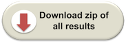
PDB header: transferaseChain: C: PDB Molecule: sucrose synthase 1;PDBTitle: the crystal structure of sucrose synthase-1 from arabidopsis thaliana2 and its functional implications.
2 c3oy2A_
100.0
13
PDB header: viral protein,transferaseChain: A: PDB Molecule: glycosyltransferase b736l;PDBTitle: crystal structure of a putative glycosyltransferase from paramecium2 bursaria chlorella virus ny2a
3 c2r60A_
100.0
12
PDB header: transferaseChain: A: PDB Molecule: glycosyl transferase, group 1;PDBTitle: structure of apo sucrose phosphate synthase (sps) of2 halothermothrix orenii
4 c3c4vB_
100.0
13
PDB header: transferaseChain: B: PDB Molecule: predicted glycosyltransferases;PDBTitle: structure of the retaining glycosyltransferase msha:the2 first step in mycothiol biosynthesis. organism:3 corynebacterium glutamicum : complex with udp and 1l-ins-1-4 p.
5 c3okaA_
100.0
13
PDB header: transferaseChain: A: PDB Molecule: gdp-mannose-dependent alpha-(1-6)-phosphatidylinositolPDBTitle: crystal structure of corynebacterium glutamicum pimb' in complex with2 gdp-man (triclinic crystal form)
6 c2jjmH_
100.0
13
PDB header: transferaseChain: H: PDB Molecule: glycosyl transferase, group 1 family protein;PDBTitle: crystal structure of a family gt4 glycosyltransferase from2 bacillus anthracis orf ba1558.
7 d2bisa1
100.0
14
Fold: UDP-Glycosyltransferase/glycogen phosphorylaseSuperfamily: UDP-Glycosyltransferase/glycogen phosphorylaseFamily: Glycosyl transferases group 18 c2qzsA_
100.0
14
PDB header: transferaseChain: A: PDB Molecule: glycogen synthase;PDBTitle: crystal structure of wild-type e.coli gs in complex with adp2 and glucose(wtgsb)
9 c2xmpB_
100.0
14
PDB header: sugar binding proteinChain: B: PDB Molecule: trehalose-synthase tret;PDBTitle: crystal structure of trehalose synthase tret mutant e326a2 from p.horishiki in complex with udp
10 c2x6rA_
100.0
14
PDB header: isomeraseChain: A: PDB Molecule: trehalose-synthase tret;PDBTitle: crystal structure of trehalose synthase tret from p.2 horikoshi produced by soaking in trehalose
11 c2gejA_
100.0
13
PDB header: transferaseChain: A: PDB Molecule: phosphatidylinositol mannosyltransferase (pima);PDBTitle: crystal structure of phosphatidylinositol mannosyltransferase (pima)2 from mycobacterium smegmatis in complex with gdp-man
12 c2x0dA_
100.0
15
PDB header: transferaseChain: A: PDB Molecule: wsaf;PDBTitle: apo structure of wsaf
13 c1uquB_
100.0
12
PDB header: synthaseChain: B: PDB Molecule: alpha, alpha-trehalose-phosphate synthase;PDBTitle: trehalose-6-phosphate from e. coli bound with udp-glucose.
14 d1rzua_
100.0
14
Fold: UDP-Glycosyltransferase/glycogen phosphorylaseSuperfamily: UDP-Glycosyltransferase/glycogen phosphorylaseFamily: Glycosyl transferases group 115 d2iw1a1
100.0
12
Fold: UDP-Glycosyltransferase/glycogen phosphorylaseSuperfamily: UDP-Glycosyltransferase/glycogen phosphorylaseFamily: Glycosyl transferases group 116 d1uqta_
100.0
14
Fold: UDP-Glycosyltransferase/glycogen phosphorylaseSuperfamily: UDP-Glycosyltransferase/glycogen phosphorylaseFamily: Trehalose-6-phosphate synthase, OtsA17 c3o3cD_
100.0
13
PDB header: transferaseChain: D: PDB Molecule: glycogen [starch] synthase isoform 2;PDBTitle: glycogen synthase basal state udp complex
18 c3nb0A_
100.0
12
PDB header: transferaseChain: A: PDB Molecule: glycogen [starch] synthase isoform 2;PDBTitle: glucose-6-phosphate activated form of yeast glycogen synthase
19 c3rhzB_
100.0
9
PDB header: transferaseChain: B: PDB Molecule: nucleotide sugar synthetase-like protein;PDBTitle: structure and functional analysis of a new subfamily of2 glycosyltransferases required for glycosylation of serine-rich3 streptococcal adhesions
20 c3dzcA_
100.0
12
PDB header: isomeraseChain: A: PDB Molecule: udp-n-acetylglucosamine 2-epimerase;PDBTitle: 2.35 angstrom resolution structure of wecb (vc0917), a udp-n-2 acetylglucosamine 2-epimerase from vibrio cholerae.
21 c3ot5D_
not modelled
100.0
12
PDB header: isomeraseChain: D: PDB Molecule: udp-n-acetylglucosamine 2-epimerase;PDBTitle: 2.2 angstrom resolution crystal structure of putative udp-n-2 acetylglucosamine 2-epimerase from listeria monocytogenes
22 d1f6da_
not modelled
100.0
11
Fold: UDP-Glycosyltransferase/glycogen phosphorylaseSuperfamily: UDP-Glycosyltransferase/glycogen phosphorylaseFamily: UDP-N-acetylglucosamine 2-epimerase23 d1v4va_
not modelled
100.0
12
Fold: UDP-Glycosyltransferase/glycogen phosphorylaseSuperfamily: UDP-Glycosyltransferase/glycogen phosphorylaseFamily: UDP-N-acetylglucosamine 2-epimerase24 c2iv3B_
not modelled
100.0
10
PDB header: transferaseChain: B: PDB Molecule: glycosyltransferase;PDBTitle: crystal structure of avigt4, a glycosyltransferase involved2 in avilamycin a biosynthesis
25 c2q6vA_
not modelled
100.0
11
PDB header: transferaseChain: A: PDB Molecule: glucuronosyltransferase gumk;PDBTitle: crystal structure of gumk in complex with udp
26 c2xcuC_
not modelled
100.0
13
PDB header: transferaseChain: C: PDB Molecule: 3-deoxy-d-manno-2-octulosonic acid transferase;PDBTitle: membrane-embedded monofunctional glycosyltransferase waaa of aquifex2 aeolicus, comlex with cmp
27 d2f9fa1
not modelled
99.9
20
Fold: UDP-Glycosyltransferase/glycogen phosphorylaseSuperfamily: UDP-Glycosyltransferase/glycogen phosphorylaseFamily: Glycosyl transferases group 128 d1o6ca_
not modelled
99.9
12
Fold: UDP-Glycosyltransferase/glycogen phosphorylaseSuperfamily: UDP-Glycosyltransferase/glycogen phosphorylaseFamily: UDP-N-acetylglucosamine 2-epimerase29 c3qhpB_
not modelled
99.9
15
PDB header: transferaseChain: B: PDB Molecule: type 1 capsular polysaccharide biosynthesis protein jPDBTitle: crystal structure of the catalytic domain of cholesterol-alpha-2 glucosyltransferase from helicobacter pylori
30 d1f0ka_
not modelled
99.8
9
Fold: UDP-Glycosyltransferase/glycogen phosphorylaseSuperfamily: UDP-Glycosyltransferase/glycogen phosphorylaseFamily: Peptidoglycan biosynthesis glycosyltransferase MurG31 c2vsnB_
not modelled
99.8
11
PDB header: transferaseChain: B: PDB Molecule: xcogt;PDBTitle: structure and topological arrangement of an o-glcnac2 transferase homolog: insight into molecular control of3 intracellular glycosylation
32 d2bfwa1
not modelled
99.8
16
Fold: UDP-Glycosyltransferase/glycogen phosphorylaseSuperfamily: UDP-Glycosyltransferase/glycogen phosphorylaseFamily: Glycosyl transferases group 133 c3ia7A_
not modelled
99.8
4
PDB header: transferaseChain: A: PDB Molecule: calg4;PDBTitle: crystal structure of calg4, the calicheamicin glycosyltransferase
34 c3othB_
not modelled
99.8
8
PDB header: transferase/antibioticChain: B: PDB Molecule: calg1;PDBTitle: crystal structure of calg1, calicheamicin glycostyltransferase, tdp2 and calicheamicin alpha3i bound form
35 c3iaaB_
not modelled
99.8
10
PDB header: transferaseChain: B: PDB Molecule: calg2;PDBTitle: crystal structure of calg2, calicheamicin glycosyltransferase, tdp2 bound form
36 c3pe3D_
not modelled
99.7
7
PDB header: transferaseChain: D: PDB Molecule: udp-n-acetylglucosamine--peptide n-PDBTitle: structure of human o-glcnac transferase and its complex with a peptide2 substrate
37 c2iyaB_
not modelled
99.7
6
PDB header: transferaseChain: B: PDB Molecule: oleandomycin glycosyltransferase;PDBTitle: the crystal structure of macrolide glycosyltransferases: a2 blueprint for antibiotic engineering
38 c2iyfA_
not modelled
99.7
7
PDB header: transferaseChain: A: PDB Molecule: oleandomycin glycosyltransferase;PDBTitle: the crystal structure of macrolide glycosyltransferases: a2 blueprint for antibiotic engineering
39 c2p6pB_
not modelled
99.6
6
PDB header: transferaseChain: B: PDB Molecule: glycosyl transferase;PDBTitle: x-ray crystal structure of c-c bond-forming dtdp-d-olivose-transferase2 urdgt2
40 d1iira_
not modelled
99.6
9
Fold: UDP-Glycosyltransferase/glycogen phosphorylaseSuperfamily: UDP-Glycosyltransferase/glycogen phosphorylaseFamily: Gtf glycosyltransferase41 d1pn3a_
not modelled
99.5
8
Fold: UDP-Glycosyltransferase/glycogen phosphorylaseSuperfamily: UDP-Glycosyltransferase/glycogen phosphorylaseFamily: Gtf glycosyltransferase42 c3d0qB_
not modelled
99.4
10
PDB header: transferaseChain: B: PDB Molecule: protein calg3;PDBTitle: crystal structure of calg3 from micromonospora echinospora determined2 in space group i222
43 d1rrva_
not modelled
99.3
10
Fold: UDP-Glycosyltransferase/glycogen phosphorylaseSuperfamily: UDP-Glycosyltransferase/glycogen phosphorylaseFamily: Gtf glycosyltransferase44 c3hbmA_
not modelled
98.7
11
PDB header: hydrolaseChain: A: PDB Molecule: udp-sugar hydrolase;PDBTitle: crystal structure of pseg from campylobacter jejuni
45 c3l7mC_
not modelled
98.4
9
PDB header: structural proteinChain: C: PDB Molecule: teichoic acid biosynthesis protein f;PDBTitle: structure of the wall teichoic acid polymerase tagf, h548a
46 d2c1xa1
not modelled
98.0
7
Fold: UDP-Glycosyltransferase/glycogen phosphorylaseSuperfamily: UDP-Glycosyltransferase/glycogen phosphorylaseFamily: UDPGT-like47 d2acva1
not modelled
97.8
11
Fold: UDP-Glycosyltransferase/glycogen phosphorylaseSuperfamily: UDP-Glycosyltransferase/glycogen phosphorylaseFamily: UDPGT-like48 d1ygpa_
not modelled
97.7
16
Fold: UDP-Glycosyltransferase/glycogen phosphorylaseSuperfamily: UDP-Glycosyltransferase/glycogen phosphorylaseFamily: Oligosaccharide phosphorylase49 d2gj4a1
not modelled
97.7
16
Fold: UDP-Glycosyltransferase/glycogen phosphorylaseSuperfamily: UDP-Glycosyltransferase/glycogen phosphorylaseFamily: Oligosaccharide phosphorylase50 c2c4mA_
not modelled
97.6
17
PDB header: transferaseChain: A: PDB Molecule: glycogen phosphorylase;PDBTitle: starch phosphorylase: structural studies explain oxyanion-2 dependent kinetic stability and regulatory control.
51 c3ddsB_
not modelled
97.6
16
PDB header: transferaseChain: B: PDB Molecule: glycogen phosphorylase, liver form;PDBTitle: crystal structure of glycogen phosphorylase complexed with an2 anthranilimide based inhibitor gsk261
52 d1l5wa_
not modelled
97.5
19
Fold: UDP-Glycosyltransferase/glycogen phosphorylaseSuperfamily: UDP-Glycosyltransferase/glycogen phosphorylaseFamily: Oligosaccharide phosphorylase53 c3tovB_
not modelled
97.5
11
PDB header: transferaseChain: B: PDB Molecule: glycosyl transferase family 9;PDBTitle: the crystal structure of the glycosyl transferase family 9 from2 veillonella parvula dsm 2008
54 d2pq6a1
not modelled
97.5
12
Fold: UDP-Glycosyltransferase/glycogen phosphorylaseSuperfamily: UDP-Glycosyltransferase/glycogen phosphorylaseFamily: UDPGT-like55 c2o6lA_
not modelled
97.5
11
PDB header: transferaseChain: A: PDB Molecule: udp-glucuronosyltransferase 2b7;PDBTitle: crystal structure of the udp-glucuronic acid binding domain2 of the human drug metabolizing udp-glucuronosyltransferase3 2b7
56 d2atia1
not modelled
97.3
16
Fold: UDP-Glycosyltransferase/glycogen phosphorylaseSuperfamily: UDP-Glycosyltransferase/glycogen phosphorylaseFamily: Oligosaccharide phosphorylase57 d2vcha1
not modelled
97.2
7
Fold: UDP-Glycosyltransferase/glycogen phosphorylaseSuperfamily: UDP-Glycosyltransferase/glycogen phosphorylaseFamily: UDPGT-like58 c3hbjA_
not modelled
97.1
10
PDB header: transferaseChain: A: PDB Molecule: flavonoid 3-o-glucosyltransferase;PDBTitle: structure of ugt78g1 complexed with udp
59 c3q3hA_
not modelled
97.1
9
PDB header: transferaseChain: A: PDB Molecule: hmw1c-like glycosyltransferase;PDBTitle: crystal structure of the actinobacillus pleuropneumoniae hmw1c2 glycosyltransferase in complex with udp-glc
60 c2h1fB_
not modelled
96.8
13
PDB header: transferaseChain: B: PDB Molecule: lipopolysaccharide heptosyltransferase-1;PDBTitle: e. coli heptosyltransferase waac with adp
61 d1pswa_
not modelled
96.3
11
Fold: UDP-Glycosyltransferase/glycogen phosphorylaseSuperfamily: UDP-Glycosyltransferase/glycogen phosphorylaseFamily: ADP-heptose LPS heptosyltransferase II62 c2jzcA_
not modelled
94.6
8
PDB header: transferaseChain: A: PDB Molecule: udp-n-acetylglucosamine transferase subunitPDBTitle: nmr solution structure of alg13: the sugar donor subunit of2 a yeast n-acetylglucosamine transferase. northeast3 structural genomics consortium target yg1
63 c2iz6A_
not modelled
85.8
9
PDB header: metal transportChain: A: PDB Molecule: molybdenum cofactor carrier protein;PDBTitle: structure of the chlamydomonas rheinhardtii moco carrier2 protein
64 d1d4aa_
not modelled
79.9
18
Fold: Flavodoxin-likeSuperfamily: FlavoproteinsFamily: Quinone reductase65 c2yy7B_
not modelled
71.0
11
PDB header: oxidoreductaseChain: B: PDB Molecule: l-threonine dehydrogenase;PDBTitle: crystal structure of thermolabile l-threonine dehydrogenase from2 flavobacterium frigidimaris kuc-1
66 c1drwA_
not modelled
55.2
16
PDB header: oxidoreductaseChain: A: PDB Molecule: dihydrodipicolinate reductase;PDBTitle: escherichia coli dhpr/nhdh complex
67 d2qwxa1
not modelled
53.8
11
Fold: Flavodoxin-likeSuperfamily: FlavoproteinsFamily: Quinone reductase68 d1vi6a_
not modelled
52.3
20
Fold: Flavodoxin-likeSuperfamily: Ribosomal protein S2Family: Ribosomal protein S269 c2qsjB_
not modelled
50.2
9
PDB header: transcriptionChain: B: PDB Molecule: dna-binding response regulator, luxr family;PDBTitle: crystal structure of a luxr family dna-binding response2 regulator from silicibacter pomeroyi
70 c3rysA_
not modelled
49.7
14
PDB header: hydrolaseChain: A: PDB Molecule: adenosine deaminase 1;PDBTitle: the crystal structure of adenine deaminase (aaur1117) from2 arthrobacter aurescens
71 c3bbnB_
not modelled
45.1
16
PDB header: ribosomeChain: B: PDB Molecule: ribosomal protein s2;PDBTitle: homology model for the spinach chloroplast 30s subunit2 fitted to 9.4a cryo-em map of the 70s chlororibosome.
72 c2zkqb_
not modelled
41.8
10
PDB header: ribosomal protein/rnaChain: B: PDB Molecule: rna expansion segment es3;PDBTitle: structure of a mammalian ribosomal 40s subunit within an2 80s complex obtained by docking homology models of the rna3 and proteins into an 8.7 a cryo-em map
73 d2gy9b1
not modelled
40.5
17
Fold: Flavodoxin-likeSuperfamily: Ribosomal protein S2Family: Ribosomal protein S274 d1r8ka_
not modelled
40.4
17
Fold: Isocitrate/Isopropylmalate dehydrogenase-likeSuperfamily: Isocitrate/Isopropylmalate dehydrogenase-likeFamily: PdxA-like75 c2xznB_
not modelled
40.4
12
PDB header: ribosomeChain: B: PDB Molecule: rps0e;PDBTitle: crystal structure of the eukaryotic 40s ribosomal2 subunit in complex with initiation factor 1. this file3 contains the 40s subunit and initiation factor for4 molecule 2
76 c3bchA_
not modelled
39.2
16
PDB header: cell adhesion, ribosomal proteinChain: A: PDB Molecule: 40s ribosomal protein sa;PDBTitle: crystal structure of the human laminin receptor precursor
77 c3b2nA_
not modelled
36.1
9
PDB header: transcriptionChain: A: PDB Molecule: uncharacterized protein q99uf4;PDBTitle: crystal structure of dna-binding response regulator, luxr family, from2 staphylococcus aureus
78 c3ktoA_
not modelled
33.0
14
PDB header: transcription regulatorChain: A: PDB Molecule: response regulator receiver protein;PDBTitle: crystal structure of response regulator receiver protein2 from pseudoalteromonas atlantica
79 d1vfla1
not modelled
32.8
11
Fold: TIM beta/alpha-barrelSuperfamily: Metallo-dependent hydrolasesFamily: Adenosine/AMP deaminase80 d1e6ca_
not modelled
32.7
27
Fold: P-loop containing nucleoside triphosphate hydrolasesSuperfamily: P-loop containing nucleoside triphosphate hydrolasesFamily: Shikimate kinase (AroK)81 d1oi7a2
not modelled
32.5
12
Fold: Flavodoxin-likeSuperfamily: Succinyl-CoA synthetase domainsFamily: Succinyl-CoA synthetase domains82 c3izbA_
not modelled
31.6
18
PDB header: ribosomeChain: A: PDB Molecule: 40s ribosomal protein rps0 (s2p);PDBTitle: localization of the small subunit ribosomal proteins into a 6.1 a2 cryo-em map of saccharomyces cerevisiae translating 80s ribosome
83 d1ny5a1
not modelled
29.4
6
Fold: Flavodoxin-likeSuperfamily: CheY-likeFamily: CheY-related84 c3sc6F_
not modelled
29.2
17
PDB header: oxidoreductaseChain: F: PDB Molecule: dtdp-4-dehydrorhamnose reductase;PDBTitle: 2.65 angstrom resolution crystal structure of dtdp-4-dehydrorhamnose2 reductase (rfbd) from bacillus anthracis str. ames in complex with3 nadp
85 c2h29A_
not modelled
28.6
9
PDB header: transferaseChain: A: PDB Molecule: probable nicotinate-nucleotidePDBTitle: crystal structure of nicotinic acid mononucleotide2 adenylyltransferase from staphylococcus aureus: product3 bound form 1
86 c3pg8B_
not modelled
27.5
17
PDB header: transferaseChain: B: PDB Molecule: phospho-2-dehydro-3-deoxyheptonate aldolase;PDBTitle: truncated form of 3-deoxy-d-arabino-heptulosonate 7-phosphate synthase2 from thermotoga maritima
87 c3cm0A_
not modelled
27.4
12
PDB header: transferaseChain: A: PDB Molecule: adenylate kinase;PDBTitle: crystal structure of adenylate kinase from thermus2 thermophilus hb8
88 d1ys7a2
not modelled
27.4
16
Fold: Flavodoxin-likeSuperfamily: CheY-likeFamily: CheY-related89 d1rcua_
not modelled
26.0
12
Fold: MCP/YpsA-likeSuperfamily: MCP/YpsA-likeFamily: MoCo carrier protein-like90 c1rcuB_
not modelled
26.0
12
PDB header: structural genomics, unknown functionChain: B: PDB Molecule: conserved hypothetical protein vt76;PDBTitle: x-ray structure of tm1055 northeast structural genomics2 consortium target vt76
91 c2qx7A_
not modelled
26.0
5
PDB header: plant proteinChain: A: PDB Molecule: eugenol synthase 1;PDBTitle: structure of eugenol synthase from ocimum basilicum
92 d2uubb1
not modelled
23.9
10
Fold: Flavodoxin-likeSuperfamily: Ribosomal protein S2Family: Ribosomal protein S293 d1a4ma_
not modelled
23.9
11
Fold: TIM beta/alpha-barrelSuperfamily: Metallo-dependent hydrolasesFamily: Adenosine/AMP deaminase94 d1diha1
not modelled
23.1
16
Fold: NAD(P)-binding Rossmann-fold domainsSuperfamily: NAD(P)-binding Rossmann-fold domainsFamily: Glyceraldehyde-3-phosphate dehydrogenase-like, N-terminal domain95 d1k66a_
not modelled
22.6
16
Fold: Flavodoxin-likeSuperfamily: CheY-likeFamily: CheY-related96 c1y6gB_
not modelled
22.5
12
PDB header: transferase/dnaChain: B: PDB Molecule: dna alpha-glucosyltransferase;PDBTitle: alpha-glucosyltransferase in complex with udp and a 13_mer2 dna containing a hmu base at 2.8 a resolution
97 c3luaA_
not modelled
22.3
13
PDB header: transcription regulatorChain: A: PDB Molecule: response regulator receiver protein;PDBTitle: crystal structure of a signal receiver domain of two component signal2 transduction (histidine kinase) from clostridium thermocellum
98 d1a04a2
not modelled
22.0
10
Fold: Flavodoxin-likeSuperfamily: CheY-likeFamily: CheY-related99 c3crnA_
not modelled
21.8
11
PDB header: signaling proteinChain: A: PDB Molecule: response regulator receiver domain protein, chey-like;PDBTitle: crystal structure of response regulator receiver domain protein (chey-2 like) from methanospirillum hungatei jf-1
100 c3hzhA_
not modelled
21.4
13
PDB header: signaling proteinChain: A: PDB Molecule: chemotaxis response regulator (chey-3);PDBTitle: crystal structure of the chex-chey-bef3-mg+2 complex from2 borrelia burgdorferi
101 c2j48A_
not modelled
20.7
15
PDB header: transferaseChain: A: PDB Molecule: two-component sensor kinase;PDBTitle: nmr structure of the pseudo-receiver domain of the cika2 protein.
102 c3cu5B_
not modelled
20.6
9
PDB header: transcription regulatorChain: B: PDB Molecule: two component transcriptional regulator, arac family;PDBTitle: crystal structure of a two component transcriptional regulator arac2 from clostridium phytofermentans isdg
103 c3h5iA_
not modelled
20.5
8
PDB header: transcriptionChain: A: PDB Molecule: response regulator/sensory box protein/ggdefPDBTitle: crystal structure of the n-terminal domain of a response2 regulator/sensory box/ggdef 3-domain protein from3 carboxydothermus hydrogenoformans
104 c1yxoB_
not modelled
20.3
14
PDB header: oxidoreductaseChain: B: PDB Molecule: 4-hydroxythreonine-4-phosphate dehydrogenase 1;PDBTitle: crystal structure of pyridoxal phosphate biosynthetic protein pdxa2 pa0593