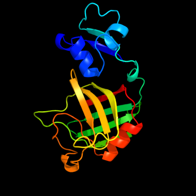
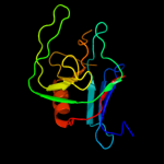
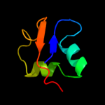
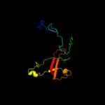
1 c1y7mB_
100.0
38
PDB header: structural genomics, unknown functionChain: B: PDB Molecule: hypothetical protein bsu14040;PDBTitle: crystal structure of the b. subtilis ykud protein at 2 a2 resolution
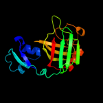
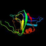
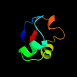
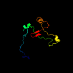
2 d1y7ma1
100.0
39
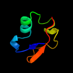
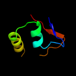
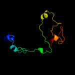
Fold: L,D-transpeptidase catalytic domain-likeSuperfamily: L,D-transpeptidase catalytic domain-likeFamily: L,D-transpeptidase catalytic domain-like3 c2hklB_
100.0
29
PDB header: transferaseChain: B: PDB Molecule: l,d-transpeptidase;PDBTitle: crystal structure of enterococcus faecium l,d-2 transpeptidase c442s mutant
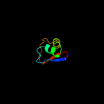
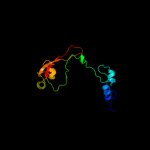
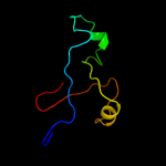
4 d1zata1
100.0
30
Fold: L,D-transpeptidase catalytic domain-likeSuperfamily: L,D-transpeptidase catalytic domain-likeFamily: L,D-transpeptidase catalytic domain-like5 d1y7ma2
98.1
36
Fold: LysM domainSuperfamily: LysM domainFamily: LysM domain6 c2l9yA_
98.0
39
PDB header: sugar binding proteinChain: A: PDB Molecule: cvnh-lysm lectin;PDBTitle: solution structure of the mocvnh-lysm module from the rice blast2 fungus magnaporthe oryzae protein (mgg_03307)
7 d1e0ga_
97.6
31
Fold: LysM domainSuperfamily: LysM domainFamily: LysM domain8 c2djpA_
97.6
21
PDB header: structural genomics, unknown functionChain: A: PDB Molecule: hypothetical protein sb145;PDBTitle: the solution structure of the lysm domain of human2 hypothetical protein sb145
9 c2gu1A_
91.3
18
PDB header: hydrolaseChain: A: PDB Molecule: zinc peptidase;PDBTitle: crystal structure of a zinc containing peptidase from2 vibrio cholerae
10 c1h5nC_
87.9
19
PDB header: oxidoreductaseChain: C: PDB Molecule: dmso reductase;PDBTitle: dmso reductase modified by the presence of dms and air
11 c1y5iA_
86.3
19
PDB header: oxidoreductaseChain: A: PDB Molecule: respiratory nitrate reductase 1 alpha chain;PDBTitle: the crystal structure of the narghi mutant nari-k86a
12 c1eu1A_
49.9
16
PDB header: oxidoreductaseChain: A: PDB Molecule: dimethyl sulfoxide reductase;PDBTitle: the crystal structure of rhodobacter sphaeroides dimethylsulfoxide2 reductase reveals two distinct molybdenum coordination environments.
13 c3mcaB_
44.8
29
PDB header: translation regulation/hydrolaseChain: B: PDB Molecule: protein dom34;PDBTitle: structure of the dom34-hbs1 complex and implications for its role in2 no-go decay
14 d1wjja_
43.1
23
Fold: OB-foldSuperfamily: Nucleic acid-binding proteinsFamily: Single strand DNA-binding domain, SSB15 d1y5ia1
39.9
18
Fold: Double psi beta-barrelSuperfamily: ADC-likeFamily: Formate dehydrogenase/DMSO reductase, C-terminal domain16 c1kqgA_
37.7
14
PDB header: oxidoreductaseChain: A: PDB Molecule: formate dehydrogenase, nitrate-inducible, major subunit;PDBTitle: formate dehydrogenase n from e. coli
17 c2e7zA_
34.7
15
PDB header: lyaseChain: A: PDB Molecule: acetylene hydratase ahy;PDBTitle: acetylene hydratase from pelobacter acetylenicus
18 c1tmoA_
31.9
16
PDB header: oxidoreductaseChain: A: PDB Molecule: trimethylamine n-oxide reductase;PDBTitle: trimethylamine n-oxide reductase from shewanella massilia
19 d1eu1a1
27.0
14
Fold: Double psi beta-barrelSuperfamily: ADC-likeFamily: Formate dehydrogenase/DMSO reductase, C-terminal domain20 c2iv2X_
26.3
22
PDB header: oxidoreductaseChain: X: PDB Molecule: formate dehydrogenase h;PDBTitle: reinterpretation of reduced form of formate dehydrogenase h2 from e. coli
21 c1bmv2_
not modelled
20.8
17
PDB header: virus/rnaChain: 2: PDB Molecule: protein (icosahedral virus - b and c domain);PDBTitle: protein-rna interactions in an icosahedral virus at 3.02 angstroms resolution
22 c2kkeA_
not modelled
16.9
37
PDB header: structural genomics, unknown functionChain: A: PDB Molecule: uncharacterized protein;PDBTitle: solution nmr structure of a dimeric protein of unknown2 function from methanobacterium thermoautotrophicum,3 northeast structural genomics consortium target tr5
23 d1ogya1
not modelled
16.4
20
Fold: Double psi beta-barrelSuperfamily: ADC-likeFamily: Formate dehydrogenase/DMSO reductase, C-terminal domain24 d2jioa1
not modelled
16.4
15
Fold: Double psi beta-barrelSuperfamily: ADC-likeFamily: Formate dehydrogenase/DMSO reductase, C-terminal domain25 c1vlfQ_
not modelled
15.1
13
PDB header: oxidoreductaseChain: Q: PDB Molecule: pyrogallol hydroxytransferase large subunit;PDBTitle: crystal structure of pyrogallol-phloroglucinol2 transhydroxylase from pelobacter acidigallici complexed3 with inhibitor 1,2,4,5-tetrahydroxy-benzene
26 c2k50A_
not modelled
13.9
11
PDB header: structural genomics, unknown functionChain: A: PDB Molecule: replication factor a related protein;PDBTitle: solution nmr structure of the replication factor a related2 protein from methanobacterium thermoautotrophicum.3 northeast structural genomics target tr91a.
27 d2hthb1
not modelled
13.0
16
Fold: PH domain-like barrelSuperfamily: PH domain-likeFamily: VPS36 N-terminal domain-like28 d1kqfa1
not modelled
13.0
12
Fold: Double psi beta-barrelSuperfamily: ADC-likeFamily: Formate dehydrogenase/DMSO reductase, C-terminal domain29 c2ivfA_
not modelled
11.4
21
PDB header: oxidoreductaseChain: A: PDB Molecule: ethylbenzene dehydrogenase alpha-subunit;PDBTitle: ethylbenzene dehydrogenase from aromatoleum aromaticum
30 d2c42a3
not modelled
11.0
11
Fold: TK C-terminal domain-likeSuperfamily: TK C-terminal domain-likeFamily: Pyruvate-ferredoxin oxidoreductase, PFOR, domain II31 d2vgna1
not modelled
10.3
12
Fold: Sm-like foldSuperfamily: Dom34/Pelota N-terminal domain-likeFamily: Dom34/Pelota N-terminal domain-like32 d1h0ha1
not modelled
10.3
11
Fold: Double psi beta-barrelSuperfamily: ADC-likeFamily: Formate dehydrogenase/DMSO reductase, C-terminal domain33 d1g8ka1
not modelled
8.4
25
Fold: Double psi beta-barrelSuperfamily: ADC-likeFamily: Formate dehydrogenase/DMSO reductase, C-terminal domain34 d1k78a1
not modelled
8.1
71
Fold: DNA/RNA-binding 3-helical bundleSuperfamily: Homeodomain-likeFamily: Paired domain35 c3pjvD_
not modelled
7.9
12
PDB header: lyaseChain: D: PDB Molecule: cyclic dimeric gmp binding protein;PDBTitle: structure of pseudomonas fluorescence lapd periplasmic domain
36 d1vlfm1
not modelled
7.8
15
Fold: Double psi beta-barrelSuperfamily: ADC-likeFamily: Formate dehydrogenase/DMSO reductase, C-terminal domain37 d2qi2a1
not modelled
7.5
16
Fold: Sm-like foldSuperfamily: Dom34/Pelota N-terminal domain-likeFamily: Dom34/Pelota N-terminal domain-like38 d6paxa1
not modelled
6.8
57
Fold: DNA/RNA-binding 3-helical bundleSuperfamily: Homeodomain-likeFamily: Paired domain39 d1qvpa_
not modelled
6.7
21
Fold: SH3-like barrelSuperfamily: C-terminal domain of transcriptional repressorsFamily: FeoA-like40 d1qvca_
not modelled
6.7
13
Fold: OB-foldSuperfamily: Nucleic acid-binding proteinsFamily: Single strand DNA-binding domain, SSB41 d1t3la1
not modelled
6.1
17
Fold: SH3-like barrelSuperfamily: SH3-domainFamily: SH3-domain42 c2vw9B_
not modelled
5.9
13
PDB header: dna-binding proteinChain: B: PDB Molecule: single-stranded dna binding protein;PDBTitle: single stranded dna binding protein complex from2 helicobacter pylori
43 c2iheA_
not modelled
5.4
13
PDB header: dna binding proteinChain: A: PDB Molecule: single-stranded dna-binding protein;PDBTitle: crystal structure of wild-type single-stranded dna binding protein2 from thermus aquaticus
44 c2bfuL_
not modelled
5.2
33
PDB header: virusChain: L: PDB Molecule: cowpea mosaic virus, large (l) subunit;PDBTitle: x-ray structure of cpmv top component
45 c2inpE_
not modelled
5.2
9
PDB header: oxidoreductaseChain: E: PDB Molecule: phenol hydroxylase component pho;PDBTitle: structure of the phenol hydroxylase-regulatory protein2 complex
46 d1e0ta1
not modelled
5.2
36
Fold: PK beta-barrel domain-likeSuperfamily: PK beta-barrel domain-likeFamily: Pyruvate kinase beta-barrel domain47 c3rf1B_
not modelled
5.1
21
PDB header: ligaseChain: B: PDB Molecule: glycyl-trna synthetase alpha subunit;PDBTitle: the crystal structure of glycyl-trna synthetase subunit alpha from2 campylobacter jejuni subsp. jejuni nctc 11168