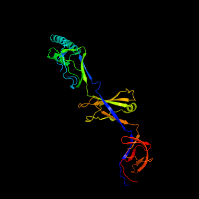
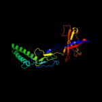
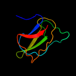
1 c3h9iB_
100.0
100
PDB header: transport proteinChain: B: PDB Molecule: cation efflux system protein cusb;PDBTitle: crystal structure of the membrane fusion protein cusb from escherichia2 coli
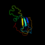
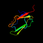
2 c2f1mA_
99.9
19
PDB header: transport proteinChain: A: PDB Molecule: acriflavine resistance protein a;PDBTitle: conformational flexibility in the multidrug efflux system protein acra
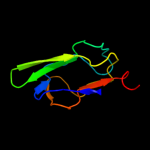
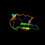
3 c3lnnB_
99.9
22
PDB header: metal transportChain: B: PDB Molecule: membrane fusion protein (mfp) heavy metal cation effluxPDBTitle: crystal structure of zneb from cupriavidus metallidurans
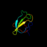
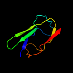
4 c3fppB_
99.9
18
PDB header: membrane proteinChain: B: PDB Molecule: macrolide-specific efflux protein maca;PDBTitle: crystal structure of e.coli maca
5 c1t5eB_
99.9
19
PDB header: transport proteinChain: B: PDB Molecule: multidrug resistance protein mexa;PDBTitle: the structure of mexa
6 d1vf7a_
99.9
19
Fold: HlyD-like secretion proteinsSuperfamily: HlyD-like secretion proteinsFamily: HlyD-like secretion proteins7 c2k33A_
99.4
17
PDB header: membrane protein, transport proteinChain: A: PDB Molecule: acra;PDBTitle: solution structure of an n-glycosylated protein using in2 vitro glycosylation
8 c2b8gA_
98.5
21
PDB header: biosynthetic proteinChain: A: PDB Molecule: biotin/lipoyl attachment protein;PDBTitle: solution structure of bacillus subtilis blap biotinylated-2 form (energy minimized mean structure)
9 d1dcza_
98.4
23
Fold: Barrel-sandwich hybridSuperfamily: Single hybrid motifFamily: Biotinyl/lipoyl-carrier proteins and domains10 c2ejgD_
98.4
27
PDB header: ligaseChain: D: PDB Molecule: 149aa long hypothetical methylmalonyl-coa decarboxylasePDBTitle: crystal structure of the biotin protein ligase (mutation r48a) and2 biotin carboxyl carrier protein complex from pyrococcus horikoshii3 ot3
11 d1o78a_
98.2
23
Fold: Barrel-sandwich hybridSuperfamily: Single hybrid motifFamily: Biotinyl/lipoyl-carrier proteins and domains12 c2ejmA_
98.1
33
PDB header: ligaseChain: A: PDB Molecule: methylcrotonoyl-coa carboxylase subunit alpha;PDBTitle: solution structure of ruh-072, an apo-biotnyl domain form2 human acetyl coenzyme a carboxylase
13 c3n6rK_
98.0
26
PDB header: ligaseChain: K: PDB Molecule: propionyl-coa carboxylase, alpha subunit;PDBTitle: crystal structure of the holoenzyme of propionyl-coa carboxylase (pcc)
14 c2dn8A_
98.0
21
PDB header: ligaseChain: A: PDB Molecule: acetyl-coa carboxylase 2;PDBTitle: solution structure of rsgi ruh-053, an apo-biotin carboxy2 carrier protein from human transcarboxylase
15 c2kccA_
97.9
21
PDB header: ligaseChain: A: PDB Molecule: acetyl-coa carboxylase 2;PDBTitle: solution structure of biotinoyl domain from human acetyl-2 coa carboxylase 2
16 d1qjoa_
97.8
26
Fold: Barrel-sandwich hybridSuperfamily: Single hybrid motifFamily: Biotinyl/lipoyl-carrier proteins and domains17 d1ghja_
97.8
25
Fold: Barrel-sandwich hybridSuperfamily: Single hybrid motifFamily: Biotinyl/lipoyl-carrier proteins and domains18 d1k8ma_
97.7
17
Fold: Barrel-sandwich hybridSuperfamily: Single hybrid motifFamily: Biotinyl/lipoyl-carrier proteins and domains19 d1bdoa_
97.7
25
Fold: Barrel-sandwich hybridSuperfamily: Single hybrid motifFamily: Biotinyl/lipoyl-carrier proteins and domains20 d1iyua_
97.7
23
Fold: Barrel-sandwich hybridSuperfamily: Single hybrid motifFamily: Biotinyl/lipoyl-carrier proteins and domains21 c2q8iB_
not modelled
97.7
21
PDB header: transferaseChain: B: PDB Molecule: dihydrolipoyllysine-residue acetyltransferase component ofPDBTitle: pyruvate dehydrogenase kinase isoform 3 in complex with antitumor drug2 radicicol
22 d1y8ob1
not modelled
97.7
22
Fold: Barrel-sandwich hybridSuperfamily: Single hybrid motifFamily: Biotinyl/lipoyl-carrier proteins and domains23 c2l5tA_
not modelled
97.5
23
PDB header: transferaseChain: A: PDB Molecule: lipoamide acyltransferase;PDBTitle: solution nmr structure of e2 lipoyl domain from thermoplasma2 acidophilum
24 c2dncA_
not modelled
97.5
32
PDB header: transferaseChain: A: PDB Molecule: pyruvate dehydrogenase protein x component;PDBTitle: solution structure of rsgi ruh-054, a lipoyl domain from2 human 2-oxoacid dehydrogenase
25 d1laba_
not modelled
97.4
25
Fold: Barrel-sandwich hybridSuperfamily: Single hybrid motifFamily: Biotinyl/lipoyl-carrier proteins and domains26 d1gjxa_
not modelled
97.3
28
Fold: Barrel-sandwich hybridSuperfamily: Single hybrid motifFamily: Biotinyl/lipoyl-carrier proteins and domains27 c2dneA_
not modelled
97.0
17
PDB header: transferaseChain: A: PDB Molecule: dihydrolipoyllysine-residue acetyltransferasePDBTitle: solution structure of rsgi ruh-058, a lipoyl domain of2 human 2-oxoacid dehydrogenase
28 d1pmra_
not modelled
96.8
15
Fold: Barrel-sandwich hybridSuperfamily: Single hybrid motifFamily: Biotinyl/lipoyl-carrier proteins and domains29 c3fmcC_
not modelled
96.0
31
PDB header: hydrolaseChain: C: PDB Molecule: putative succinylglutamate desuccinylase / aspartoacylase;PDBTitle: crystal structure of a putative succinylglutamate desuccinylase /2 aspartoacylase family protein (sama_0604) from shewanella amazonensis3 sb2b at 1.80 a resolution
30 c2qj8B_
not modelled
95.0
23
PDB header: hydrolaseChain: B: PDB Molecule: mlr6093 protein;PDBTitle: crystal structure of an aspartoacylase family protein (mlr6093) from2 mesorhizobium loti maff303099 at 2.00 a resolution
31 c3na6A_
not modelled
94.3
21
PDB header: hydrolaseChain: A: PDB Molecule: succinylglutamate desuccinylase/aspartoacylase;PDBTitle: crystal structure of a succinylglutamate desuccinylase (tm1040_2694)2 from silicibacter sp. tm1040 at 2.00 a resolution
32 d2pnrc1
not modelled
93.9
23
Fold: Barrel-sandwich hybridSuperfamily: Single hybrid motifFamily: Biotinyl/lipoyl-carrier proteins and domains33 d2tpta3
not modelled
93.3
19
Fold: alpha/beta-HammerheadSuperfamily: Pyrimidine nucleoside phosphorylase C-terminal domainFamily: Pyrimidine nucleoside phosphorylase C-terminal domain34 c2dsjA_
not modelled
92.3
21
PDB header: transferaseChain: A: PDB Molecule: pyrimidine-nucleoside (thymidine) phosphorylase;PDBTitle: crystal structure of project id tt0128 from thermus thermophilus hb8
35 c2qf7A_
not modelled
91.7
22
PDB header: ligaseChain: A: PDB Molecule: pyruvate carboxylase protein;PDBTitle: crystal structure of a complete multifunctional pyruvate carboxylase2 from rhizobium etli
36 d1uoua3
not modelled
91.5
22
Fold: alpha/beta-HammerheadSuperfamily: Pyrimidine nucleoside phosphorylase C-terminal domainFamily: Pyrimidine nucleoside phosphorylase C-terminal domain37 c1otpA_
not modelled
91.4
19
PDB header: phosphorylaseChain: A: PDB Molecule: thymidine phosphorylase;PDBTitle: structural and theoretical studies suggest domain movement produces an2 active conformation of thymidine phosphorylase
38 d1brwa3
not modelled
91.4
33
Fold: alpha/beta-HammerheadSuperfamily: Pyrimidine nucleoside phosphorylase C-terminal domainFamily: Pyrimidine nucleoside phosphorylase C-terminal domain39 c3iftA_
not modelled
89.9
24
PDB header: oxidoreductaseChain: A: PDB Molecule: glycine cleavage system h protein;PDBTitle: crystal structure of glycine cleavage system protein h from2 mycobacterium tuberculosis, using x-rays from the compact light3 source.
40 d1glaf_
not modelled
89.6
22
Fold: Barrel-sandwich hybridSuperfamily: Duplicated hybrid motifFamily: Glucose permease-like41 d2gpra_
not modelled
89.6
15
Fold: Barrel-sandwich hybridSuperfamily: Duplicated hybrid motifFamily: Glucose permease-like42 d2f3ga_
not modelled
89.2
22
Fold: Barrel-sandwich hybridSuperfamily: Duplicated hybrid motifFamily: Glucose permease-like43 c3mxuA_
not modelled
89.2
21
PDB header: oxidoreductaseChain: A: PDB Molecule: glycine cleavage system h protein;PDBTitle: crystal structure of glycine cleavage system protein h from bartonella2 henselae
44 c1yuzB_
89.0
18
PDB header: oxidoreductaseChain: B: PDB Molecule: nigerythrin;PDBTitle: partially reduced state of nigerythrin
45 c1brwB_
not modelled
88.0
33
PDB header: transferaseChain: B: PDB Molecule: protein (pyrimidine nucleoside phosphorylase);PDBTitle: the crystal structure of pyrimidine nucleoside2 phosphorylase in a closed conformation
46 c3h5qA_
not modelled
87.7
33
PDB header: transferaseChain: A: PDB Molecule: pyrimidine-nucleoside phosphorylase;PDBTitle: crystal structure of a putative pyrimidine-nucleoside phosphorylase2 from staphylococcus aureus
47 c2j0fC_
not modelled
87.2
29
PDB header: transferaseChain: C: PDB Molecule: thymidine phosphorylase;PDBTitle: structural basis for non-competitive product inhibition in2 human thymidine phosphorylase: implication for drug design
48 d1gpra_
not modelled
86.5
22
Fold: Barrel-sandwich hybridSuperfamily: Duplicated hybrid motifFamily: Glucose permease-like49 c1dvbA_
85.0
14
PDB header: electron transportChain: A: PDB Molecule: rubrerythrin;PDBTitle: rubrerythrin
50 c2jkuA_
not modelled
84.5
21
PDB header: ligaseChain: A: PDB Molecule: propionyl-coa carboxylase alpha chain,PDBTitle: crystal structure of the n-terminal region of the biotin2 acceptor domain of human propionyl-coa carboxylase
51 c2hr5B_
not modelled
83.5
23
PDB header: metal binding proteinChain: B: PDB Molecule: rubrerythrin;PDBTitle: pf1283- rubrerythrin from pyrococcus furiosus iron bound form
52 c3cdxB_
not modelled
83.0
22
PDB header: hydrolaseChain: B: PDB Molecule: succinylglutamatedesuccinylase/aspartoacylase;PDBTitle: crystal structure of2 succinylglutamatedesuccinylase/aspartoacylase from3 rhodobacter sphaeroides
53 c2hsiB_
not modelled
82.1
22
PDB header: structural genomics, unknown functionChain: B: PDB Molecule: putative peptidase m23;PDBTitle: crystal structure of putative peptidase m23 from2 pseudomonas aeruginosa, new york structural genomics3 consortium
54 c2gu1A_
not modelled
80.7
22
PDB header: hydrolaseChain: A: PDB Molecule: zinc peptidase;PDBTitle: crystal structure of a zinc containing peptidase from2 vibrio cholerae
55 d1qwya_
not modelled
76.9
22
Fold: Barrel-sandwich hybridSuperfamily: Duplicated hybrid motifFamily: Peptidoglycan hydrolase LytM56 c2aukA_
not modelled
74.3
21
PDB header: transferaseChain: A: PDB Molecule: dna-directed rna polymerase beta' chain;PDBTitle: structure of e. coli rna polymerase beta' g/g' insert
57 d1o4ua2
not modelled
67.7
10
Fold: alpha/beta-HammerheadSuperfamily: Nicotinate/Quinolinate PRTase N-terminal domain-likeFamily: NadC N-terminal domain-like58 c2b44A_
not modelled
67.7
22
PDB header: hydrolaseChain: A: PDB Molecule: glycyl-glycine endopeptidase lytm;PDBTitle: truncated s. aureus lytm, p 32 2 1 crystal form
59 c3d4rE_
not modelled
66.4
29
PDB header: unknown functionChain: E: PDB Molecule: domain of unknown function from the pfam-b_34464 family;PDBTitle: crystal structure of a duf2118 family protein (mmp0046) from2 methanococcus maripaludis at 2.20 a resolution
60 c2xhaB_
not modelled
65.9
28
PDB header: transcriptionChain: B: PDB Molecule: transcription antitermination protein nusg;PDBTitle: crystal structure of domain 2 of thermotoga maritima n-utilization2 substance g (nusg)
61 d1yuja_
not modelled
65.2
18
Fold: beta-beta-alpha zinc fingersSuperfamily: beta-beta-alpha zinc fingersFamily: Classic zinc finger, C2H262 d1e2wa2
not modelled
64.1
31
Fold: Barrel-sandwich hybridSuperfamily: Rudiment single hybrid motifFamily: Cytochrome f, small domain63 d1ci3m2
not modelled
64.1
25
Fold: Barrel-sandwich hybridSuperfamily: Rudiment single hybrid motifFamily: Cytochrome f, small domain64 d1onla_
not modelled
63.3
27
Fold: Barrel-sandwich hybridSuperfamily: Single hybrid motifFamily: Biotinyl/lipoyl-carrier proteins and domains65 d1qpoa2
not modelled
62.8
21
Fold: alpha/beta-HammerheadSuperfamily: Nicotinate/Quinolinate PRTase N-terminal domain-likeFamily: NadC N-terminal domain-like66 c2edgA_
not modelled
62.5
23
PDB header: biosynthetic proteinChain: A: PDB Molecule: glycine cleavage system h protein;PDBTitle: solution structure of the gcv_h domain from mouse glycine
67 c3it5B_
not modelled
61.3
24
PDB header: hydrolaseChain: B: PDB Molecule: protease lasa;PDBTitle: crystal structure of the lasa virulence factor from pseudomonas2 aeruginosa
68 c1ctmA_
not modelled
60.7
16
PDB header: electron transport(cytochrome)Chain: A: PDB Molecule: cytochrome f;PDBTitle: crystal structure of chloroplast cytochrome f reveals a2 novel cytochrome fold and unexpected heme ligation
69 d1qapa2
not modelled
59.5
20
Fold: alpha/beta-HammerheadSuperfamily: Nicotinate/Quinolinate PRTase N-terminal domain-likeFamily: NadC N-terminal domain-like70 d1yuza2
not modelled
59.0
18
Fold: Rubredoxin-likeSuperfamily: Rubredoxin-likeFamily: Rubredoxin71 c2xhcA_
not modelled
58.8
17
PDB header: transcriptionChain: A: PDB Molecule: transcription antitermination protein nusg;PDBTitle: crystal structure of thermotoga maritima n-utilization substance g2 (nusg)
72 d1nnqa2
not modelled
56.5
19
Fold: Rubredoxin-likeSuperfamily: Rubredoxin-likeFamily: Rubredoxin73 d1hpca_
not modelled
55.9
23
Fold: Barrel-sandwich hybridSuperfamily: Single hybrid motifFamily: Biotinyl/lipoyl-carrier proteins and domains74 c3a8jF_
not modelled
53.6
23
PDB header: transferase/transport proteinChain: F: PDB Molecule: glycine cleavage system h protein;PDBTitle: crystal structure of et-ehred complex
75 d1dgsa1
53.1
33
Fold: SAM domain-likeSuperfamily: RuvA domain 2-likeFamily: NAD+-dependent DNA ligase, domain 376 d1ep3b1
not modelled
49.2
20
Fold: Reductase/isomerase/elongation factor common domainSuperfamily: Riboflavin synthase domain-likeFamily: Ferredoxin reductase FAD-binding domain-like77 c3nyyA_
not modelled
48.6
31
PDB header: hydrolaseChain: A: PDB Molecule: putative glycyl-glycine endopeptidase lytm;PDBTitle: crystal structure of a putative glycyl-glycine endopeptidase lytm2 (rumgna_02482) from ruminococcus gnavus atcc 29149 at 1.60 a3 resolution
78 c2aujD_
not modelled
48.5
44
PDB header: transferaseChain: D: PDB Molecule: dna-directed rna polymerase beta' chain;PDBTitle: structure of thermus aquaticus rna polymerase beta'-subunit2 insert
79 d2cu8a1
not modelled
43.0
40
Fold: Glucocorticoid receptor-like (DNA-binding domain)Superfamily: Glucocorticoid receptor-like (DNA-binding domain)Family: LIM domain80 d1lkoa2
not modelled
42.9
19
Fold: Rubredoxin-likeSuperfamily: Rubredoxin-likeFamily: Rubredoxin81 c1e2vB_
not modelled
42.5
31
PDB header: electron transport proteinsChain: B: PDB Molecule: cytochrome f;PDBTitle: n153q mutant of cytochrome f from chlamydomonas reinhardtii
82 c2jxmB_
not modelled
41.4
38
PDB header: electron transportChain: B: PDB Molecule: cytochrome f;PDBTitle: ensemble of twenty structures of the prochlorothrix2 hollandica plastocyanin- cytochrome f complex
83 c3iynM_
not modelled
41.0
22
PDB header: virusChain: M: PDB Molecule: penton base protein;PDBTitle: 3.6-angstrom cryoem structure of human adenovirus type 5
84 d2i4sa1
not modelled
40.7
12
Fold: PDZ domain-likeSuperfamily: PDZ domain-likeFamily: EpsC C-terminal domain-like85 c2bldD_
not modelled
40.6
22
PDB header: virusChain: D: PDB Molecule: penton protein;PDBTitle: the quasi-atomic model of human adenovirus type 52 capsid (part 1)
86 c2zpmA_
not modelled
40.5
16
PDB header: hydrolaseChain: A: PDB Molecule: regulator of sigma e protease;PDBTitle: crystal structure analysis of pdz domain b
87 d1krha1
not modelled
40.5
17
Fold: Reductase/isomerase/elongation factor common domainSuperfamily: Riboflavin synthase domain-likeFamily: Ferredoxin reductase FAD-binding domain-like88 c3csqC_
not modelled
39.8
33
PDB header: hydrolaseChain: C: PDB Molecule: morphogenesis protein 1;PDBTitle: crystal and cryoem structural studies of a cell wall2 degrading enzyme in the bacteriophage phi29 tail
89 c2owoA_
not modelled
38.9
25
PDB header: ligase/dnaChain: A: PDB Molecule: dna ligase;PDBTitle: last stop on the road to repair: structure of e.coli dna ligase bound2 to nicked dna-adenylate
90 d2j01q1
not modelled
38.5
10
Fold: alpha/beta-HammerheadSuperfamily: Ribosomal protein L16p/L10eFamily: Ribosomal protein L16p91 c2kjpA_
not modelled
38.0
12
PDB header: structural genomics, unknown functionChain: A: PDB Molecule: uncharacterized protein ylbl;PDBTitle: solution structure of protein ylbl (bsu15050) from bacillus2 subtilis, northeast structural genomics consortium target3 sr713a
92 c3ndjA_
not modelled
37.8
7
PDB header: transferaseChain: A: PDB Molecule: methyltransferase;PDBTitle: x-ray structure of a c-3'-methyltransferase in complex with s-2 adenosyl-l-homocysteine and sugar product
93 c1v9pB_
not modelled
37.4
33
PDB header: ligaseChain: B: PDB Molecule: dna ligase;PDBTitle: crystal structure of nad+-dependent dna ligase
94 c1q90A_
not modelled
36.7
31
PDB header: photosynthesisChain: A: PDB Molecule: apocytochrome f;PDBTitle: structure of the cytochrome b6f (plastohydroquinone : plastocyanin2 oxidoreductase) from chlamydomonas reinhardtii
95 c1tu2B_
not modelled
36.7
25
PDB header: electron transportChain: B: PDB Molecule: apocytochrome f;PDBTitle: the complex of nostoc cytochrome f and plastocyanin determin with2 paramagnetic nmr. based on the structures of cytochrome f and3 plastocyanin, 10 structures
96 d1pvma3
not modelled
36.2
24
Fold: Rubredoxin-likeSuperfamily: Hypothetical protein Ta0289 C-terminal domainFamily: Hypothetical protein Ta0289 C-terminal domain97 c1dgsB_
not modelled
35.0
33
PDB header: ligaseChain: B: PDB Molecule: dna ligase;PDBTitle: crystal structure of nad+-dependent dna ligase from t.2 filiformis
98 d2i6va1
not modelled
33.8
15
Fold: PDZ domain-likeSuperfamily: PDZ domain-likeFamily: EpsC C-terminal domain-like99 d2gmga1
not modelled
33.1
25
Fold: DNA/RNA-binding 3-helical bundleSuperfamily: "Winged helix" DNA-binding domainFamily: PF0610-like100 c2e75C_
not modelled
32.8
25
PDB header: photosynthesisChain: C: PDB Molecule: apocytochrome f;PDBTitle: crystal structure of the cytochrome b6f complex with 2-nonyl-4-2 hydroxyquinoline n-oxide (nqno) from m.laminosus
101 c2ftcI_
not modelled
31.6
15
PDB header: ribosomeChain: I: PDB Molecule: mitochondrial ribosomal protein l16;PDBTitle: structural model for the large subunit of the mammalian mitochondrial2 ribosome
102 d1qfja1
not modelled
29.9
15
Fold: Reductase/isomerase/elongation factor common domainSuperfamily: Riboflavin synthase domain-likeFamily: Ferredoxin reductase FAD-binding domain-like103 d1hf2a1
not modelled
29.7
13
Fold: Single-stranded right-handed beta-helixSuperfamily: Cell-division inhibitor MinC, C-terminal domainFamily: Cell-division inhibitor MinC, C-terminal domain104 d1tu2b2
not modelled
29.4
25
Fold: Barrel-sandwich hybridSuperfamily: Rudiment single hybrid motifFamily: Cytochrome f, small domain105 c3bboO_
not modelled
29.2
14
PDB header: ribosomeChain: O: PDB Molecule: ribosomal protein l16;PDBTitle: homology model for the spinach chloroplast 50s subunit2 fitted to 9.4a cryo-em map of the 70s chlororibosome
106 d1sota1
not modelled
29.1
15
Fold: PDZ domain-likeSuperfamily: PDZ domain-likeFamily: HtrA-like serine proteases107 c2kl1A_
not modelled
26.9
12
PDB header: protein bindingChain: A: PDB Molecule: ylbl protein;PDBTitle: solution structure of gtr34c from geobacillus thermodenitrificans.2 northeast structural genomics consortium target gtr34c
108 c2k5cA_
not modelled
26.4
33
PDB header: metal binding proteinChain: A: PDB Molecule: uncharacterized protein pf0385;PDBTitle: nmr structure for pf0385
109 d1akya2
not modelled
25.5
20
Fold: Rubredoxin-likeSuperfamily: Microbial and mitochondrial ADK, insert "zinc finger" domainFamily: Microbial and mitochondrial ADK, insert "zinc finger" domain110 d2ak3a2
not modelled
25.3
23
Fold: Rubredoxin-likeSuperfamily: Microbial and mitochondrial ADK, insert "zinc finger" domainFamily: Microbial and mitochondrial ADK, insert "zinc finger" domain111 c3i18A_
not modelled
25.0
9
PDB header: structural genomics, unknown functionChain: A: PDB Molecule: lmo2051 protein;PDBTitle: crystal structure of the pdz domain of the sdrc-like protein2 (lmo2051) from listeria monocytogenes, northeast structural3 genomics consortium target lmr166b
112 d1hcza2
not modelled
24.7
19
Fold: Barrel-sandwich hybridSuperfamily: Rudiment single hybrid motifFamily: Cytochrome f, small domain113 d1jj2h_
not modelled
24.5
4
Fold: alpha/beta-HammerheadSuperfamily: Ribosomal protein L16p/L10eFamily: Ribosomal protein L10e114 d1e4va2
not modelled
24.4
23
Fold: Rubredoxin-likeSuperfamily: Microbial and mitochondrial ADK, insert "zinc finger" domainFamily: Microbial and mitochondrial ADK, insert "zinc finger" domain115 d1ky9a1
not modelled
24.4
20
Fold: PDZ domain-likeSuperfamily: PDZ domain-likeFamily: HtrA-like serine proteases116 d1j1la_
not modelled
23.7
17
Fold: Double-stranded beta-helixSuperfamily: RmlC-like cupinsFamily: Pirin-like117 c1w8xP_
not modelled
22.9
19
PDB header: virusChain: P: PDB Molecule: protein p16;PDBTitle: structural analysis of prd1
118 d2epqa1
not modelled
22.7
30
Fold: beta-beta-alpha zinc fingersSuperfamily: beta-beta-alpha zinc fingersFamily: Classic zinc finger, C2H2119 d1ffkf_
not modelled
21.4
0
Fold: alpha/beta-HammerheadSuperfamily: Ribosomal protein L16p/L10eFamily: Ribosomal protein L10e120 d1uwfa1
not modelled
21.1
18
Fold: Common fold of diphtheria toxin/transcription factors/cytochrome fSuperfamily: Bacterial adhesinsFamily: Pilus subunits