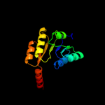
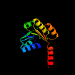
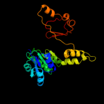
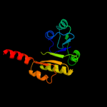
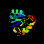
1 c3eywA_
100.0
100
PDB header: transport proteinChain: A: PDB Molecule: c-terminal domain of glutathione-regulated potassium-effluxPDBTitle: crystal structure of the c-terminal domain of e. coli kefc in complex2 with keff
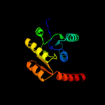
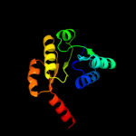
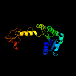
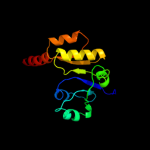
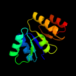
2 c1zcdA_
100.0
18
PDB header: membrane proteinChain: A: PDB Molecule: na(+)/h(+) antiporter 1;PDBTitle: crystal structure of the na+/h+ antiporter nhaa
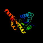
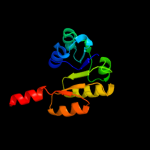
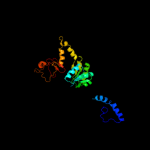
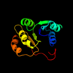
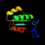
3 c3fwzA_
99.9
22
PDB header: membrane proteinChain: A: PDB Molecule: inner membrane protein ybal;PDBTitle: crystal structure of trka-n domain of inner membrane protein ybal from2 escherichia coli
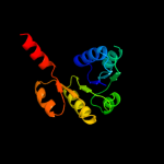
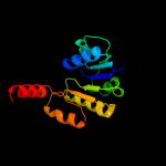
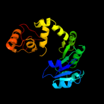
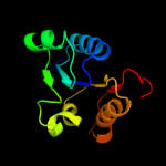
4 c3c85A_
99.9
23
PDB header: transport proteinChain: A: PDB Molecule: putative glutathione-regulated potassium-efflux systemPDBTitle: crystal structure of trka domain of putative glutathione-regulated2 potassium-efflux kefb from vibrio parahaemolyticus
5 d1id1a_
99.9
15
Fold: NAD(P)-binding Rossmann-fold domainsSuperfamily: NAD(P)-binding Rossmann-fold domainsFamily: Potassium channel NAD-binding domain6 d1lssa_
99.9
26
Fold: NAD(P)-binding Rossmann-fold domainsSuperfamily: NAD(P)-binding Rossmann-fold domainsFamily: Potassium channel NAD-binding domain7 d2hmva1
99.8
19
Fold: NAD(P)-binding Rossmann-fold domainsSuperfamily: NAD(P)-binding Rossmann-fold domainsFamily: Potassium channel NAD-binding domain8 c3llvA_
99.8
17
PDB header: nad(p) binding proteinChain: A: PDB Molecule: exopolyphosphatase-related protein;PDBTitle: the crystal structure of the nad(p)-binding domain of an2 exopolyphosphatase-related protein from archaeoglobus fulgidus to3 1.7a
9 d2fy8a1
99.8
23
Fold: NAD(P)-binding Rossmann-fold domainsSuperfamily: NAD(P)-binding Rossmann-fold domainsFamily: Potassium channel NAD-binding domain10 c2g1uA_
99.8
24
PDB header: transport proteinChain: A: PDB Molecule: hypothetical protein tm1088a;PDBTitle: crystal structure of a putative transport protein (tm1088a) from2 thermotoga maritima at 1.50 a resolution
11 c2fy8A_
99.8
19
PDB header: transport proteinChain: A: PDB Molecule: calcium-gated potassium channel mthk;PDBTitle: crystal structure of mthk rck domain in its ligand-free gating-ring2 form
12 c3l4bG_
99.8
17
PDB header: transport proteinChain: G: PDB Molecule: trka k+ channel protien tm1088b;PDBTitle: crystal structure of an octomeric two-subunit trka k+ channel ring2 gating assembly, tm1088a:tm1088b, from thermotoga maritima
13 c1lnqC_
99.7
18
PDB header: metal transportChain: C: PDB Molecule: potassium channel related protein;PDBTitle: crystal structure of mthk at 3.3 a
14 c3u6nC_
99.4
11
PDB header: transport proteinChain: C: PDB Molecule: high-conductance ca2+-activated k+ channel protein;PDBTitle: open structure of the bk channel gating ring
15 c3mt5A_
99.3
15
PDB header: membrane protein, transport proteinChain: A: PDB Molecule: potassium large conductance calcium-activated channel,PDBTitle: crystal structure of the human bk gating apparatus
16 c3nafA_
98.8
14
PDB header: ion transportChain: A: PDB Molecule: calcium-activated potassium channel subunit alpha-1;PDBTitle: structure of the intracellular gating ring from the human high-2 conductance ca2+ gated k+ channel (bk channel)
17 d1e5qa1
98.5
15
Fold: NAD(P)-binding Rossmann-fold domainsSuperfamily: NAD(P)-binding Rossmann-fold domainsFamily: Glyceraldehyde-3-phosphate dehydrogenase-like, N-terminal domain18 d1pjqa1
98.3
17
Fold: NAD(P)-binding Rossmann-fold domainsSuperfamily: NAD(P)-binding Rossmann-fold domainsFamily: Siroheme synthase N-terminal domain-like19 c3ic5A_
98.2
21
PDB header: structural genomics, unknown functionChain: A: PDB Molecule: putative saccharopine dehydrogenase;PDBTitle: n-terminal domain of putative saccharopine dehydrogenase from ruegeria2 pomeroyi.
20 d2pgda2
98.0
13
Fold: NAD(P)-binding Rossmann-fold domainsSuperfamily: NAD(P)-binding Rossmann-fold domainsFamily: 6-phosphogluconate dehydrogenase-like, N-terminal domain21 c3ktdC_
not modelled
98.0
17
PDB header: oxidoreductaseChain: C: PDB Molecule: prephenate dehydrogenase;PDBTitle: crystal structure of a putative prephenate dehydrogenase (cgl0226)2 from corynebacterium glutamicum atcc 13032 at 2.60 a resolution
22 c3cumA_
not modelled
97.9
18
PDB header: oxidoreductaseChain: A: PDB Molecule: probable 3-hydroxyisobutyrate dehydrogenase;PDBTitle: crystal structure of a possible 3-hydroxyisobutyrate dehydrogenase2 from pseudomonas aeruginosa pao1
23 c1e5lA_
not modelled
97.9
17
PDB header: oxidoreductaseChain: A: PDB Molecule: saccharopine reductase;PDBTitle: apo saccharopine reductase from magnaporthe grisea
24 c3fwnB_
not modelled
97.9
9
PDB header: oxidoreductaseChain: B: PDB Molecule: 6-phosphogluconate dehydrogenase, decarboxylating;PDBTitle: dimeric 6-phosphogluconate dehydrogenase complexed with 6-2 phosphogluconate and 2'-monophosphoadenosine-5'-diphosphate
25 c2iz1C_
not modelled
97.8
10
PDB header: oxidoreductaseChain: C: PDB Molecule: 6-phosphogluconate dehydrogenase, decarboxylating;PDBTitle: 6pdh complexed with pex inhibitor synchrotron data
26 c2axqA_
not modelled
97.8
17
PDB header: oxidoreductaseChain: A: PDB Molecule: saccharopine dehydrogenase;PDBTitle: apo histidine-tagged saccharopine dehydrogenase (l-glu2 forming) from saccharomyces cerevisiae
27 d2f1ka2
not modelled
97.8
13
Fold: NAD(P)-binding Rossmann-fold domainsSuperfamily: NAD(P)-binding Rossmann-fold domainsFamily: 6-phosphogluconate dehydrogenase-like, N-terminal domain28 c3l6dB_
not modelled
97.8
21
PDB header: oxidoreductaseChain: B: PDB Molecule: putative oxidoreductase;PDBTitle: crystal structure of putative oxidoreductase from pseudomonas putida2 kt2440
29 c3d4oA_
not modelled
97.8
18
PDB header: oxidoreductaseChain: A: PDB Molecule: dipicolinate synthase subunit a;PDBTitle: crystal structure of dipicolinate synthase subunit a (np_243269.1)2 from bacillus halodurans at 2.10 a resolution
30 c3dhyC_
not modelled
97.8
19
PDB header: hydrolaseChain: C: PDB Molecule: adenosylhomocysteinase;PDBTitle: crystal structures of mycobacterium tuberculosis s-adenosyl-l-2 homocysteine hydrolase in ternary complex with substrate and3 inhibitors
31 c2rirA_
not modelled
97.7
19
PDB header: oxidoreductaseChain: A: PDB Molecule: dipicolinate synthase, a chain;PDBTitle: crystal structure of dipicolinate synthase, a chain, from bacillus2 subtilis
32 c1bg6A_
not modelled
97.7
16
PDB header: oxidoreductaseChain: A: PDB Molecule: n-(1-d-carboxylethyl)-l-norvaline dehydrogenase;PDBTitle: crystal structure of the n-(1-d-carboxylethyl)-l-norvaline2 dehydrogenase from arthrobacter sp. strain 1c
33 c1pgjA_
not modelled
97.7
17
PDB header: oxidoreductaseChain: A: PDB Molecule: 6-phosphogluconate dehydrogenase;PDBTitle: x-ray structure of 6-phosphogluconate dehydrogenase from the protozoan2 parasite t. brucei
34 c2qx7A_
not modelled
97.7
17
PDB header: plant proteinChain: A: PDB Molecule: eugenol synthase 1;PDBTitle: structure of eugenol synthase from ocimum basilicum
35 c3oneA_
not modelled
97.7
19
PDB header: hydrolase/hydrolase substrateChain: A: PDB Molecule: adenosylhomocysteinase;PDBTitle: crystal structure of lupinus luteus s-adenosyl-l-homocysteine2 hydrolase in complex with adenine
36 c2f1kD_
not modelled
97.7
14
PDB header: oxidoreductaseChain: D: PDB Molecule: prephenate dehydrogenase;PDBTitle: crystal structure of synechocystis arogenate dehydrogenase
37 d1pjca1
not modelled
97.7
20
Fold: NAD(P)-binding Rossmann-fold domainsSuperfamily: NAD(P)-binding Rossmann-fold domainsFamily: Formate/glycerate dehydrogenases, NAD-domain38 c1bxgA_
not modelled
97.7
20
PDB header: amino acid dehydrogenaseChain: A: PDB Molecule: phenylalanine dehydrogenase;PDBTitle: phenylalanine dehydrogenase structure in ternary complex2 with nad+ and beta-phenylpropionate
39 c2p4qA_
not modelled
97.7
12
PDB header: oxidoreductaseChain: A: PDB Molecule: 6-phosphogluconate dehydrogenase, decarboxylating 1;PDBTitle: crystal structure analysis of gnd1 in saccharomyces cerevisiae
40 d1c1da1
not modelled
97.7
16
Fold: NAD(P)-binding Rossmann-fold domainsSuperfamily: NAD(P)-binding Rossmann-fold domainsFamily: Aminoacid dehydrogenase-like, C-terminal domain41 c3qhaB_
not modelled
97.6
16
PDB header: oxidoreductaseChain: B: PDB Molecule: putative oxidoreductase;PDBTitle: crystal structure of a putative oxidoreductase from mycobacterium2 avium 104
42 c2ew2B_
not modelled
97.6
19
PDB header: oxidoreductaseChain: B: PDB Molecule: 2-dehydropantoate 2-reductase, putative;PDBTitle: crystal structure of the putative 2-dehydropantoate 2-reductase from2 enterococcus faecalis
43 c1v8bA_
not modelled
97.6
20
PDB header: hydrolaseChain: A: PDB Molecule: adenosylhomocysteinase;PDBTitle: crystal structure of a hydrolase
44 c1vpdA_
not modelled
97.6
20
PDB header: oxidoreductaseChain: A: PDB Molecule: tartronate semialdehyde reductase;PDBTitle: x-ray crystal structure of tartronate semialdehyde reductase2 [salmonella typhimurium lt2]
45 c3b1fA_
not modelled
97.6
19
PDB header: oxidoreductaseChain: A: PDB Molecule: putative prephenate dehydrogenase;PDBTitle: crystal structure of prephenate dehydrogenase from streptococcus2 mutans
46 c2zcuA_
not modelled
97.6
20
PDB header: oxidoreductaseChain: A: PDB Molecule: uncharacterized oxidoreductase ytfg;PDBTitle: crystal structure of a new type of nadph-dependent quinone2 oxidoreductase (qor2) from escherichia coli
47 c3ggpA_
not modelled
97.6
15
PDB header: oxidoreductaseChain: A: PDB Molecule: prephenate dehydrogenase;PDBTitle: crystal structure of prephenate dehydrogenase from a. aeolicus in2 complex with hydroxyphenyl propionate and nad+
48 c2g5cD_
not modelled
97.6
17
PDB header: oxidoreductaseChain: D: PDB Molecule: prephenate dehydrogenase;PDBTitle: crystal structure of prephenate dehydrogenase from aquifex aeolicus
49 c3d1lB_
not modelled
97.5
17
PDB header: oxidoreductaseChain: B: PDB Molecule: putative nadp oxidoreductase bf3122;PDBTitle: crystal structure of putative nadp oxidoreductase bf3122 from2 bacteroides fragilis
50 c3triB_
not modelled
97.5
13
PDB header: oxidoreductaseChain: B: PDB Molecule: pyrroline-5-carboxylate reductase;PDBTitle: structure of a pyrroline-5-carboxylate reductase (proc) from coxiella2 burnetii
51 d1pgja2
not modelled
97.5
17
Fold: NAD(P)-binding Rossmann-fold domainsSuperfamily: NAD(P)-binding Rossmann-fold domainsFamily: 6-phosphogluconate dehydrogenase-like, N-terminal domain52 d1leha1
not modelled
97.5
14
Fold: NAD(P)-binding Rossmann-fold domainsSuperfamily: NAD(P)-binding Rossmann-fold domainsFamily: Aminoacid dehydrogenase-like, C-terminal domain53 c3n58D_
not modelled
97.5
17
PDB header: hydrolaseChain: D: PDB Molecule: adenosylhomocysteinase;PDBTitle: crystal structure of s-adenosyl-l-homocysteine hydrolase from brucella2 melitensis in ternary complex with nad and adenosine, orthorhombic3 form
54 c1pgqA_
not modelled
97.5
13
PDB header: oxidoreductase (choh(d)-nadp+(a))Chain: A: PDB Molecule: 6-phosphogluconate dehydrogenase;PDBTitle: crystallographic study of coenzyme, coenzyme analogue and substrate2 binding in 6-phosphogluconate dehydrogenase: implications for nadp3 specificity and the enzyme mechanism
55 c3gvpB_
not modelled
97.5
16
PDB header: hydrolaseChain: B: PDB Molecule: adenosylhomocysteinase 3;PDBTitle: human sahh-like domain of human adenosylhomocysteinase 3
56 c2vhyB_
not modelled
97.5
20
PDB header: oxidoreductaseChain: B: PDB Molecule: alanine dehydrogenase;PDBTitle: crystal structure of apo l-alanine dehydrogenase from2 mycobacterium tuberculosis
57 d1bg6a2
not modelled
97.5
14
Fold: NAD(P)-binding Rossmann-fold domainsSuperfamily: NAD(P)-binding Rossmann-fold domainsFamily: 6-phosphogluconate dehydrogenase-like, N-terminal domain58 c3hwrA_
not modelled
97.5
16
PDB header: oxidoreductaseChain: A: PDB Molecule: 2-dehydropantoate 2-reductase;PDBTitle: crystal structure of pane/apba family ketopantoate reductase2 (yp_299159.1) from ralstonia eutropha jmp134 at 2.15 a resolution
59 c3g0oA_
not modelled
97.5
22
PDB header: oxidoreductaseChain: A: PDB Molecule: 3-hydroxyisobutyrate dehydrogenase;PDBTitle: crystal structure of 3-hydroxyisobutyrate dehydrogenase2 (ygbj) from salmonella typhimurium
60 c2we7A_
not modelled
97.4
18
PDB header: oxidoreductaseChain: A: PDB Molecule: xanthine dehydrogenase;PDBTitle: crystal structure of mycobacterium tuberculosis rv0376c2 homologue from mycobacterium smegmatis
61 c3ckyA_
not modelled
97.4
15
PDB header: oxidoreductaseChain: A: PDB Molecule: 2-hydroxymethyl glutarate dehydrogenase;PDBTitle: structural and kinetic properties of a beta-hydroxyacid dehydrogenase2 involved in nicotinate fermentation
62 c1lehB_
not modelled
97.4
15
PDB header: oxidoreductaseChain: B: PDB Molecule: leucine dehydrogenase;PDBTitle: leucine dehydrogenase from bacillus sphaericus
63 c2gf2B_
not modelled
97.4
18
PDB header: oxidoreductaseChain: B: PDB Molecule: 3-hydroxyisobutyrate dehydrogenase;PDBTitle: crystal structure of human hydroxyisobutyrate dehydrogenase
64 c3k96B_
not modelled
97.4
15
PDB header: oxidoreductaseChain: B: PDB Molecule: glycerol-3-phosphate dehydrogenase [nad(p)+];PDBTitle: 2.1 angstrom resolution crystal structure of glycerol-3-phosphate2 dehydrogenase (gpsa) from coxiella burnetii
65 c1d4fD_
not modelled
97.4
17
PDB header: hydrolaseChain: D: PDB Molecule: s-adenosylhomocysteine hydrolase;PDBTitle: crystal structure of recombinant rat-liver d244e mutant s-2 adenosylhomocysteine hydrolase
66 c3d64A_
not modelled
97.4
27
PDB header: hydrolaseChain: A: PDB Molecule: adenosylhomocysteinase;PDBTitle: crystal structure of s-adenosyl-l-homocysteine hydrolase from2 burkholderia pseudomallei
67 c3plnA_
not modelled
97.4
22
PDB header: oxidoreductaseChain: A: PDB Molecule: udp-glucose 6-dehydrogenase;PDBTitle: crystal structure of klebsiella pneumoniae udp-glucose 6-dehydrogenase2 complexed with udp-glucose
68 d1xgka_
not modelled
97.3
15
Fold: NAD(P)-binding Rossmann-fold domainsSuperfamily: NAD(P)-binding Rossmann-fold domainsFamily: Tyrosine-dependent oxidoreductases69 d1li4a1
not modelled
97.3
22
Fold: NAD(P)-binding Rossmann-fold domainsSuperfamily: NAD(P)-binding Rossmann-fold domainsFamily: Formate/glycerate dehydrogenases, NAD-domain70 c3pefA_
not modelled
97.3
16
PDB header: oxidoreductaseChain: A: PDB Molecule: 6-phosphogluconate dehydrogenase, nad-binding;PDBTitle: crystal structure of gamma-hydroxybutyrate dehydrogenase from2 geobacter metallireducens in complex with nadp+
71 d1vpda2
not modelled
97.3
21
Fold: NAD(P)-binding Rossmann-fold domainsSuperfamily: NAD(P)-binding Rossmann-fold domainsFamily: 6-phosphogluconate dehydrogenase-like, N-terminal domain72 c3gg2B_
not modelled
97.3
18
PDB header: oxidoreductaseChain: B: PDB Molecule: sugar dehydrogenase, udp-glucose/gdp-mannosePDBTitle: crystal structure of udp-glucose 6-dehydrogenase from2 porphyromonas gingivalis bound to product udp-glucuronate
73 d2jfga1
not modelled
97.3
15
Fold: MurCD N-terminal domainSuperfamily: MurCD N-terminal domainFamily: MurCD N-terminal domain74 d3cuma2
not modelled
97.3
19
Fold: NAD(P)-binding Rossmann-fold domainsSuperfamily: NAD(P)-binding Rossmann-fold domainsFamily: 6-phosphogluconate dehydrogenase-like, N-terminal domain75 c2y0dB_
not modelled
97.3
25
PDB header: oxidoreductaseChain: B: PDB Molecule: udp-glucose dehydrogenase;PDBTitle: bcec mutation y10k
76 d1qyda_
not modelled
97.3
24
Fold: NAD(P)-binding Rossmann-fold domainsSuperfamily: NAD(P)-binding Rossmann-fold domainsFamily: Tyrosine-dependent oxidoreductases77 d1l7da1
not modelled
97.3
18
Fold: NAD(P)-binding Rossmann-fold domainsSuperfamily: NAD(P)-binding Rossmann-fold domainsFamily: Formate/glycerate dehydrogenases, NAD-domain78 c3ojlA_
not modelled
97.3
18
PDB header: oxidoreductaseChain: A: PDB Molecule: cap5o;PDBTitle: native structure of the udp-n-acetyl-mannosamine dehydrogenase cap5o2 from staphylococcus aureus
79 c2o3jC_
not modelled
97.3
14
PDB header: oxidoreductaseChain: C: PDB Molecule: udp-glucose 6-dehydrogenase;PDBTitle: structure of caenorhabditis elegans udp-glucose dehydrogenase
80 c2pv7B_
not modelled
97.3
17
PDB header: isomerase, oxidoreductaseChain: B: PDB Molecule: t-protein [includes: chorismate mutase (ec 5.4.99.5) (cm)PDBTitle: crystal structure of chorismate mutase / prephenate dehydrogenase2 (tyra) (1574749) from haemophilus influenzae rd at 2.00 a resolution
81 c2z2vA_
not modelled
97.3
18
PDB header: oxidoreductaseChain: A: PDB Molecule: hypothetical protein ph1688;PDBTitle: crystal structure of l-lysine dehydrogenase from2 hyperthermophilic archaeon pyrococcus horikoshii
82 c1mv8A_
not modelled
97.3
17
PDB header: oxidoreductaseChain: A: PDB Molecule: gdp-mannose 6-dehydrogenase;PDBTitle: 1.55 a crystal structure of a ternary complex of gdp-mannose2 dehydrogenase from psuedomonas aeruginosa
83 d1p3da1
not modelled
97.3
20
Fold: MurCD N-terminal domainSuperfamily: MurCD N-terminal domainFamily: MurCD N-terminal domain84 c3c24A_
not modelled
97.3
16
PDB header: oxidoreductaseChain: A: PDB Molecule: putative oxidoreductase;PDBTitle: crystal structure of a putative oxidoreductase (yp_511008.1) from2 jannaschia sp. ccs1 at 1.62 a resolution
85 c2vrcD_
not modelled
97.2
19
PDB header: oxidoreductaseChain: D: PDB Molecule: triphenylmethane reductase;PDBTitle: crystal structure of the citrobacter sp. triphenylmethane2 reductase complexed with nadp(h)
86 d1v8ba1
not modelled
97.2
20
Fold: NAD(P)-binding Rossmann-fold domainsSuperfamily: NAD(P)-binding Rossmann-fold domainsFamily: Formate/glycerate dehydrogenases, NAD-domain87 d9ldta1
not modelled
97.2
9
Fold: NAD(P)-binding Rossmann-fold domainsSuperfamily: NAD(P)-binding Rossmann-fold domainsFamily: LDH N-terminal domain-like88 c2ag8A_
not modelled
97.2
19
PDB header: oxidoreductaseChain: A: PDB Molecule: pyrroline-5-carboxylate reductase;PDBTitle: nadp complex of pyrroline-5-carboxylate reductase from neisseria2 meningitidis
89 c2uyyD_
not modelled
97.2
15
PDB header: cytokineChain: D: PDB Molecule: n-pac protein;PDBTitle: structure of the cytokine-like nuclear factor n-pac
90 c2x4gA_
not modelled
97.2
15
PDB header: isomeraseChain: A: PDB Molecule: nucleoside-diphosphate-sugar epimerase;PDBTitle: crystal structure of pa4631, a nucleoside-diphosphate-sugar2 epimerase from pseudomonas aeruginosa
91 c3c1oA_
not modelled
97.2
24
PDB header: oxidoreductaseChain: A: PDB Molecule: eugenol synthase;PDBTitle: the multiple phenylpropene synthases in both clarkia2 breweri and petunia hybrida represent two distinct lineages
92 c1pjtB_
not modelled
97.2
22
PDB header: transferase/oxidoreductase/lyaseChain: B: PDB Molecule: siroheme synthase;PDBTitle: the structure of the ser128ala point-mutant variant of cysg,2 the multifunctional3 methyltransferase/dehydrogenase/ferrochelatase for4 siroheme synthesis
93 c3hn2A_
not modelled
97.2
23
PDB header: oxidoreductaseChain: A: PDB Molecule: 2-dehydropantoate 2-reductase;PDBTitle: crystal structure of 2-dehydropantoate 2-reductase from geobacter2 metallireducens gs-15
94 d1np3a2
not modelled
97.1
22
Fold: NAD(P)-binding Rossmann-fold domainsSuperfamily: NAD(P)-binding Rossmann-fold domainsFamily: 6-phosphogluconate dehydrogenase-like, N-terminal domain95 c1np3B_
not modelled
97.1
20
PDB header: oxidoreductaseChain: B: PDB Molecule: ketol-acid reductoisomerase;PDBTitle: crystal structure of class i acetohydroxy acid isomeroreductase from2 pseudomonas aeruginosa
96 c2vq3B_
not modelled
97.1
27
PDB header: oxidoreductaseChain: B: PDB Molecule: metalloreductase steap3;PDBTitle: crystal structure of the membrane proximal oxidoreductase2 domain of human steap3, the dominant ferric reductase of3 the erythroid transferrin cycle
97 c3d0oA_
not modelled
97.1
15
PDB header: oxidoreductaseChain: A: PDB Molecule: l-lactate dehydrogenase 1;PDBTitle: crystal structure of lactate dehydrogenase from2 staphylococcus aureus
98 d1ldma1
not modelled
97.1
10
Fold: NAD(P)-binding Rossmann-fold domainsSuperfamily: NAD(P)-binding Rossmann-fold domainsFamily: LDH N-terminal domain-like99 c2dwcB_
not modelled
97.1
17
PDB header: transferaseChain: B: PDB Molecule: 433aa long hypothetical phosphoribosylglycinamide formylPDBTitle: crystal structure of probable phosphoribosylglycinamide formyl2 transferase from pyrococcus horikoshii ot3 complexed with adp
100 c3i5mA_
not modelled
97.1
20
PDB header: oxidoreductaseChain: A: PDB Molecule: putative leucoanthocyanidin reductase 1;PDBTitle: structure of the apo form of leucoanthocyanidin reductase from vitis2 vinifera
101 c2gasA_
not modelled
97.1
17
PDB header: oxidoreductaseChain: A: PDB Molecule: isoflavone reductase;PDBTitle: crystal structure of isoflavone reductase
102 d1i10a1
not modelled
97.1
13
Fold: NAD(P)-binding Rossmann-fold domainsSuperfamily: NAD(P)-binding Rossmann-fold domainsFamily: LDH N-terminal domain-like103 c1i36A_
not modelled
97.1
20
PDB header: structural genomics, unknown functionChain: A: PDB Molecule: conserved hypothetical protein mth1747;PDBTitle: structure of conserved protein mth1747 of unknown function2 reveals structural similarity with 3-hydroxyacid3 dehydrogenases
104 c3e48B_
not modelled
97.1
11
PDB header: structural genomics, unknown functionChain: B: PDB Molecule: putative nucleoside-diphosphate-sugar epimerase;PDBTitle: crystal structure of a nucleoside-diphosphate-sugar epimerase2 (sav0421) from staphylococcus aureus, northeast structural genomics3 consortium target zr319
105 c1ur5C_
not modelled
97.1
17
PDB header: oxidoreductaseChain: C: PDB Molecule: malate dehydrogenase;PDBTitle: stabilization of a tetrameric malate dehydrogenase by2 introduction of a disulfide bridge at the dimer/dimer3 interface
106 c2ep9A_
not modelled
97.1
16
PDB header: oxidoreductaseChain: A: PDB Molecule: l-gulonate 3-dehydrogenase;PDBTitle: crystal structure of the rabbit l-gulonate 3-dehydrogenase2 (nadh form)
107 d2cvza2
not modelled
97.1
15
Fold: NAD(P)-binding Rossmann-fold domainsSuperfamily: NAD(P)-binding Rossmann-fold domainsFamily: 6-phosphogluconate dehydrogenase-like, N-terminal domain108 d1i0za1
not modelled
97.1
11
Fold: NAD(P)-binding Rossmann-fold domainsSuperfamily: NAD(P)-binding Rossmann-fold domainsFamily: LDH N-terminal domain-like109 d1uxja1
not modelled
97.0
18
Fold: NAD(P)-binding Rossmann-fold domainsSuperfamily: NAD(P)-binding Rossmann-fold domainsFamily: LDH N-terminal domain-like110 c3egoB_
not modelled
97.0
15
PDB header: oxidoreductaseChain: B: PDB Molecule: probable 2-dehydropantoate 2-reductase;PDBTitle: crystal structure of probable 2-dehydropantoate 2-reductase2 pane from bacillus subtilis
111 c3dfzB_
not modelled
97.0
17
PDB header: oxidoreductaseChain: B: PDB Molecule: precorrin-2 dehydrogenase;PDBTitle: sirc, precorrin-2 dehydrogenase
112 c2hjrK_
not modelled
97.0
17
PDB header: oxidoreductaseChain: K: PDB Molecule: malate dehydrogenase;PDBTitle: crystal structure of cryptosporidium parvum malate2 dehydrogenase
113 d1wdka3
not modelled
97.0
16
Fold: NAD(P)-binding Rossmann-fold domainsSuperfamily: NAD(P)-binding Rossmann-fold domainsFamily: 6-phosphogluconate dehydrogenase-like, N-terminal domain114 c1pjcA_
97.0
19
PDB header: oxidoreductaseChain: A: PDB Molecule: protein (l-alanine dehydrogenase);PDBTitle: l-alanine dehydrogenase complexed with nad
115 c3dzbA_
not modelled
97.0
19
PDB header: biosynthetic proteinChain: A: PDB Molecule: prephenate dehydrogenase;PDBTitle: crystal structure of prephenate dehydrogenase from streptococcus2 thermophilus
116 c3fpjA_
not modelled
97.0
24
PDB header: biosynthetic protein, transferaseChain: A: PDB Molecule: putative uncharacterized protein;PDBTitle: crystal structure of e81q mutant of mtnas in complex with s-2 adenosylmethionine
117 c3e18A_
not modelled
97.0
14
PDB header: oxidoreductaseChain: A: PDB Molecule: oxidoreductase;PDBTitle: crystal structure of nad-binding protein from listeria innocua
118 c3dojA_
not modelled
97.0
20
PDB header: oxidoreductaseChain: A: PDB Molecule: dehydrogenase-like protein;PDBTitle: structure of glyoxylate reductase 1 from arabidopsis2 (atglyr1)
119 c3ghyA_
not modelled
97.0
21
PDB header: oxidoreductaseChain: A: PDB Molecule: ketopantoate reductase protein;PDBTitle: crystal structure of a putative ketopantoate reductase from ralstonia2 solanacearum molk2
120 c2ho3D_
not modelled
97.0
10
PDB header: oxidoreductaseChain: D: PDB Molecule: oxidoreductase, gfo/idh/moca family;PDBTitle: crystal structure of oxidoreductase, gfo/idh/moca family from2 streptococcus pneumoniae