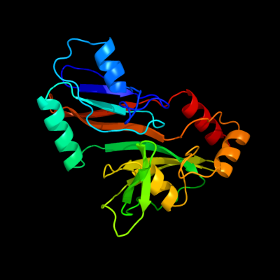
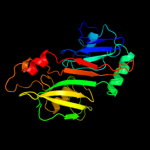
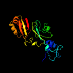
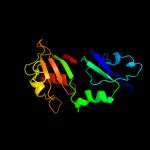
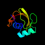
| 1 | d1v9ka_
|
|
 |
100.0 |
98 |
Fold:Pseudouridine synthase
Superfamily:Pseudouridine synthase
Family:Pseudouridine synthase RsuA/RluD |
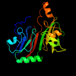
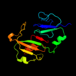
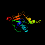
| 2 | d1v9fa_
|
|
 |
100.0 |
31 |
Fold:Pseudouridine synthase
Superfamily:Pseudouridine synthase
Family:Pseudouridine synthase RsuA/RluD |
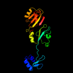
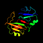
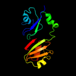
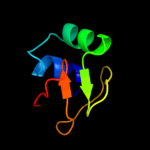
| 3 | c1v9fA_
|
|
 |
100.0 |
31 |
PDB header:lyase
Chain: A: PDB Molecule:ribosomal large subunit pseudouridine synthase d;
PDBTitle: crystal structure of catalytic domain of pseudouridine2 synthase rlud from escherichia coli
|
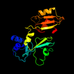
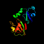
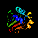
| 4 | c1qyuA_
|
|
 |
100.0 |
31 |
PDB header:lyase
Chain: A: PDB Molecule:ribosomal large subunit pseudouridine synthase d;
PDBTitle: structure of the catalytic domain of 23s rrna pseudouridine2 synthase rlud
|
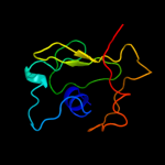
| 5 | c2i82D_
|
|
 |
100.0 |
38 |
PDB header:lyase/rna
Chain: D: PDB Molecule:ribosomal large subunit pseudouridine synthase a;
PDBTitle: crystal structure of pseudouridine synthase rlua: indirect2 sequence readout through protein-induced rna structure
|
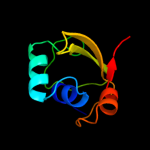
| 6 | c1kskA_
|
|
 |
100.0 |
24 |
PDB header:lyase
Chain: A: PDB Molecule:ribosomal small subunit pseudouridine synthase a;
PDBTitle: structure of rsua
|
| 7 | c1vioA_
|
|
 |
100.0 |
21 |
PDB header:lyase
Chain: A: PDB Molecule:ribosomal small subunit pseudouridine synthase a;
PDBTitle: crystal structure of pseudouridylate synthase
|
| 8 | c3dh3C_
|
|
 |
100.0 |
19 |
PDB header:isomerase/rna
Chain: C: PDB Molecule:ribosomal large subunit pseudouridine synthase f;
PDBTitle: crystal structure of rluf in complex with a 22 nucleotide2 rna substrate
|
| 9 | c2omlA_
|
|
 |
100.0 |
17 |
PDB header:isomerase
Chain: A: PDB Molecule:ribosomal large subunit pseudouridine synthase e;
PDBTitle: crystal structure of e. coli pseudouridine synthase rlue
|
| 10 | c2olwB_
|
|
 |
100.0 |
17 |
PDB header:isomerase
Chain: B: PDB Molecule:ribosomal large subunit pseudouridine synthase e;
PDBTitle: crystal structure of e. coli pseudouridine synthase rlue
|
| 11 | d1kska4
|
|
 |
100.0 |
23 |
Fold:Pseudouridine synthase
Superfamily:Pseudouridine synthase
Family:Pseudouridine synthase RsuA/RluD |
| 12 | d1vioa1
|
|
 |
100.0 |
21 |
Fold:Pseudouridine synthase
Superfamily:Pseudouridine synthase
Family:Pseudouridine synthase RsuA/RluD |
| 13 | c2gmlA_
|
|
 |
100.0 |
18 |
PDB header:isomerase
Chain: A: PDB Molecule:ribosomal large subunit pseudouridine synthase f;
PDBTitle: crystal structure of catalytic domain of e.coli rluf
|
| 14 | c2k6pA_
|
|
 |
99.5 |
19 |
PDB header:unknown function
Chain: A: PDB Molecule:uncharacterized protein hp_1423;
PDBTitle: solution structure of hypothetical protein, hp1423
|
| 15 | d1dm9a_
|
|
 |
99.0 |
20 |
Fold:Alpha-L RNA-binding motif
Superfamily:Alpha-L RNA-binding motif
Family:Heat shock protein 15 kD |
| 16 | c1dm9A_
|
|
 |
99.0 |
20 |
PDB header:structural genomics
Chain: A: PDB Molecule:hypothetical 15.5 kd protein in mrca-pcka
PDBTitle: heat shock protein 15 kd
|
| 17 | c3bbnD_
|
|
 |
98.9 |
19 |
PDB header:ribosome
Chain: D: PDB Molecule:ribosomal protein s4;
PDBTitle: homology model for the spinach chloroplast 30s subunit2 fitted to 9.4a cryo-em map of the 70s chlororibosome.
|
| 18 | d1vioa2
|
|
 |
98.8 |
20 |
Fold:Alpha-L RNA-binding motif
Superfamily:Alpha-L RNA-binding motif
Family:Pseudouridine synthase RsuA N-terminal domain |
| 19 | d1c06a_
|
|
 |
98.6 |
12 |
Fold:Alpha-L RNA-binding motif
Superfamily:Alpha-L RNA-binding motif
Family:Ribosomal protein S4 |
| 20 | d1p9ka_
|
|
 |
98.5 |
14 |
Fold:Alpha-L RNA-binding motif
Superfamily:Alpha-L RNA-binding motif
Family:YbcJ-like |
| 21 | d2uubd1 |
|
not modelled |
98.4 |
19 |
Fold:Alpha-L RNA-binding motif
Superfamily:Alpha-L RNA-binding motif
Family:Ribosomal protein S4 |
| 22 | d2gy9d1 |
|
not modelled |
98.2 |
14 |
Fold:Alpha-L RNA-binding motif
Superfamily:Alpha-L RNA-binding motif
Family:Ribosomal protein S4 |
| 23 | c2cqjA_ |
|
not modelled |
97.9 |
18 |
PDB header:rna binding protein
Chain: A: PDB Molecule:u3 small nucleolar ribonucleoprotein protein
PDBTitle: solution structure of the s4 domain of u3 small nucleolar2 ribonucleoprotein protein imp3 homolog
|
| 24 | d1k8wa5 |
|
not modelled |
97.7 |
20 |
Fold:Pseudouridine synthase
Superfamily:Pseudouridine synthase
Family:Pseudouridine synthase II TruB |
| 25 | d2ey4a2 |
|
not modelled |
97.6 |
27 |
Fold:Pseudouridine synthase
Superfamily:Pseudouridine synthase
Family:Pseudouridine synthase II TruB |
| 26 | d2apoa2 |
|
not modelled |
97.6 |
20 |
Fold:Pseudouridine synthase
Superfamily:Pseudouridine synthase
Family:Pseudouridine synthase II TruB |
| 27 | d1sgva2 |
|
not modelled |
97.5 |
27 |
Fold:Pseudouridine synthase
Superfamily:Pseudouridine synthase
Family:Pseudouridine synthase II TruB |
| 28 | c2ey4A_ |
|
not modelled |
97.5 |
27 |
PDB header:isomerase/biosynthetic protein
Chain: A: PDB Molecule:probable trna pseudouridine synthase b;
PDBTitle: crystal structure of a cbf5-nop10-gar1 complex
|
| 29 | d1r3ea2 |
|
not modelled |
97.5 |
24 |
Fold:Pseudouridine synthase
Superfamily:Pseudouridine synthase
Family:Pseudouridine synthase II TruB |
| 30 | c3uaiA_ |
|
not modelled |
97.3 |
17 |
PDB header:isomerase/chaperone
Chain: A: PDB Molecule:h/aca ribonucleoprotein complex subunit 4;
PDBTitle: structure of the shq1-cbf5-nop10-gar1 complex from saccharomyces2 cerevisiae
|
| 31 | c3hp7A_ |
|
not modelled |
97.3 |
25 |
PDB header:structural genomics, unknown function
Chain: A: PDB Molecule:hemolysin, putative;
PDBTitle: putative hemolysin from streptococcus thermophilus.
|
| 32 | c2apoA_ |
|
not modelled |
97.3 |
23 |
PDB header:isomerase/rna binding protein
Chain: A: PDB Molecule:probable trna pseudouridine synthase b;
PDBTitle: crystal structure of the methanococcus jannaschii cbf52 nop10 complex
|
| 33 | c1k8wA_ |
|
not modelled |
97.3 |
20 |
PDB header:lyase/rna
Chain: A: PDB Molecule:trna pseudouridine synthase b;
PDBTitle: crystal structure of the e. coli pseudouridine synthase2 trub bound to a t stem-loop rna
|
| 34 | c1sgvA_ |
|
not modelled |
97.3 |
27 |
PDB header:lyase
Chain: A: PDB Molecule:trna pseudouridine synthase b;
PDBTitle: structure of trna psi55 pseudouridine synthase (trub)
|
| 35 | d1kska3 |
|
not modelled |
97.1 |
27 |
Fold:Alpha-L RNA-binding motif
Superfamily:Alpha-L RNA-binding motif
Family:Pseudouridine synthase RsuA N-terminal domain |
| 36 | c1s1hD_ |
|
not modelled |
96.9 |
11 |
PDB header:ribosome
Chain: D: PDB Molecule:40s ribosomal protein s9-a;
PDBTitle: structure of the ribosomal 80s-eef2-sordarin complex from2 yeast obtained by docking atomic models for rna and protein3 components into a 11.7 a cryo-em map. this file, 1s1h,4 contains 40s subunit. the 60s ribosomal subunit is in file5 1s1i.
|
| 37 | c2xzmD_ |
|
not modelled |
96.8 |
10 |
PDB header:ribosome
Chain: D: PDB Molecule:ribosomal protein s4 containing protein;
PDBTitle: crystal structure of the eukaryotic 40s ribosomal2 subunit in complex with initiation factor 1. this file3 contains the 40s subunit and initiation factor for4 molecule 1
|
| 38 | d1h3fa2 |
|
not modelled |
96.6 |
11 |
Fold:Alpha-L RNA-binding motif
Superfamily:Alpha-L RNA-binding motif
Family:Tyrosyl-tRNA synthetase (TyrRS), C-terminal domain |
| 39 | d1jh3a_ |
|
not modelled |
95.7 |
15 |
Fold:Alpha-L RNA-binding motif
Superfamily:Alpha-L RNA-binding motif
Family:Tyrosyl-tRNA synthetase (TyrRS), C-terminal domain |
| 40 | c1ze2B_ |
|
not modelled |
95.7 |
28 |
PDB header:lyase/rna
Chain: B: PDB Molecule:trna pseudouridine synthase b;
PDBTitle: conformational change of pseudouridine 55 synthase upon its2 association with rna substrate
|
| 41 | c3kbgA_ |
|
not modelled |
95.5 |
27 |
PDB header:ribosomal protein
Chain: A: PDB Molecule:30s ribosomal protein s4e;
PDBTitle: crystal structure of the 30s ribosomal protein s4e from2 thermoplasma acidophilum. northeast structural genomics3 consortium target tar28.
|
| 42 | c1h3eA_ |
|
not modelled |
95.1 |
13 |
PDB header:ligase
Chain: A: PDB Molecule:tyrosyl-trna synthetase;
PDBTitle: tyrosyl-trna synthetase from thermus thermophilus complexed2 with wild-type trnatyr(gua) and with atp and tyrosinol
|
| 43 | c2xzmW_ |
|
not modelled |
94.9 |
25 |
PDB header:ribosome
Chain: W: PDB Molecule:40s ribosomal protein s4;
PDBTitle: crystal structure of the eukaryotic 40s ribosomal2 subunit in complex with initiation factor 1. this file3 contains the 40s subunit and initiation factor for4 molecule 1
|
| 44 | c2janD_ |
|
not modelled |
93.9 |
22 |
PDB header:ligase
Chain: D: PDB Molecule:tyrosyl-trna synthetase;
PDBTitle: tyrosyl-trna synthetase from mycobacterium tuberculosis in2 unliganded state
|
| 45 | c3iz6D_ |
|
not modelled |
93.9 |
28 |
PDB header:ribosome
Chain: D: PDB Molecule:40s ribosomal protein s4 (s4e);
PDBTitle: localization of the small subunit ribosomal proteins into a 5.5 a2 cryo-em map of triticum aestivum translating 80s ribosome
|
| 46 | c3izbD_ |
|
not modelled |
93.6 |
25 |
PDB header:ribosome
Chain: D: PDB Molecule:40s ribosomal protein rps4 (s4e);
PDBTitle: localization of the small subunit ribosomal proteins into a 6.1 a2 cryo-em map of saccharomyces cerevisiae translating 80s ribosome
|
| 47 | d1fm0d_ |
|
not modelled |
85.2 |
19 |
Fold:beta-Grasp (ubiquitin-like)
Superfamily:MoaD/ThiS
Family:MoaD |
| 48 | c2qieB_ |
|
not modelled |
83.7 |
19 |
PDB header:transferase
Chain: B: PDB Molecule:molybdopterin synthase small subunit;
PDBTitle: staphylococcus aureus molybdopterin synthase in complex2 with precursor z
|
| 49 | c3po0A_ |
|
not modelled |
82.4 |
19 |
PDB header:protein binding
Chain: A: PDB Molecule:small archaeal modifier protein 1;
PDBTitle: crystal structure of samp1 from haloferax volcanii
|
| 50 | d1vjka_ |
|
not modelled |
82.3 |
13 |
Fold:beta-Grasp (ubiquitin-like)
Superfamily:MoaD/ThiS
Family:MoaD |
| 51 | c3iz6C_ |
|
not modelled |
81.1 |
10 |
PDB header:ribosome
Chain: C: PDB Molecule:40s ribosomal protein s9 (s4p);
PDBTitle: localization of the small subunit ribosomal proteins into a 5.5 a2 cryo-em map of triticum aestivum translating 80s ribosome
|
| 52 | d2g1la1 |
|
not modelled |
78.5 |
24 |
Fold:SMAD/FHA domain
Superfamily:SMAD/FHA domain
Family:FHA domain |
| 53 | d1wgka_ |
|
not modelled |
77.7 |
19 |
Fold:beta-Grasp (ubiquitin-like)
Superfamily:MoaD/ThiS
Family:C9orf74 homolog |
| 54 | c2g1eA_ |
|
not modelled |
75.4 |
15 |
PDB header:transferase
Chain: A: PDB Molecule:hypothetical protein ta0895;
PDBTitle: solution structure of ta0895
|
| 55 | c3fm8A_ |
|
not modelled |
71.0 |
20 |
PDB header:transport protein/hydrolase activator
Chain: A: PDB Molecule:kinesin-like protein kif13b;
PDBTitle: crystal structure of full length centaurin alpha-1 bound with the fha2 domain of kif13b (capri target)
|
| 56 | d1xo3a_ |
|
not modelled |
70.6 |
19 |
Fold:beta-Grasp (ubiquitin-like)
Superfamily:MoaD/ThiS
Family:C9orf74 homolog |
| 57 | c2eh0A_ |
|
not modelled |
69.0 |
28 |
PDB header:transport protein
Chain: A: PDB Molecule:kinesin-like protein kif1b;
PDBTitle: solution structure of the fha domain from human kinesin-2 like protein kif1b
|
| 58 | c2kmmA_ |
|
not modelled |
67.9 |
28 |
PDB header:hydrolase
Chain: A: PDB Molecule:guanosine-3',5'-bis(diphosphate) 3'-
PDBTitle: solution nmr structure of the tgs domain of pg1808 from2 porphyromonas gingivalis. northeast structural genomics3 consortium target pgr122a (418-481)
|
| 59 | c3hvzB_ |
|
not modelled |
66.4 |
19 |
PDB header:structural genomics, unknown function
Chain: B: PDB Molecule:uncharacterized protein;
PDBTitle: crystal structure of the tgs domain of the clolep_03100 protein from2 clostridium leptum, northeast structural genomics consortium target3 qlr13a
|
| 60 | c2qjlA_ |
|
not modelled |
66.1 |
14 |
PDB header:signaling protein
Chain: A: PDB Molecule:ubiquitin-related modifier 1;
PDBTitle: crystal structure of urm1
|
| 61 | c1v8cA_ |
|
not modelled |
66.1 |
21 |
PDB header:protein binding
Chain: A: PDB Molecule:moad related protein;
PDBTitle: crystal structure of moad related protein from thermus2 thermophilus hb8
|
| 62 | d2affa1 |
|
not modelled |
63.3 |
26 |
Fold:SMAD/FHA domain
Superfamily:SMAD/FHA domain
Family:FHA domain |
| 63 | c1r21A_ |
|
not modelled |
61.1 |
26 |
PDB header:cell cycle
Chain: A: PDB Molecule:antigen ki-67;
PDBTitle: solution structure of human ki67 fha domain
|
| 64 | c3dwmA_ |
|
not modelled |
59.7 |
28 |
PDB header:transferase
Chain: A: PDB Molecule:9.5 kda culture filtrate antigen cfp10a;
PDBTitle: crystal structure of mycobacterium tuberculosis cyso, an antigen
|
| 65 | d1wlna1 |
|
not modelled |
59.0 |
24 |
Fold:SMAD/FHA domain
Superfamily:SMAD/FHA domain
Family:FHA domain |
| 66 | d1v8ca1 |
|
not modelled |
59.0 |
15 |
Fold:beta-Grasp (ubiquitin-like)
Superfamily:MoaD/ThiS
Family:MoaD |
| 67 | c3rpfC_ |
|
not modelled |
57.9 |
14 |
PDB header:transferase
Chain: C: PDB Molecule:molybdopterin converting factor, subunit 1 (moad);
PDBTitle: protein-protein complex of subunit 1 and 2 of molybdopterin-converting2 factor from helicobacter pylori 26695
|
| 68 | d1tkea1 |
|
not modelled |
57.3 |
24 |
Fold:beta-Grasp (ubiquitin-like)
Superfamily:TGS-like
Family:TGS domain |
| 69 | d1rwsa_ |
|
not modelled |
54.7 |
29 |
Fold:beta-Grasp (ubiquitin-like)
Superfamily:MoaD/ThiS
Family:ThiS |
| 70 | c2hj1A_ |
|
not modelled |
53.2 |
17 |
PDB header:structural genomics, unknown function
Chain: A: PDB Molecule:hypothetical protein;
PDBTitle: crystal structure of a 3d domain-swapped dimer of protein hi0395 from2 haemophilus influenzae
|
| 71 | d2hj1a1 |
|
not modelled |
53.2 |
17 |
Fold:beta-Grasp (ubiquitin-like)
Superfamily:MoaD/ThiS
Family:HI0395-like |
| 72 | d2ff4a3 |
|
not modelled |
52.3 |
36 |
Fold:SMAD/FHA domain
Superfamily:SMAD/FHA domain
Family:FHA domain |
| 73 | c2k9xA_ |
|
not modelled |
50.2 |
26 |
PDB header:unknown function
Chain: A: PDB Molecule:uncharacterized protein;
PDBTitle: solution structure of urm1 from trypanosoma brucei
|
| 74 | d1wxqa2 |
|
not modelled |
46.5 |
32 |
Fold:beta-Grasp (ubiquitin-like)
Superfamily:TGS-like
Family:G domain-linked domain |
| 75 | c1yj5C_ |
|
not modelled |
42.2 |
27 |
PDB header:transferase
Chain: C: PDB Molecule:5' polynucleotide kinase-3' phosphatase fha domain;
PDBTitle: molecular architecture of mammalian polynucleotide kinase, a dna2 repair enzyme
|
| 76 | c3elsA_ |
|
not modelled |
38.1 |
24 |
PDB header:splicing
Chain: A: PDB Molecule:pre-mrna leakage protein 1;
PDBTitle: crystal structure of yeast pml1p, residues 51-204
|
| 77 | c3gqsB_ |
|
not modelled |
37.8 |
28 |
PDB header:structural genomics, unknown function
Chain: B: PDB Molecule:adenylate cyclase-like protein;
PDBTitle: crystal structure of the fha domain of ct664 protein from chlamydia2 trachomatis
|
| 78 | c2jqlA_ |
|
not modelled |
37.7 |
24 |
PDB header:cell cycle
Chain: A: PDB Molecule:dna damage response protein kinase dun1;
PDBTitle: nmr structure of the yeast dun1 fha domain in complex with2 a doubly phosphorylated (pt) peptide derived from rad533 scd1
|
| 79 | d1nyra2 |
|
not modelled |
37.5 |
19 |
Fold:beta-Grasp (ubiquitin-like)
Superfamily:TGS-like
Family:TGS domain |
| 80 | c3kt9A_ |
|
not modelled |
36.3 |
17 |
PDB header:hydrolase
Chain: A: PDB Molecule:aprataxin;
PDBTitle: aprataxin fha domain
|
| 81 | c3zrhA_ |
|
not modelled |
35.8 |
19 |
PDB header:hydrolase
Chain: A: PDB Molecule:ubiquitin thioesterase zranb1;
PDBTitle: crystal structure of the lys29, lys33-linkage-specific trabid otu2 deubiquitinase domain reveals an ankyrin-repeat ubiquitin binding3 domain (ankubd)
|
| 82 | c3hx1B_ |
|
not modelled |
34.3 |
25 |
PDB header:structural genomics, unknown function
Chain: B: PDB Molecule:slr1951 protein;
PDBTitle: crystal structure of the slr1951 protein from synechocystis sp.2 northeast structural genomics consortium target sgr167a
|
| 83 | c2l52A_ |
|
not modelled |
33.0 |
32 |
PDB header:protein binding
Chain: A: PDB Molecule:methanosarcina acetivorans samp1 homolog;
PDBTitle: solution structure of the small archaeal modifier protein 1 (samp1)2 from methanosarcina acetivorans
|
| 84 | c3poaA_ |
|
not modelled |
31.1 |
29 |
PDB header:peptide binding protein
Chain: A: PDB Molecule:putative uncharacterized protein tb39.8;
PDBTitle: structural and functional analysis of phosphothreonine-dependent fha2 domain interactions
|
| 85 | d1lgpa_ |
|
not modelled |
30.6 |
21 |
Fold:SMAD/FHA domain
Superfamily:SMAD/FHA domain
Family:FHA domain |
| 86 | d1g6ga_ |
|
not modelled |
28.3 |
23 |
Fold:SMAD/FHA domain
Superfamily:SMAD/FHA domain
Family:FHA domain |
| 87 | d1zud21 |
|
not modelled |
27.4 |
14 |
Fold:beta-Grasp (ubiquitin-like)
Superfamily:MoaD/ThiS
Family:ThiS |
| 88 | d2piea1 |
|
not modelled |
26.4 |
24 |
Fold:SMAD/FHA domain
Superfamily:SMAD/FHA domain
Family:FHA domain |
| 89 | c2kklA_ |
|
not modelled |
25.8 |
33 |
PDB header:structural genomics, unknown function
Chain: A: PDB Molecule:uncharacterized protein mb1858;
PDBTitle: solution nmr structure of fha domain of mb1858 from2 mycobacterium bovis. northeast structural genomics3 consortium target mbr243c (24-155).
|
| 90 | c1gxcA_ |
|
not modelled |
25.8 |
16 |
PDB header:phosphoprotein-binding domain
Chain: A: PDB Molecule:serine/threonine-protein kinase chk2;
PDBTitle: fha domain from human chk2 kinase in complex with a2 synthetic phosphopeptide
|
| 91 | d1gxca_ |
|
not modelled |
25.8 |
16 |
Fold:SMAD/FHA domain
Superfamily:SMAD/FHA domain
Family:FHA domain |
| 92 | c3cwiA_ |
|
not modelled |
23.1 |
23 |
PDB header:biosynthetic protein
Chain: A: PDB Molecule:thiamine-biosynthesis protein this;
PDBTitle: crystal structure of thiamine biosynthesis protein (this)2 from geobacter metallireducens. northeast structural3 genomics consortium target gmr137
|
| 93 | c2jkdB_ |
|
not modelled |
23.1 |
24 |
PDB header:gene regulation
Chain: B: PDB Molecule:pre-mrna leakage protein 1;
PDBTitle: structure of the yeast pml1 splicing factor and its2 integration into the res complex
|
| 94 | c2h89B_ |
|
not modelled |
21.2 |
10 |
PDB header:oxidoreductase
Chain: B: PDB Molecule:succinate dehydrogenase ip subunit;
PDBTitle: avian respiratory complex ii with malonate bound
|
| 95 | c2zodB_ |
|
not modelled |
20.1 |
21 |
PDB header:transferase
Chain: B: PDB Molecule:selenide, water dikinase;
PDBTitle: crystal structure of selenophosphate synthetase from2 aquifex aeolicus
|
| 96 | c2ekiA_ |
|
not modelled |
20.0 |
20 |
PDB header:signaling protein
Chain: A: PDB Molecule:developmentally-regulated gtp-binding protein 1;
PDBTitle: solution structures of the tgs domain of human2 developmentally-regulated gtp-binding protein 1
|
| 97 | d1k25a2 |
|
not modelled |
19.9 |
7 |
Fold:Penicillin-binding protein 2x (pbp-2x), c-terminal domain
Superfamily:Penicillin-binding protein 2x (pbp-2x), c-terminal domain
Family:Penicillin-binding protein 2x (pbp-2x), c-terminal domain |
| 98 | d1g3ga_ |
|
not modelled |
19.8 |
23 |
Fold:SMAD/FHA domain
Superfamily:SMAD/FHA domain
Family:FHA domain |
| 99 | d1c1za5 |
|
not modelled |
19.7 |
29 |
Fold:Complement control module/SCR domain
Superfamily:Complement control module/SCR domain
Family:Complement control module/SCR domain |