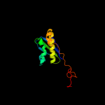
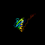
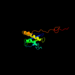
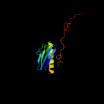
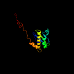
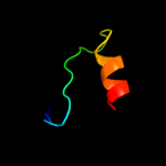
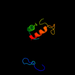
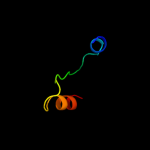
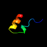
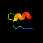
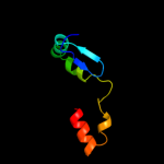
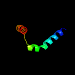
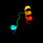
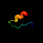
1 d2gy9i1
100.0
100
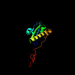
Fold: Ribosomal protein S5 domain 2-likeSuperfamily: Ribosomal protein S5 domain 2-likeFamily: Translational machinery components2 d2vqei1
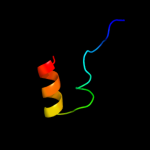
100.0
54
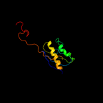
Fold: Ribosomal protein S5 domain 2-likeSuperfamily: Ribosomal protein S5 domain 2-likeFamily: Translational machinery components3 c3bbnI_
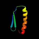
100.0
46
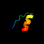
PDB header: ribosomeChain: I: PDB Molecule: ribosomal protein s9;PDBTitle: homology model for the spinach chloroplast 30s subunit2 fitted to 9.4a cryo-em map of the 70s chlororibosome.
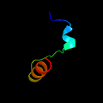
4 c2xzmI_
100.0
25
PDB header: ribosomeChain: I: PDB Molecule: rps16e;PDBTitle: crystal structure of the eukaryotic 40s ribosomal2 subunit in complex with initiation factor 1. this file3 contains the 40s subunit and initiation factor for4 molecule 1
5 c2zkqi_
100.0
33
PDB header: ribosomal protein/rnaChain: I: PDB Molecule: PDBTitle: structure of a mammalian ribosomal 40s subunit within an2 80s complex obtained by docking homology models of the rna3 and proteins into an 8.7 a cryo-em map
6 c1s1hI_
100.0
36
PDB header: ribosomeChain: I: PDB Molecule: 40s ribosomal protein s16;PDBTitle: structure of the ribosomal 80s-eef2-sordarin complex from2 yeast obtained by docking atomic models for rna and protein3 components into a 11.7 a cryo-em map. this file, 1s1h,4 contains 40s subunit. the 60s ribosomal subunit is in file5 1s1i.
7 c3iz6I_
100.0
36
PDB header: ribosomeChain: I: PDB Molecule: 40s ribosomal protein s16 (s9p);PDBTitle: localization of the small subunit ribosomal proteins into a 5.5 a2 cryo-em map of triticum aestivum translating 80s ribosome
8 c1s3bB_
54.6
50
PDB header: oxidoreductaseChain: B: PDB Molecule: amine oxidase [flavin-containing] b;PDBTitle: crystal structure of maob in complex with n-methyl-n-2 propargyl-1(r)-aminoindan
9 d1d5ta1
50.8
20
Fold: FAD/NAD(P)-binding domainSuperfamily: FAD/NAD(P)-binding domainFamily: GDI-like N domain10 c1v0jB_
46.8
38
PDB header: isomeraseChain: B: PDB Molecule: udp-galactopyranose mutase;PDBTitle: udp-galactopyranose mutase from mycobacterium tuberculosis
11 c2rrlA_
42.5
15
PDB header: protein transportChain: A: PDB Molecule: flagellar hook-length control protein;PDBTitle: solution structure of the c-terminal domain of the flik
12 c1ltxR_
39.5
20
PDB header: transferase/protein bindingChain: R: PDB Molecule: rab escort protein 1;PDBTitle: structure of rab escort protein-1 in complex with rab2 geranylgeranyl transferase and isoprenoid
13 c2hydB_
35.6
21
PDB header: transport proteinChain: B: PDB Molecule: abc transporter homolog;PDBTitle: multidrug abc transporter sav1866
14 d1hyua1
35.2
30
Fold: FAD/NAD(P)-binding domainSuperfamily: FAD/NAD(P)-binding domainFamily: FAD/NAD-linked reductases, N-terminal and central domains15 c3cp8C_
33.7
32
PDB header: oxidoreductaseChain: C: PDB Molecule: trna uridine 5-carboxymethylaminomethylPDBTitle: crystal structure of gida from chlorobium tepidum
16 c2uzzD_
33.2
38
PDB header: oxidoreductaseChain: D: PDB Molecule: n-methyl-l-tryptophan oxidase;PDBTitle: x-ray structure of n-methyl-l-tryptophan oxidase (mtox)
17 c2v6oA_
33.1
12
PDB header: oxidoreductaseChain: A: PDB Molecule: thioredoxin glutathione reductase;PDBTitle: structure of schistosoma mansoni thioredoxin-glutathione2 reductase (smtgr)
18 c3jskN_
32.8
24
PDB header: biosynthetic proteinChain: N: PDB Molecule: cypbp37 protein;PDBTitle: thiazole synthase from neurospora crassa
19 d2bcgg1
32.7
21
Fold: FAD/NAD(P)-binding domainSuperfamily: FAD/NAD(P)-binding domainFamily: GDI-like N domain20 d1gesa1
30.9
32
Fold: FAD/NAD(P)-binding domainSuperfamily: FAD/NAD(P)-binding domainFamily: FAD/NAD-linked reductases, N-terminal and central domains21 c3k7tB_
not modelled
30.7
45
PDB header: oxidoreductaseChain: B: PDB Molecule: 6-hydroxy-l-nicotine oxidase;PDBTitle: crystal structure of apo-form 6-hydroxy-l-nicotine oxidase,2 crystal form p3121
22 d1rbla1
not modelled
30.4
25
Fold: TIM beta/alpha-barrelSuperfamily: RuBisCo, C-terminal domainFamily: RuBisCo, large subunit, C-terminal domain23 d1wdda1
not modelled
28.9
22
Fold: TIM beta/alpha-barrelSuperfamily: RuBisCo, C-terminal domainFamily: RuBisCo, large subunit, C-terminal domain24 c2vvlD_
not modelled
28.5
36
PDB header: oxidoreductaseChain: D: PDB Molecule: monoamine oxidase n;PDBTitle: the structure of mao-n-d3, a variant of monoamine oxidase2 from aspergillus niger.
25 c2yl4A_
not modelled
27.8
26
PDB header: membrane proteinChain: A: PDB Molecule: atp-binding cassette sub-family b member 10,PDBTitle: structure of the human mitochondrial abc transporter, abcb10
26 c4a1eU_
not modelled
27.0
29
PDB header: ribosomeChain: U: PDB Molecule: rpl35;PDBTitle: t.thermophila 60s ribosomal subunit in complex with2 initiation factor 6. this file contains 5s rrna, 5.8s rrna3 and proteins of molecule 1
27 c2zxiC_
not modelled
26.7
33
PDB header: fad-binding proteinChain: C: PDB Molecule: trna uridine 5-carboxymethylaminomethylPDBTitle: structure of aquifex aeolicus gida in the form ii crystal
28 c3cgvA_
not modelled
25.7
36
PDB header: structural genomics, unknown functionChain: A: PDB Molecule: geranylgeranyl reductase related protein;PDBTitle: crystal structure of geranylgeranyl bacteriochlorophyll reductase-like2 fixc homolog (np_393992.1) from thermoplasma acidophilum at 1.60 a3 resolution
29 c3rhaA_
not modelled
23.8
28
PDB header: oxidoreductaseChain: A: PDB Molecule: putrescine oxidase;PDBTitle: the crystal structure of oxidoreductase from arthrobacter aurescens
30 c1ryiB_
not modelled
23.4
38
PDB header: oxidoreductaseChain: B: PDB Molecule: glycine oxidase;PDBTitle: structure of glycine oxidase with bound inhibitor glycolate
31 d1rrea_
not modelled
22.5
25
Fold: Ribosomal protein S5 domain 2-likeSuperfamily: Ribosomal protein S5 domain 2-likeFamily: ATP-dependent protease Lon (La), catalytic domain32 c3g05B_
not modelled
21.7
30
PDB header: rna binding proteinChain: B: PDB Molecule: trna uridine 5-carboxymethylaminomethyl modification enzymePDBTitle: crystal structure of n-terminal domain (2-550) of e.coli mnmg
33 c2olnA_
not modelled
21.6
38
PDB header: oxidoreductaseChain: A: PDB Molecule: nikd protein;PDBTitle: nikd, an unusual amino acid oxidase essential for2 nikkomycin biosynthesis: closed form at 1.15 a resolution
34 c3m6aC_
not modelled
21.2
25
PDB header: hydrolaseChain: C: PDB Molecule: atp-dependent protease la 1;PDBTitle: crystal structure of bacillus subtilis lon c-terminal domain
35 c2w0hA_
not modelled
21.0
22
PDB header: oxidoreductaseChain: A: PDB Molecule: trypanothione reductase;PDBTitle: x ray structure of leishmania infantum trypanothione2 reductase in complex with antimony and nadph
36 c2qaeA_
not modelled
19.3
32
PDB header: oxidoreductaseChain: A: PDB Molecule: dihydrolipoyl dehydrogenase;PDBTitle: crystal structure analysis of trypanosoma cruzi lipoamide2 dehydrogenase
37 c1dxlC_
not modelled
19.3
30
PDB header: oxidoreductaseChain: C: PDB Molecule: dihydrolipoamide dehydrogenase;PDBTitle: dihydrolipoamide dehydrogenase of glycine decarboxylase2 from pisum sativum
38 c3uo9B_
not modelled
17.8
29
PDB header: hydrolase/hydrolase inhibitorChain: B: PDB Molecule: glutaminase kidney isoform, mitochondrial;PDBTitle: crystal structure of human gac in complex with glutamate and bptes
39 c2nvkX_
not modelled
17.3
36
PDB header: oxidoreductaseChain: X: PDB Molecule: thioredoxin reductase;PDBTitle: crystal structure of thioredoxin reductase from drosophila2 melanogaster
40 d3lada1
not modelled
17.0
35
Fold: FAD/NAD(P)-binding domainSuperfamily: FAD/NAD(P)-binding domainFamily: FAD/NAD-linked reductases, N-terminal and central domains41 d1dxla1
not modelled
16.9
30
Fold: FAD/NAD(P)-binding domainSuperfamily: FAD/NAD(P)-binding domainFamily: FAD/NAD-linked reductases, N-terminal and central domains42 d1d4ca2
not modelled
15.8
16
Fold: FAD/NAD(P)-binding domainSuperfamily: FAD/NAD(P)-binding domainFamily: Succinate dehydrogenase/fumarate reductase flavoprotein N-terminal domain43 d2bj7a1
not modelled
15.5
21
Fold: Ribbon-helix-helixSuperfamily: Ribbon-helix-helixFamily: CopG-like44 d1reoa1
not modelled
15.4
24
Fold: FAD/NAD(P)-binding domainSuperfamily: FAD/NAD(P)-binding domainFamily: FAD-linked reductases, N-terminal domain45 c1f8sA_
not modelled
15.4
13
PDB header: oxidoreductaseChain: A: PDB Molecule: l-amino acid oxidase;PDBTitle: crystal structure of l-amino acid oxidase from calloselasma2 rhodostoma, complexed with three molecules of o-aminobenzoate.
46 c3b5xB_
not modelled
15.3
31
PDB header: membrane proteinChain: B: PDB Molecule: lipid a export atp-binding/permease protein msba;PDBTitle: crystal structure of msba from vibrio cholerae
47 c2r9zB_
not modelled
15.2
30
PDB header: oxidoreductaseChain: B: PDB Molecule: glutathione amide reductase;PDBTitle: glutathione amide reductase from chromatium gracile
48 c3ihgA_
not modelled
14.9
20
PDB header: flavoprotein, oxidoreductaseChain: A: PDB Molecule: rdme;PDBTitle: crystal structure of a ternary complex of aklavinone-112 hydroxylase with fad and aklavinone
49 c2e1mA_
not modelled
14.8
26
PDB header: oxidoreductaseChain: A: PDB Molecule: l-glutamate oxidase;PDBTitle: crystal structure of l-glutamate oxidase from streptomyces sp. x-119-6
50 c3dmeB_
not modelled
14.6
33
PDB header: structural genomics, unknown functionChain: B: PDB Molecule: conserved exported protein;PDBTitle: crystal structure of conserved exported protein from2 bordetella pertussis. northeast structural genomics target3 ber141
51 c1tytA_
not modelled
14.6
25
PDB header: oxidoreductaseChain: A: PDB Molecule: trypanothione reductase, oxidized form;PDBTitle: crystal and molecular structure of crithidia fasciculata2 trypanothione reductase at 2.6 angstroms resolution
52 c3ss4C_
not modelled
14.5
29
PDB header: hydrolaseChain: C: PDB Molecule: glutaminase c;PDBTitle: crystal structure of mouse glutaminase c, phosphate-bound form
53 d1kifa1
not modelled
14.4
25
Fold: Nucleotide-binding domainSuperfamily: Nucleotide-binding domainFamily: D-aminoacid oxidase, N-terminal domain54 d1h6va1
not modelled
14.1
32
Fold: FAD/NAD(P)-binding domainSuperfamily: FAD/NAD(P)-binding domainFamily: FAD/NAD-linked reductases, N-terminal and central domains55 d1chua2
not modelled
14.0
19
Fold: FAD/NAD(P)-binding domainSuperfamily: FAD/NAD(P)-binding domainFamily: Succinate dehydrogenase/fumarate reductase flavoprotein N-terminal domain56 c3p4rM_
not modelled
13.9
20
PDB header: oxidoreductaseChain: M: PDB Molecule: fumarate reductase flavoprotein subunit;PDBTitle: crystal structure of menaquinol:fumarate oxidoreductase in complex2 with glutarate
57 c3nyeA_
not modelled
13.8
24
PDB header: oxidoreductaseChain: A: PDB Molecule: d-arginine dehydrogenase;PDBTitle: crystal structure of pseudomonas aeruginosa d-arginine dehydrogenase2 in complex with imino-arginine
58 d1seza1
not modelled
13.8
37
Fold: FAD/NAD(P)-binding domainSuperfamily: FAD/NAD(P)-binding domainFamily: FAD-linked reductases, N-terminal domain59 c3g5uB_
not modelled
13.7
26
PDB header: membrane proteinChain: B: PDB Molecule: multidrug resistance protein 1a;PDBTitle: structure of p-glycoprotein reveals a molecular basis for2 poly-specific drug binding
60 c1ndaD_
not modelled
13.5
30
PDB header: oxidoreductaseChain: D: PDB Molecule: trypanothione oxidoreductase;PDBTitle: the structure of trypanosoma cruzi trypanothione reductase2 in the oxidized and nadph reduced state
61 d2gf3a1
not modelled
13.3
24
Fold: FAD/NAD(P)-binding domainSuperfamily: FAD/NAD(P)-binding domainFamily: FAD-linked reductases, N-terminal domain62 d1v59a1
not modelled
13.2
29
Fold: FAD/NAD(P)-binding domainSuperfamily: FAD/NAD(P)-binding domainFamily: FAD/NAD-linked reductases, N-terminal and central domains63 d1pj5a2
not modelled
13.1
33
Fold: FAD/NAD(P)-binding domainSuperfamily: FAD/NAD(P)-binding domainFamily: FAD-linked reductases, N-terminal domain64 c1kf6A_
not modelled
13.0
20
PDB header: oxidoreductaseChain: A: PDB Molecule: fumarate reductase flavoprotein;PDBTitle: e. coli quinol-fumarate reductase with bound inhibitor hqno
65 d2dw4a2
not modelled
12.8
32
Fold: FAD/NAD(P)-binding domainSuperfamily: FAD/NAD(P)-binding domainFamily: FAD-linked reductases, N-terminal domain66 d2bi7a1
not modelled
12.8
24
Fold: Nucleotide-binding domainSuperfamily: Nucleotide-binding domainFamily: UDP-galactopyranose mutase, N-terminal domain67 d2gmha1
not modelled
12.7
24
Fold: FAD/NAD(P)-binding domainSuperfamily: FAD/NAD(P)-binding domainFamily: FAD-linked reductases, N-terminal domain68 d1lpfa1
not modelled
12.6
39
Fold: FAD/NAD(P)-binding domainSuperfamily: FAD/NAD(P)-binding domainFamily: FAD/NAD-linked reductases, N-terminal and central domains69 d2hzaa1
not modelled
12.3
30
Fold: Ribbon-helix-helixSuperfamily: Ribbon-helix-helixFamily: CopG-like70 c2cfyB_
not modelled
12.3
30
PDB header: oxidoreductaseChain: B: PDB Molecule: thioredoxin reductase 1;PDBTitle: crystal structure of human thioredoxin reductase 1
71 c3bnuA_
not modelled
12.2
26
PDB header: oxidoreductaseChain: A: PDB Molecule: polyamine oxidase fms1;PDBTitle: crystal structure of polyamine oxidase fms1 from2 saccharomyces cerevisiae in complex with bis-(3s,3's)-3 methylated spermine
72 d2i0za1
not modelled
12.1
36
Fold: FAD/NAD(P)-binding domainSuperfamily: FAD/NAD(P)-binding domainFamily: HI0933 N-terminal domain-like73 c2r4jA_
not modelled
11.9
43
PDB header: oxidoreductaseChain: A: PDB Molecule: aerobic glycerol-3-phosphate dehydrogenase;PDBTitle: crystal structure of escherichia coli semet substituted2 glycerol-3-phosphate dehydrogenase in complex with dhap
74 c3bjrA_
not modelled
11.7
23
PDB header: hydrolaseChain: A: PDB Molecule: putative carboxylesterase;PDBTitle: crystal structure of a putative carboxylesterase (lp_1002) from2 lactobacillus plantarum wcfs1 at 2.09 a resolution
75 c1h83A_
not modelled
11.6
39
PDB header: oxidoreductaseChain: A: PDB Molecule: polyamine oxidase;PDBTitle: structure of polyamine oxidase in complex with2 1,8-diaminooctane
76 c3i6dA_
not modelled
11.5
37
PDB header: oxidoreductaseChain: A: PDB Molecule: protoporphyrinogen oxidase;PDBTitle: crystal structure of ppo from bacillus subtilis with af
77 d1rp0a1
not modelled
11.5
24
Fold: FAD/NAD(P)-binding domainSuperfamily: FAD/NAD(P)-binding domainFamily: Thi4-like78 d1vg0a1
not modelled
11.3
24
Fold: FAD/NAD(P)-binding domainSuperfamily: FAD/NAD(P)-binding domainFamily: GDI-like N domain79 c3l8kB_
not modelled
11.3
43
PDB header: oxidoreductaseChain: B: PDB Molecule: dihydrolipoyl dehydrogenase;PDBTitle: crystal structure of a dihydrolipoyl dehydrogenase from2 sulfolobus solfataricus
80 c2gqfA_
not modelled
11.2
22
PDB header: structural genomics, unknown functionChain: A: PDB Molecule: hypothetical protein hi0933;PDBTitle: crystal structure of flavoprotein hi0933 from haemophilus influenzae2 rd
81 c3hdqI_
not modelled
11.0
26
PDB header: isomeraseChain: I: PDB Molecule: udp-galactopyranose mutase;PDBTitle: crystal structure of udp-galactopyranose mutase (oxidized2 form) in complex with substrate
82 c2aczA_
not modelled
10.9
24
PDB header: oxidoreductase/electron transportChain: A: PDB Molecule: succinate dehydrogenase flavoprotein subunit;PDBTitle: complex ii (succinate dehydrogenase) from e. coli with atpenin a52 inhibitor co-crystallized at the ubiquinone binding site
83 c2yg4B_
not modelled
10.9
32
PDB header: oxidoreductaseChain: B: PDB Molecule: putrescine oxidase;PDBTitle: structure-based redesign of cofactor binding in putrescine2 oxidase: wild type bound to putrescine
84 c3gyxA_
not modelled
10.6
17
PDB header: oxidoreductaseChain: A: PDB Molecule: adenylylsulfate reductase;PDBTitle: crystal structure of adenylylsulfate reductase from2 desulfovibrio gigas
85 c3r9uA_
not modelled
10.6
29
PDB header: oxidoreductaseChain: A: PDB Molecule: thioredoxin reductase;PDBTitle: thioredoxin-disulfide reductase from campylobacter jejuni.
86 d1r7la_
not modelled
10.4
20
Fold: Bacillus phage proteinSuperfamily: Bacillus phage proteinFamily: Bacillus phage protein87 c3o0hA_
not modelled
10.4
30
PDB header: oxidoreductaseChain: A: PDB Molecule: glutathione reductase;PDBTitle: crystal structure of glutathione reductase from bartonella henselae
88 c3nixF_
not modelled
10.2
25
PDB header: oxidoreductaseChain: F: PDB Molecule: flavoprotein/dehydrogenase;PDBTitle: crystal structure of flavoprotein/dehydrogenase from cytophaga2 hutchinsonii. northeast structural genomics consortium target chr43.
89 c1geuA_
not modelled
10.1
36
PDB header: oxidoreductase(flavoenzyme)Chain: A: PDB Molecule: glutathione reductase;PDBTitle: anatomy of an engineered nad-binding site
90 c2gmhA_
not modelled
10.0
24
PDB header: oxidoreductaseChain: A: PDB Molecule: electron transfer flavoprotein-ubiquinonePDBTitle: structure of porcine electron transfer flavoprotein-2 ubiquinone oxidoreductase in complexed with ubiquinone
91 d2gjca1
not modelled
10.0
24
Fold: FAD/NAD(P)-binding domainSuperfamily: FAD/NAD(P)-binding domainFamily: Thi4-like92 d1onfa1
not modelled
9.9
45
Fold: FAD/NAD(P)-binding domainSuperfamily: FAD/NAD(P)-binding domainFamily: FAD/NAD-linked reductases, N-terminal and central domains93 c1yvvB_
not modelled
9.8
30
PDB header: oxidoreductaseChain: B: PDB Molecule: amine oxidase, flavin-containing;PDBTitle: x-ray structurure of p. syringae q888a4 oxidoreductase at2 resolution 2.5a. northeast structural genomics consortium3 target psr10.
94 d1rhya2
not modelled
9.8
15
Fold: Ribosomal protein S5 domain 2-likeSuperfamily: Ribosomal protein S5 domain 2-likeFamily: Imidazole glycerol phosphate dehydratase95 c2hqmB_
not modelled
9.8
45
PDB header: oxidoreductaseChain: B: PDB Molecule: glutathione reductase;PDBTitle: crystal structure of glutathione reductase glr1 from the yeast2 saccharomyces cerevisiae
96 c2qa2A_
not modelled
9.7
29
PDB header: oxidoreductaseChain: A: PDB Molecule: polyketide oxygenase cabe;PDBTitle: crystal structure of cabe, an aromatic hydroxylase from angucycline2 biosynthesis, determined to 2.7 a resolution
97 c1kifE_
not modelled
9.7
26
PDB header: flavoproteinChain: E: PDB Molecule: d-amino acid oxidase;PDBTitle: d-amino acid oxidase from pig kidney
98 c2b9yA_
not modelled
9.7
29
PDB header: isomeraseChain: A: PDB Molecule: putative aminooxidase;PDBTitle: crystal structure of cla-producing fatty acid isomerase2 from p. acnes
99 d1jnra2
not modelled
9.4
32
Fold: FAD/NAD(P)-binding domainSuperfamily: FAD/NAD(P)-binding domainFamily: Succinate dehydrogenase/fumarate reductase flavoprotein N-terminal domain


