| 1 | c3ddsB_
|
|
 |
100.0 |
50 |
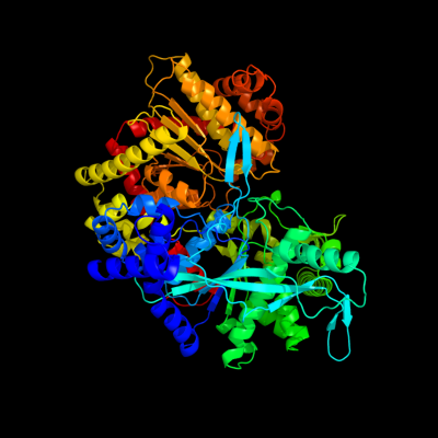
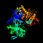
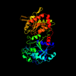
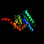
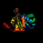
PDB header:transferase
Chain: B: PDB Molecule:glycogen phosphorylase, liver form;
PDBTitle: crystal structure of glycogen phosphorylase complexed with an2 anthranilimide based inhibitor gsk261
|
| 2 | d2gj4a1
|
|
 |
100.0 |
50 |
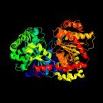
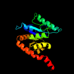
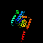
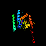
Fold:UDP-Glycosyltransferase/glycogen phosphorylase
Superfamily:UDP-Glycosyltransferase/glycogen phosphorylase
Family:Oligosaccharide phosphorylase |
| 3 | d2atia1
|
|
 |
100.0 |
51 |
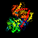
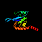
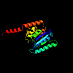
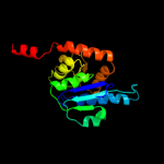
Fold:UDP-Glycosyltransferase/glycogen phosphorylase
Superfamily:UDP-Glycosyltransferase/glycogen phosphorylase
Family:Oligosaccharide phosphorylase |
| 4 | d1ygpa_
|
|
 |
100.0 |
47 |
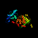
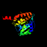
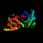
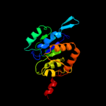
Fold:UDP-Glycosyltransferase/glycogen phosphorylase
Superfamily:UDP-Glycosyltransferase/glycogen phosphorylase
Family:Oligosaccharide phosphorylase |
| 5 | c2c4mA_
|
|
 |
100.0 |
42 |
PDB header:transferase
Chain: A: PDB Molecule:glycogen phosphorylase;
PDBTitle: starch phosphorylase: structural studies explain oxyanion-2 dependent kinetic stability and regulatory control.
|
| 6 | d1l5wa_
|
|
 |
100.0 |
46 |
Fold:UDP-Glycosyltransferase/glycogen phosphorylase
Superfamily:UDP-Glycosyltransferase/glycogen phosphorylase
Family:Oligosaccharide phosphorylase |
| 7 | c3nb0A_
|
|
 |
99.4 |
18 |
PDB header:transferase
Chain: A: PDB Molecule:glycogen [starch] synthase isoform 2;
PDBTitle: glucose-6-phosphate activated form of yeast glycogen synthase
|
| 8 | c3o3cD_
|
|
 |
99.3 |
20 |
PDB header:transferase
Chain: D: PDB Molecule:glycogen [starch] synthase isoform 2;
PDBTitle: glycogen synthase basal state udp complex
|
| 9 | d1rzua_
|
|
 |
98.7 |
16 |
Fold:UDP-Glycosyltransferase/glycogen phosphorylase
Superfamily:UDP-Glycosyltransferase/glycogen phosphorylase
Family:Glycosyl transferases group 1 |
| 10 | c2x6rA_
|
|
 |
98.6 |
19 |
PDB header:isomerase
Chain: A: PDB Molecule:trehalose-synthase tret;
PDBTitle: crystal structure of trehalose synthase tret from p.2 horikoshi produced by soaking in trehalose
|
| 11 | c2xmpB_
|
|
 |
98.3 |
17 |
PDB header:sugar binding protein
Chain: B: PDB Molecule:trehalose-synthase tret;
PDBTitle: crystal structure of trehalose synthase tret mutant e326a2 from p.horishiki in complex with udp
|
| 12 | c2qzsA_
|
|
 |
98.3 |
18 |
PDB header:transferase
Chain: A: PDB Molecule:glycogen synthase;
PDBTitle: crystal structure of wild-type e.coli gs in complex with adp2 and glucose(wtgsb)
|
| 13 | c2r60A_
|
|
 |
98.0 |
20 |
PDB header:transferase
Chain: A: PDB Molecule:glycosyl transferase, group 1;
PDBTitle: structure of apo sucrose phosphate synthase (sps) of2 halothermothrix orenii
|
| 14 | d1uqta_
|
|
 |
97.9 |
13 |
Fold:UDP-Glycosyltransferase/glycogen phosphorylase
Superfamily:UDP-Glycosyltransferase/glycogen phosphorylase
Family:Trehalose-6-phosphate synthase, OtsA |
| 15 | c1uquB_
|
|
 |
97.9 |
11 |
PDB header:synthase
Chain: B: PDB Molecule:alpha, alpha-trehalose-phosphate synthase;
PDBTitle: trehalose-6-phosphate from e. coli bound with udp-glucose.
|
| 16 | d2bisa1
|
|
 |
97.8 |
17 |
Fold:UDP-Glycosyltransferase/glycogen phosphorylase
Superfamily:UDP-Glycosyltransferase/glycogen phosphorylase
Family:Glycosyl transferases group 1 |
| 17 | c3oy2A_
|
|
 |
97.7 |
16 |
PDB header:viral protein,transferase
Chain: A: PDB Molecule:glycosyltransferase b736l;
PDBTitle: crystal structure of a putative glycosyltransferase from paramecium2 bursaria chlorella virus ny2a
|
| 18 | c3s29C_
|
|
 |
97.7 |
17 |
PDB header:transferase
Chain: C: PDB Molecule:sucrose synthase 1;
PDBTitle: the crystal structure of sucrose synthase-1 from arabidopsis thaliana2 and its functional implications.
|
| 19 | c3c4vB_
|
|
 |
97.5 |
18 |
PDB header:transferase
Chain: B: PDB Molecule:predicted glycosyltransferases;
PDBTitle: structure of the retaining glycosyltransferase msha:the2 first step in mycothiol biosynthesis. organism:3 corynebacterium glutamicum : complex with udp and 1l-ins-1-4 p.
|
| 20 | c3okaA_
|
|
 |
97.0 |
18 |
PDB header:transferase
Chain: A: PDB Molecule:gdp-mannose-dependent alpha-(1-6)-phosphatidylinositol
PDBTitle: crystal structure of corynebacterium glutamicum pimb' in complex with2 gdp-man (triclinic crystal form)
|
| 21 | d2bfwa1 |
|
not modelled |
96.8 |
16 |
Fold:UDP-Glycosyltransferase/glycogen phosphorylase
Superfamily:UDP-Glycosyltransferase/glycogen phosphorylase
Family:Glycosyl transferases group 1 |
| 22 | c2gejA_ |
|
not modelled |
96.4 |
13 |
PDB header:transferase
Chain: A: PDB Molecule:phosphatidylinositol mannosyltransferase (pima);
PDBTitle: crystal structure of phosphatidylinositol mannosyltransferase (pima)2 from mycobacterium smegmatis in complex with gdp-man
|
| 23 | c2jjmH_ |
|
not modelled |
96.4 |
19 |
PDB header:transferase
Chain: H: PDB Molecule:glycosyl transferase, group 1 family protein;
PDBTitle: crystal structure of a family gt4 glycosyltransferase from2 bacillus anthracis orf ba1558.
|
| 24 | d2f9fa1 |
|
not modelled |
96.0 |
15 |
Fold:UDP-Glycosyltransferase/glycogen phosphorylase
Superfamily:UDP-Glycosyltransferase/glycogen phosphorylase
Family:Glycosyl transferases group 1 |
| 25 | d2iw1a1 |
|
not modelled |
95.3 |
15 |
Fold:UDP-Glycosyltransferase/glycogen phosphorylase
Superfamily:UDP-Glycosyltransferase/glycogen phosphorylase
Family:Glycosyl transferases group 1 |
| 26 | c2x0dA_ |
|
not modelled |
93.4 |
11 |
PDB header:transferase
Chain: A: PDB Molecule:wsaf;
PDBTitle: apo structure of wsaf
|
| 27 | c2vsnB_ |
|
not modelled |
93.3 |
15 |
PDB header:transferase
Chain: B: PDB Molecule:xcogt;
PDBTitle: structure and topological arrangement of an o-glcnac2 transferase homolog: insight into molecular control of3 intracellular glycosylation
|
| 28 | c3ot5D_ |
|
not modelled |
93.2 |
12 |
PDB header:isomerase
Chain: D: PDB Molecule:udp-n-acetylglucosamine 2-epimerase;
PDBTitle: 2.2 angstrom resolution crystal structure of putative udp-n-2 acetylglucosamine 2-epimerase from listeria monocytogenes
|
| 29 | d1f6da_ |
|
not modelled |
90.5 |
17 |
Fold:UDP-Glycosyltransferase/glycogen phosphorylase
Superfamily:UDP-Glycosyltransferase/glycogen phosphorylase
Family:UDP-N-acetylglucosamine 2-epimerase |
| 30 | c3pe3D_ |
|
not modelled |
90.2 |
18 |
PDB header:transferase
Chain: D: PDB Molecule:udp-n-acetylglucosamine--peptide n-
PDBTitle: structure of human o-glcnac transferase and its complex with a peptide2 substrate
|
| 31 | c3dzcA_ |
|
not modelled |
84.0 |
11 |
PDB header:isomerase
Chain: A: PDB Molecule:udp-n-acetylglucosamine 2-epimerase;
PDBTitle: 2.35 angstrom resolution structure of wecb (vc0917), a udp-n-2 acetylglucosamine 2-epimerase from vibrio cholerae.
|
| 32 | c3rhzB_ |
|
not modelled |
79.6 |
13 |
PDB header:transferase
Chain: B: PDB Molecule:nucleotide sugar synthetase-like protein;
PDBTitle: structure and functional analysis of a new subfamily of2 glycosyltransferases required for glycosylation of serine-rich3 streptococcal adhesions
|
| 33 | c2iv3B_ |
|
not modelled |
62.4 |
20 |
PDB header:transferase
Chain: B: PDB Molecule:glycosyltransferase;
PDBTitle: crystal structure of avigt4, a glycosyltransferase involved2 in avilamycin a biosynthesis
|
| 34 | c3qhpB_ |
|
not modelled |
61.2 |
15 |
PDB header:transferase
Chain: B: PDB Molecule:type 1 capsular polysaccharide biosynthesis protein j
PDBTitle: crystal structure of the catalytic domain of cholesterol-alpha-2 glucosyltransferase from helicobacter pylori
|
| 35 | c1yz4A_ |
|
not modelled |
59.2 |
14 |
PDB header:hydrolase
Chain: A: PDB Molecule:dual specificity phosphatase-like 15 isoform a;
PDBTitle: crystal structure of dusp15
|
| 36 | c2g6zB_ |
|
not modelled |
47.5 |
22 |
PDB header:hydrolase
Chain: B: PDB Molecule:dual specificity protein phosphatase 5;
PDBTitle: crystal structure of human dusp5
|
| 37 | d1w36c1 |
|
not modelled |
45.6 |
18 |
Fold:P-loop containing nucleoside triphosphate hydrolases
Superfamily:P-loop containing nucleoside triphosphate hydrolases
Family:Tandem AAA-ATPase domain |
| 38 | c2z8tX_ |
|
not modelled |
41.3 |
15 |
PDB header:hydrolase
Chain: X: PDB Molecule:protein-glutaminase;
PDBTitle: crystal structure of protein-glutaminase of c.proteolyticum2 strain 9670
|
| 39 | d1m3ga_ |
|
not modelled |
38.4 |
16 |
Fold:(Phosphotyrosine protein) phosphatases II
Superfamily:(Phosphotyrosine protein) phosphatases II
Family:Dual specificity phosphatase-like |
| 40 | c1wrmA_ |
|
not modelled |
37.1 |
21 |
PDB header:hydrolase
Chain: A: PDB Molecule:dual specificity phosphatase 22;
PDBTitle: crystal structure of jsp-1
|
| 41 | c2e0tA_ |
|
not modelled |
37.0 |
24 |
PDB header:hydrolase
Chain: A: PDB Molecule:dual specificity phosphatase 26;
PDBTitle: crystal structure of catalytic domain of dual specificity phosphatase2 26, ms0830 from homo sapiens
|
| 42 | d1x99a_ |
|
not modelled |
33.1 |
44 |
Fold:Cytolysin/lectin
Superfamily:Cytolysin/lectin
Family:Fungal fruit body lectin |
| 43 | d1y2ta_ |
|
not modelled |
32.6 |
44 |
Fold:Cytolysin/lectin
Superfamily:Cytolysin/lectin
Family:Fungal fruit body lectin |
| 44 | c2oudA_ |
|
not modelled |
31.2 |
20 |
PDB header:hydrolase
Chain: A: PDB Molecule:dual specificity protein phosphatase 10;
PDBTitle: crystal structure of the catalytic domain of human mkp5
|
| 45 | d1vhra_ |
|
not modelled |
30.4 |
19 |
Fold:(Phosphotyrosine protein) phosphatases II
Superfamily:(Phosphotyrosine protein) phosphatases II
Family:Dual specificity phosphatase-like |
| 46 | c3rgqA_ |
|
not modelled |
29.2 |
13 |
PDB header:hydrolase
Chain: A: PDB Molecule:protein-tyrosine phosphatase mitochondrial 1;
PDBTitle: crystal structure of ptpmt1 in complex with pi(5)p
|
| 47 | c2npiB_ |
|
not modelled |
27.9 |
14 |
PDB header:transcription
Chain: B: PDB Molecule:protein clp1;
PDBTitle: clp1-atp-pcf11 complex
|
| 48 | c2ofeA_ |
|
not modelled |
27.8 |
44 |
PDB header:sugar binding protein
Chain: A: PDB Molecule:sclerotium rolfsii lectin;
PDBTitle: the crystal structure of sclerotium rolfsii lectin in complex with n-2 acetyl-d-glucosamine
|
| 49 | c2wgpA_ |
|
not modelled |
25.8 |
16 |
PDB header:hydrolase
Chain: A: PDB Molecule:dual specificity protein phosphatase 14;
PDBTitle: crystal structure of human dual specificity phosphatase 14
|
| 50 | c2hcmA_ |
|
not modelled |
25.7 |
13 |
PDB header:hydrolase
Chain: A: PDB Molecule:dual specificity protein phosphatase;
PDBTitle: crystal structure of mouse putative dual specificity phosphatase2 complexed with zinc tungstate, new york structural genomics3 consortium
|
| 51 | d3etja2 |
|
not modelled |
24.7 |
38 |
Fold:PreATP-grasp domain
Superfamily:PreATP-grasp domain
Family:BC N-terminal domain-like |
| 52 | c3se4B_ |
|
not modelled |
24.5 |
25 |
PDB header:immune system receptor
Chain: B: PDB Molecule:interferon omega-1;
PDBTitle: human ifnw-ifnar ternary complex
|
| 53 | c2y96A_ |
|
not modelled |
22.8 |
22 |
PDB header:hydrolase
Chain: A: PDB Molecule:dual specificity phosphatase dupd1;
PDBTitle: structure of human dual-specificity phosphatase 27
|
| 54 | c1yhuA_ |
|
not modelled |
22.6 |
16 |
PDB header:oxygen storage/transport
Chain: A: PDB Molecule:hemoglobin a1 chain;
PDBTitle: crystal structure of riftia pachyptila c1 hemoglobin reveals novel2 assembly of 24 subunits.
|
| 55 | c2imgA_ |
|
not modelled |
22.5 |
11 |
PDB header:hydrolase
Chain: A: PDB Molecule:dual specificity protein phosphatase 23;
PDBTitle: crystal structure of dual specificity protein phosphatase2 23 from homo sapiens in complex with ligand malate ion
|
| 56 | c3emuA_ |
|
not modelled |
22.0 |
18 |
PDB header:hydrolase
Chain: A: PDB Molecule:leucine rich repeat and phosphatase domain
PDBTitle: crystal structure of a leucine rich repeat and phosphatase2 domain containing protein from entamoeba histolytica
|
| 57 | c2j17A_ |
|
not modelled |
21.1 |
24 |
PDB header:hydrolase
Chain: A: PDB Molecule:tyrosine-protein phosphatase yil113w;
PDBTitle: ptyr bound form of sdp-1
|
| 58 | c3prjB_ |
|
not modelled |
21.0 |
22 |
PDB header:oxidoreductase
Chain: B: PDB Molecule:udp-glucose 6-dehydrogenase;
PDBTitle: role of packing defects in the evolution of allostery and induced fit2 in human udp-glucose dehydrogenase.
|
| 59 | d1m1nb_ |
|
not modelled |
20.9 |
19 |
Fold:Chelatase-like
Superfamily:"Helical backbone" metal receptor
Family:Nitrogenase iron-molybdenum protein |
| 60 | c2nt2C_ |
|
not modelled |
20.8 |
18 |
PDB header:hydrolase
Chain: C: PDB Molecule:protein phosphatase slingshot homolog 2;
PDBTitle: crystal structure of slingshot phosphatase 2
|
| 61 | c3piwA_ |
|
not modelled |
19.5 |
24 |
PDB header:cytokine
Chain: A: PDB Molecule:type i interferon 2;
PDBTitle: zebrafish interferon 2
|
| 62 | d1u3da1 |
|
not modelled |
19.4 |
26 |
Fold:Cryptochrome/photolyase FAD-binding domain
Superfamily:Cryptochrome/photolyase FAD-binding domain
Family:Cryptochrome/photolyase FAD-binding domain |
| 63 | d1mv8a1 |
|
not modelled |
19.4 |
17 |
Fold:6-phosphogluconate dehydrogenase C-terminal domain-like
Superfamily:6-phosphogluconate dehydrogenase C-terminal domain-like
Family:UDP-glucose/GDP-mannose dehydrogenase dimerisation domain |
| 64 | d1dlja1 |
|
not modelled |
19.1 |
28 |
Fold:6-phosphogluconate dehydrogenase C-terminal domain-like
Superfamily:6-phosphogluconate dehydrogenase C-terminal domain-like
Family:UDP-glucose/GDP-mannose dehydrogenase dimerisation domain |
| 65 | c2gwoC_ |
|
not modelled |
19.0 |
13 |
PDB header:hydrolase
Chain: C: PDB Molecule:dual specificity protein phosphatase 13;
PDBTitle: crystal structure of tmdp
|
| 66 | c3bdkB_ |
|
not modelled |
18.8 |
17 |
PDB header:lyase
Chain: B: PDB Molecule:d-mannonate dehydratase;
PDBTitle: crystal structure of streptococcus suis mannonate2 dehydratase complexed with substrate analogue
|
| 67 | d1v3aa_ |
|
not modelled |
18.8 |
27 |
Fold:(Phosphotyrosine protein) phosphatases II
Superfamily:(Phosphotyrosine protein) phosphatases II
Family:Dual specificity phosphatase-like |
| 68 | c3pn1A_ |
|
not modelled |
18.5 |
19 |
PDB header:ligase/ligase inhibitor
Chain: A: PDB Molecule:dna ligase;
PDBTitle: novel bacterial nad+-dependent dna ligase inhibitors with broad2 spectrum potency and antibacterial efficacy in vivo
|
| 69 | d1v9pa3 |
|
not modelled |
18.0 |
13 |
Fold:ATP-grasp
Superfamily:DNA ligase/mRNA capping enzyme, catalytic domain
Family:Adenylation domain of NAD+-dependent DNA ligase |
| 70 | c2a6aB_ |
|
not modelled |
17.7 |
16 |
PDB header:hydrolase
Chain: B: PDB Molecule:hypothetical protein tm0874;
PDBTitle: crystal structure of glycoprotein endopeptidase (tm0874) from2 thermotoga maritima at 2.50 a resolution
|
| 71 | c2r0bA_ |
|
not modelled |
17.6 |
22 |
PDB header:hydrolase
Chain: A: PDB Molecule:serine/threonine/tyrosine-interacting protein;
PDBTitle: crystal structure of human tyrosine phosphatase-like2 serine/threonine/tyrosine-interacting protein
|
| 72 | d2f8la1 |
|
not modelled |
17.4 |
20 |
Fold:S-adenosyl-L-methionine-dependent methyltransferases
Superfamily:S-adenosyl-L-methionine-dependent methyltransferases
Family:N-6 DNA Methylase-like |
| 73 | d1o58a_ |
|
not modelled |
17.1 |
16 |
Fold:Tryptophan synthase beta subunit-like PLP-dependent enzymes
Superfamily:Tryptophan synthase beta subunit-like PLP-dependent enzymes
Family:Tryptophan synthase beta subunit-like PLP-dependent enzymes |
| 74 | d1ta8a_ |
|
not modelled |
16.9 |
26 |
Fold:ATP-grasp
Superfamily:DNA ligase/mRNA capping enzyme, catalytic domain
Family:Adenylation domain of NAD+-dependent DNA ligase |
| 75 | d3buxb2 |
|
not modelled |
16.6 |
20 |
Fold:N-cbl like
Superfamily:N-terminal domain of cbl (N-cbl)
Family:N-terminal domain of cbl (N-cbl) |
| 76 | d1b5la_ |
|
not modelled |
15.4 |
24 |
Fold:4-helical cytokines
Superfamily:4-helical cytokines
Family:Interferons/interleukin-10 (IL-10) |
| 77 | d1v4va_ |
|
not modelled |
15.2 |
14 |
Fold:UDP-Glycosyltransferase/glycogen phosphorylase
Superfamily:UDP-Glycosyltransferase/glycogen phosphorylase
Family:UDP-N-acetylglucosamine 2-epimerase |
| 78 | d1r30a_ |
|
not modelled |
15.0 |
14 |
Fold:TIM beta/alpha-barrel
Superfamily:Radical SAM enzymes
Family:Biotin synthase |
| 79 | c1r30A_ |
|
not modelled |
15.0 |
14 |
PDB header:transferase
Chain: A: PDB Molecule:biotin synthase;
PDBTitle: the crystal structure of biotin synthase, an s-2 adenosylmethionine-dependent radical enzyme
|
| 80 | c3bacA_ |
|
not modelled |
14.7 |
19 |
PDB header:ligase
Chain: A: PDB Molecule:dna ligase;
PDBTitle: structural basis for the inhibition of bacterial nad+2 dependent dna ligase
|
| 81 | c2y0dB_ |
|
not modelled |
14.5 |
13 |
PDB header:oxidoreductase
Chain: B: PDB Molecule:udp-glucose dehydrogenase;
PDBTitle: bcec mutation y10k
|
| 82 | c2k29A_ |
|
not modelled |
14.2 |
27 |
PDB header:transcription
Chain: A: PDB Molecule:antitoxin relb;
PDBTitle: structure of the dbd domain of e. coli antitoxin relb
|
| 83 | c1eysC_ |
|
not modelled |
13.8 |
18 |
PDB header:electron transport
Chain: C: PDB Molecule:photosynthetic reaction center;
PDBTitle: crystal structure of photosynthetic reaction center from a2 thermophilic bacterium, thermochromatium tepidum
|
| 84 | d1eysc_ |
|
not modelled |
13.8 |
18 |
Fold:Multiheme cytochromes
Superfamily:Multiheme cytochromes
Family:Photosynthetic reaction centre (cytochrome subunit) |
| 85 | d1mkpa_ |
|
not modelled |
13.5 |
13 |
Fold:(Phosphotyrosine protein) phosphatases II
Superfamily:(Phosphotyrosine protein) phosphatases II
Family:Dual specificity phosphatase-like |
| 86 | c3f4yF_ |
|
not modelled |
13.4 |
64 |
PDB header:viral protein
Chain: F: PDB Molecule:mutant peptide derived from hiv gp41 chr domain;
PDBTitle: hiv gp41 six-helix bundle containing a mutant chr alpha-2 peptide sequence
|
| 87 | c2o3jC_ |
|
not modelled |
13.3 |
19 |
PDB header:oxidoreductase
Chain: C: PDB Molecule:udp-glucose 6-dehydrogenase;
PDBTitle: structure of caenorhabditis elegans udp-glucose dehydrogenase
|
| 88 | d1ohea2 |
|
not modelled |
12.9 |
13 |
Fold:(Phosphotyrosine protein) phosphatases II
Superfamily:(Phosphotyrosine protein) phosphatases II
Family:Dual specificity phosphatase-like |
| 89 | c2esbA_ |
|
not modelled |
12.7 |
16 |
PDB header:hydrolase
Chain: A: PDB Molecule:dual specificity protein phosphatase 18;
PDBTitle: crystal structure of human dusp18
|
| 90 | c3rz2B_ |
|
not modelled |
12.6 |
28 |
PDB header:hydrolase
Chain: B: PDB Molecule:protein tyrosine phosphatase type iva 1;
PDBTitle: crystal of prl-1 complexed with peptide
|
| 91 | d1au1a_ |
|
not modelled |
12.6 |
10 |
Fold:4-helical cytokines
Superfamily:4-helical cytokines
Family:Interferons/interleukin-10 (IL-10) |
| 92 | d1qkia1 |
|
not modelled |
12.6 |
15 |
Fold:NAD(P)-binding Rossmann-fold domains
Superfamily:NAD(P)-binding Rossmann-fold domains
Family:Glyceraldehyde-3-phosphate dehydrogenase-like, N-terminal domain |
| 93 | d1rh2a_ |
|
not modelled |
12.6 |
29 |
Fold:4-helical cytokines
Superfamily:4-helical cytokines
Family:Interferons/interleukin-10 (IL-10) |
| 94 | c3r6mD_ |
|
not modelled |
12.5 |
25 |
PDB header:hydrolase
Chain: D: PDB Molecule:yeaz, resuscitation promoting factor;
PDBTitle: crystal structure of vibrio parahaemolyticus yeaz
|
| 95 | c3cvyA_ |
|
not modelled |
12.3 |
23 |
PDB header:lyase/dna
Chain: A: PDB Molecule:re11660p;
PDBTitle: drosophila melanogaster (6-4) photolyase bound to repaired2 ds dna
|
| 96 | d1di1a_ |
|
not modelled |
12.3 |
13 |
Fold:Terpenoid synthases
Superfamily:Terpenoid synthases
Family:Aristolochene/pentalenene synthase |
| 97 | c1fbvA_ |
|
not modelled |
12.2 |
20 |
PDB header:ligase
Chain: A: PDB Molecule:signal transduction protein cbl;
PDBTitle: structure of a cbl-ubch7 complex: ring domain function in2 ubiquitin-protein ligases
|
| 98 | d1i9sa_ |
|
not modelled |
12.1 |
14 |
Fold:(Phosphotyrosine protein) phosphatases II
Superfamily:(Phosphotyrosine protein) phosphatases II
Family:Dual specificity phosphatase-like |
| 99 | d1wu3i_ |
|
not modelled |
12.1 |
10 |
Fold:4-helical cytokines
Superfamily:4-helical cytokines
Family:Interferons/interleukin-10 (IL-10) |